
Abstract
There have been significant advancements in the understanding of maternal–fetal disease over the past century. This narrative review summarizes the landmark studies that have advanced the understanding of thyroid pathophysiology and thyroid disease during preconception, pregnancy, and postpartum, written to commemorate the 100th year anniversary of the founding of the American Thyroid Association.
Introduction
The landmark studies in maternal–fetal thyroid disease reflect pivotal early experiments, noteworthy public health accomplishments, and the international collaboration of investigators studying the key questions in this field. This body of knowledge has furthered the understanding of thyroid disease management in pregnant women and their offspring. This paper is not intended to be an exhaustive review of the seminal articles in maternal–fetal thyroid disease, but highlights the stories of select interesting historical studies in the field (Fig. 1), focusing primarily on human studies published in the past several decades.
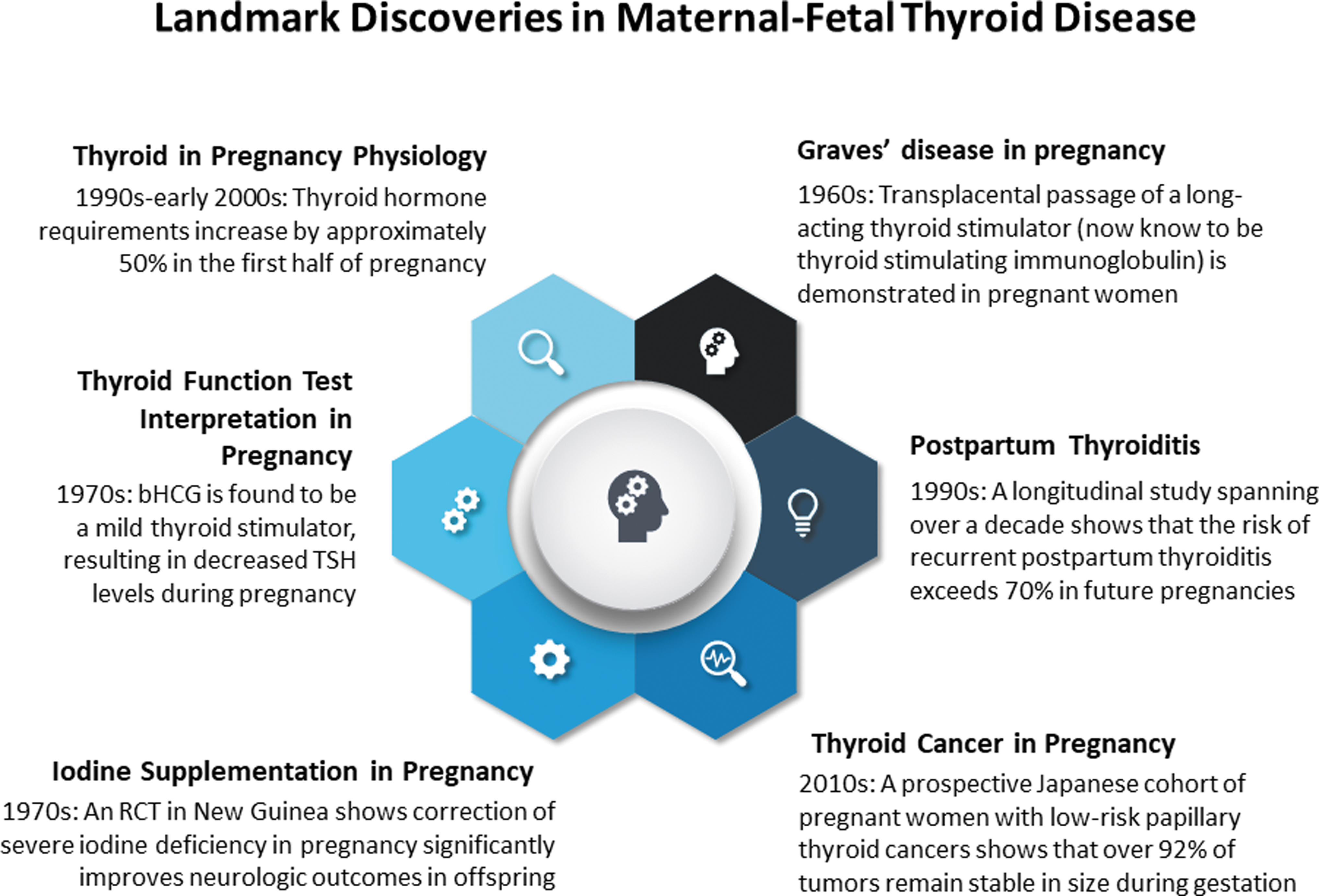
Highlighted discoveries in maternal–fetal thyroid disease over the past century.
Thyroid Physiology During Pregnancy
Changes in thyroid protein binding
The vast key literature in thyroid and pregnancy over the past century illustrates the advancements that have been made to better understand the changes in thyroid physiology unique to this life stage. Although thyroxine (T4) had been isolated in 1915, early serum T4 measurements were based on circulating protein-bound iodine (PBI) levels that had emerged in the early 1900s as a diagnostic test of estimating thyroid function. Heineman et al had observed in the 1940s that serum PBI levels are increased during pregnancy, 1 heralding the observation that gestation influences thyroid status.
Shortly after the discovery of thyroid binding globulin (TBG) in 1952, Dowling et al reported that circulating TBG-bound T4 levels were higher in pregnant women than in nonpregnant women, 2 which was hypothesized to be due to an increase of at least one of the alpha globulin binding proteins induced by pregnancy. Robbins, Nelson, and others in the 1950s showed that thyroid binding increases during pregnancy. 3 The pregnancy-associated increase in TBG was later shown to be due to estrogen, which prolongs the circulating half-life of TBG. 4 Glinoer et al in the 1990s characterized the multitude of changes in thyroid economy that occur during pregnancy, including the increased T4-bound TBG levels seen in early gestation. 5
Interpretation of serum thyroid function tests
Pregnancy is associated with unique alterations of thyroid physiology that impact the interpretation of serum thyroid function tests. As biochemical hyperthyroidism had been observed in patients with choriocarcinomas and hydatidiform moles, Hershman et al hypothesized that the normal placenta likely contained a thyroid stimulator. 6 The first clinical reports arose from studies in the 1960s showing that women with human chorionic gonadotropin (hCG)-secreting placental tumors (e.g., hydatidiform moles and choriocarcinomas) were also hyperthyroid. 7
Later work during the 1970s to early 1990s by Braunstein and Hershman, 8 Harada et al, 9 Glinoer et al 5 using more sensitive thyrotropin (TSH) assays provided compelling evidence that even in normal pregnancy, hCG directly stimulated T4 production from the thyroid, resulting in gestational transient thyrotoxicosis in some. These early studies laid the groundwork for the current American Thyroid Association (ATA) recommendations supporting a lower physiological reference limit or population-based trimester-specific reference ranges for serum TSH, primarily in the first trimester of pregnancy. 10
Increased thyroid hormone requirements
In addition to the higher TBG-bound thyroid hormone levels induced by the rise of estrogen, other advances have further informed the need for increased thyroid hormone during pregnancy. Work by Roti et al 11 and Galton et al 12 showed that the placenta is abundant in the type III deiodinase that is responsible for converting T4 and triiodothyronine (T3) to inactive metabolites, 13 leading to faster degradation of the maternal thyroid hormone supply that is available to the fetus. Pregnancy is also associated with renal hyperfiltration and increased iodine clearance, 14 which would be exacerbated in regions of endemic iodine deficiency, 15 resulting in higher iodine needs to sustain normal thyroid hormone production for the developing fetus.
Among women with autoimmune thyroid disease, Glinoer et al showed that serum TSH levels are higher starting in the first few weeks of pregnancy and continue to rise more than in euthyroid controls throughout the entire course of gestation. 16 Seminal studies by Mandel et al in 199017 and Alexander et al in 2004 quantified the increased thyroid hormone requirements during pregnancy, 18 key practice-changing concepts that were championed by Larsen. 19 Thus, in order to meet the demands for a greater total body T4 pool during pregnancy, current guidelines by the ATA advise an ∼30% higher dose of levothyroxine (LT4) in women treated for hypothyroidism. 10,18
Iodine Nutrition in Preconception, Pregnancy, and Postpartum
Iodine deficiency during pregnancy
The possibility that severe iodine deficiency during pregnancy may be associated with adverse obstetric outcomes was observed in the early 1900s. Administration of iodide tincture to colts, calves, and pigs for 2 months before delivery prevented the nearly 90% postpartum mortality rate seen in animals of iodine-deficient regions. 20 In 1939, Kemp reported the anecdotal experience of several groups who had observed the benefit of prophylactically administering iodine and iron in pregnant women to decrease rates of stillbirth. 20
Stronger scientific evidence of this association was not available until 1971, when the seminal study by Pharoah et al was published. 21 This randomized controlled trial in New Guinea, including women of childbearing age and/or pregnant women, showed a significant decrease of endemic severe iodine deficiency and associated mortality among the offspring born to families who were treated with iodized oil, compared with placebo. 21 The benefits of early iodine supplementation during pregnancy were later demonstrated by Cao et al in a pivotal cohort study performed in a severely iodine-deficient remote province of China in 1994, in which offspring of women administered iodine supplementation before the third trimester had significantly lower risk of neurological abnormalities, compared with those born to women treated during later pregnancy. 22
Increased iodine requirements during pregnancy and lactation
A large collective of work has shown that because of increased iodine and thyroid hormone needs for the developing fetus, as well as increased maternal renal iodine excretion, 23 dietary iodine needs are higher during pregnancy than they are for nonpregnant adults. In the 1970s, it was presumed that the iodine content in prenatal multivitamins was adequate for the increased iodide requirements during pregnancy. 24 However, data over the past decade have since shown that only ∼50–60% of prenatal vitamin formulations in the United States contain any iodine, let alone optimal iodine content for gestation. 25,26
Risks of iodine excess during pregnancy
In 1971, Senior and Chernoff described an infant with a goiter born to a mother who had received large doses of iodide throughout pregnancy. 27 Although Braverman and Ingbar had shown in the early 1960s that the normal adult thyroid in most cases is able to adjust to iodine excess, 28 the mechanism for this adaptation to the Wolff-Chaikoff effect 29 was not known until 1999, when Eng et al showed that iodine excess is associated with a temporary downregulation of the sodium iodide/symporter. 30 However, as the fetal thyroid gland is relatively immature, it cannot easily escape from the acute Wolff-Chaikoff effect, 31 thus posing the risk of fetal hypothyroidism upon an acute iodine load.
Fetal Development and Thyroid Disease
Role of maternal iodine and thyroid hormone during pregnancy
Before the recognition that adequate iodine status plays a crucial role in early growth and neurodevelopment, the diagnosis of neonatal severe iodine deficiency (termed cretinism in older texts) was challenging, with difficulties with feeding or gaining weight as the most common manifestations. Pioneering work by Stanbury et al in iodine metabolism during the 1950s 32 had established the important basis for their work during the 1950s and 1960s in understanding of the effects of endemic iodine deficiency.
A 1958 study of 49 children with cretinism reported that severe iodine deficiency was diagnosed at a mean age of 12 months, 33 with untreated children showing severe growth impairment, delayed closure of the fontanelles, periorbital edema, large protruding tongue, and cognitive impairments. The diagnosis of severe iodine deficiency during these early days was able to be made only crudely by the measurement of low serum PBI concentrations. 24
Further studies during the 1960s were able to show that colloid formation, iodide concentration, and synthesis of thyroglobulin and T4 in the human fetal thyroid gland begin at 11 weeks gestation. 34 In addition, the fetal thyroid gland has an ∼20–50 greater affinity for iodine than the maternal thyroid gland, as well as significantly increased thyroidal iodine turnover. 35 In 1969, Gitlin and Biasucci showed that fetal TSH appears as early as 14 weeks gestation, 36 but Greenberg et al shortly thereafter provided evidence that TSH is detectable and responsive to free T4 (fT4) even at 11 weeks gestation. 37
Although it was understood that iodine, T3, and T4 all freely cross the placenta (with T3 appearing to cross more easily than T4), 38 it remained unclear even in the 1970s whether the fetus requires its own thyroid hormone supply during pregnancy or whether maternal thyroid hormone would be sufficient. 24 During the late 1980s, key animal experiments by Morreale de Escobar et al provided evidence of the maternal transfer of T4 through the placenta to the fetal rat. 39
This was quantified by Vulsma et al, who in 1989 studied the offspring of euthyroid women with a history of underlying congenital hypothyroidism. 40 In these findings, the infants who were also affected with congenital hypothyroidism and documented to be athyreotic had ∼20–50% of the normal levels of circulating thyroid hormones at birth, which also fell in accordance with the predicted serum half-life of T4, proving that transplacental crossing from the mother was their only possible source. 40
Thyroid function newborn screening
Important studies in newborn thyroid function screening have been a significant public health accomplishment in the past century. Newborn screening programs were implemented in Switzerland in the late 1960s and rapidly became common in most developed countries during the 1970s. As it was also recognized that there is a marked increase, then fall, in neonatal serum TSH levels immediately after delivery, 41 continued refinement of strategies informed by Walfish 42 and others since has led to the adoption of regional screening algorithms.
The key report in 1979 by Fisher et al described the five oldest screening programs in North America, including a study by Dussault and colleagues in Quebec, 43 which in total had screened over one million newborns to yield an incidence rate of 1 in 3684 live births. 44 Recognition that early thyroid hormone replacement improves the cognitive and developmental impairments associated with untreated disease has virtually eliminated the adverse outcomes of congenital hypothyroidism. 45
Hypothyroidism and Hypothyroxinemia During Pregnancy
Role of thyroid hormone in early development
The association of postnatal thyroid dysfunction and neurodevelopmental disorders has long been recognized; observations in the early 1800s noted that cretins (a pejorative, outdated historic medical term used to describe individuals with stunted growth and other physical deformities) appeared to also have substantial neurocognitive impairments. 46 Seminal studies by DeLange and colleagues were pivotal in reporting the association between maternal and fetal thyroid status, particularly in areas of endemic iodine deficiency. 47,48
Later experiments during the 1960s showed that animals that had undergone thyroidectomy at birth have marked morphological changes in the central nervous system, 49 as well as behavioral abnormalities; the defects appeared to be partially or wholly reversible if thyroid hormone was started shortly after birth.
Subsequent studies have focused on the influence of maternal and fetal thyroid status at earlier timepoints (i.e., prenatally) on neurodevelopment. Following experiments initially in rat, then later mouse, models, human data in the 1990s and early 2000s confirmed thyroid hormone concentrations in the fetal cortex 50 and in cord blood. 51 However, the extent to which fetal brain development is reliant on adequate thyroid hormone was still relatively uncertain even into the very late 1990s. 52
Placental passage of thyroid hormone
For many years, the accepted thinking was that the fetal hypothalamic–pituitary thyroid development is independent of maternal thyroidal influence, even as recently as the early 1980s. 53 However, later seminal study by Obregon et al in 1984 reported that T4 and T3 were able to be measured by radioimmunoassay in rat embryos at 10–18 days gestation, 54 earlier than when the fetal rat thyroid begins to function around day 18, thus providing strong evidence that maternal thyroid hormone through placental passage is critical to fetal thyroid hormone supply. Much study has been done since to demonstrate that normal levels of thyroid hormone are essential for neuronal migration, myelination, and other structural changes of the fetal brain. 55
Thyroid hormone replacement in maternal hypothyroidism
Several studies have investigated the associations between untreated maternal hypothyroidism and risks of adverse pregnancy complications. A 1993 study by Mestman et al showed a higher rate of gestational hypertension in hypothyroid women than in the general population. 15 Pop et al published several studies, the earliest in 1999, 56 on the association between maternal hypothyroxinemia during pregnancy and impaired infant development. In 2005, Casey et al described a 3-fold increased risk of placental abruption and nearly 2-fold risk of premature delivery in women with subclinical hypothyroidism when assessed at 20 weeks gestation. 57
In one of the landmark studies in this field, Haddow et al in 1999 showed in a retrospective cohort series that untreated maternal hypothyroidism (ascertained at a mean gestational age of 17 ± 1 [standard deviation] weeks) was associated with decreased intelligent quotient (IQ) scores in offspring at age 7–9 years. 58 However, the role of maternal subclinical hypothyroidism (which may be associated with maternal thyroid antibody positivity) on these outcomes is less clear.
Higher TBG levels in normal pregnancy indicate a need for increased thyroid hormone production, but an increase in LT4 dose requirements in pregnant hypothyroid women was not observed in early studies. This may have been due to the generally higher LT4 doses used before the availability of a sensitive TSH assay to mask this need. In 1990, Mandel et al showed that 9 of 12 hypothyroid pregnant women required an increased dose of LT4 (by an average of 50%), compared with their prepregnancy dose, and returned to their prepregnancy requirement in the postpartum period. 17
A subsequent study by Alexander et al showed that this increased dose requirement occurred primarily in the first half of pregnancy and plateaued at gestational week 16. 18 A more recent study by Korevaar and colleagues has demonstrated that both high and low maternal fT4 concentrations are associated with lower childhood IQ though, suggesting that overcorrection of maternal hypothyroidism may in itself also have adverse effects. 59
Braverman et al published in 1970 their seminal article reporting that the primary source of circulating T3 in humans was the peripheral conversion of T4 to T3, leading to the recommendation of LT4 therapy for hypothyroidism, 60 including during pregnancy. It was, however, already recognized that T3 and T4 have markedly different affinities to thyroid binding proteins, suggesting that T3 may more easily cross the placenta than T4. 38
Dumitrescu et al in the early 2000s also showed that T3 and T4 transport across the blood–brain barrier occurs through the active transporter protein, monocarboxylate transporter 8. 61 Work in the past two decades has also shown that T4, but not T3, can additionally cross into the fetal brain through organic anion transporting polypeptide 1C1 (Oatp1c1), 62 but is poorly expressed in the human fetus. 63 Morreale de Escobar et al demonstrated that maternal T3 is unable to reach the fetal brain and alleviate fetal hypothyroidism. 64 Thus, any maternal use of T3-containing regimens, including desiccated thyroid extract or synthetic T3, has the potential to reduce thyroid hormone action fetal brain, but direct data are not available. 10
Hyperthyroidism During Pregnancy
Graves' disease during pregnancy
Although a case of neonatal hyperthyroidism born to a pregnant woman with Graves' disease had been described in 1912, 65 the role of maternal TSH receptor antibodies was not understood until several decades later. During the 1950s, Graves' disease was hypothesized to result from an undefined long-acting thyroid stimulator first described in 1956 by Adams and Purves. 66 The initial reports of transplacental passage of a maternal thyroid stimulating antibody were from the early 1960s 67 and supported an autoimmune basis of fetal Graves' disease. A retrospective cohort study of 18 women by Dirmikis and Munro in 1975 suggested that there must be a likely threshold of elevated maternal thyroid stimulating immunoglobulin (TSI) titers during pregnancy to pose a risk to the fetus. 68
A 1983 study of 20 women showed among those who had infants with neonatal Graves' disease, all had sera containing antibodies able to induce a >500% tissue cAMP response to TSH stimulation, 69 forming the basis for recommendations advising maternal serum TSI titers be measured during midpregnancy, to assess fetal/neonatal hyperthyroidism risk. 10
Treatment of hyperthyroidism during pregnancy
In the 1970s, it remained relatively unclear if untreated maternal thyrotoxicosis during pregnancy was associated with adverse obstetric outcomes and/or increased fetal mortality. 24 However, a landmark article by Anselmo et al in 2004 provided evidence of a direct adverse effect of high maternal thyroid hormone levels on the fetus. 70 Offspring born to women in an Azorean family with thyroid hormone resistance (i.e., offspring were unaffected) were observed to have low birthweights and suppressed TSH levels, resulting from their exposure to high maternal thyroid hormone concentrations in utero. 70
Treatment during the 1970s was primarily thioamides, titrated to serum PBI values. In 1986, a landmark study by Momotani et al demonstrated that targeting minimally elevated or high-normal maternal serum fT4 levels with antithyroid drugs is the most appropriate to maintain euthyroid status in the fetus. 71 Later important study by Andersen et al in the early 2010s showed the risks of congenital anomalies with antithyroid drug use and steered the field toward minimal use of these medications, along with consideration of their different side effect profiles, during gestation. 72
In the postpartum period, there were isolated reports of cognitive impairments and goiters in children secondary to maternal propylthiouracil use during lactation. As such, mothers receiving these drugs were initially advised to not breastfeed their infants, but Low et al in 1979 showed that propylthiouracil has a breast milk–plasma ratio of only 0.1, while that for methimazole is closer to 1.0, 73 thereby providing evidence that the breast milk content of these medications is extremely low and their use during lactation is overall safe. 10
If surgical treatment of hyperthyroidism was advised, it was understood that the thyroid surgery should be delayed until after the first trimester. It is interesting that if a total thyroidectomy was performed during pregnancy, usual postoperative thyroid hormone replacement in the 1970s was to begin three grains of DTE or its equivalent daily for the remainder of gestation, 24 advice that is no longer espoused due to the difficulties of T3 crossing the fetal blood–brain barrier. 39
Laboratory Thyroid Antibody and Thyroid Hormone Screening in Preconception and Pregnancy
The prevalence of hypothyroidism during pregnancy ranges from 0.5% (overt hypothyroidism) to 3.47% (subclinical hypothyroidism). 10,74 Several observational studies have estimated the prevalence of serum anti-TPO or anti-Tg thyroid autoantibodies (2–17% in unselected pregnant women, depending on ethnicity). 10 The consideration of serum thyroid laboratory screening during preconception or pregnancy arose from the aforementioned study, as based on potential deleterious obstetric and neonatal effects among women with hypothyroidism or hypothyroxinemia.
Other studies have also demonstrated the obstetric risks of thyroid autoimmunity per se, even in the setting of biochemical euthyroidism. A seminal study by Stagnaro-Green et al found positive serum thyroid antibodies in 19.6% of a screened first-trimester cohort and was the first to report an association between serum anti-TPO antibody positivity in pregnant women and miscarriage, 75 while more recent pooled pregnancy data by Korevaar et al in the Consortium on Thyroid and Pregnancy show positive associations between subclinical hypothyroidism, isolated hypothyroxinemia, TPO antibody positivity, and risk of preterm birth. 76
Some countries, including Spain, China, and Poland, employ universal thyroid screening during pregnancy, while the United States has adopted a more restricted case-finding approach aimed to detect women at highest risk. 10 Dosiou et al have proposed that universal screening for autoimmune thyroid dysfunction in first-trimester pregnancies is cost-effective, compared with no screening or even screening of only high-risk women. 77
The potential risks versus benefits of thyroid screening before and during pregnancy require further study, particularly regarding maternal subclinical hypothyroidism if found, 78 but a small number of randomized clinical trials in the past ∼15 years have advanced knowledge on this topic. A 2010 Italian study by Negro et al reported no differences in adverse obstetrical or neonatal outcomes in 4562 first-trimester pregnant women, whether or not they underwent universal measurement of serum TSH, FT4, and TPO Ab levels (and treated with LT4 if a TSH >2.5 with positive TPO Ab was found, or antithyroidal medication if hyperthyroid was found). 79
Similarly, the Controlled Antenatal Thyroid Screening study and a U.S. National Institutes of Health study showed no benefits to systematic second-trimester screening and correction of maternal subclinical hypothyroidism or hypothyroxinemia, if discovered, on child cognition at age 3–5 years. 80,81 In contrast, another study in China by Ma et al in 2016 had reported the beneficial association of thyroid function screening (and initiation of LT4 if subclinical hypothyroidism was found) and decreased miscarriage and macrosomia risks, compared with no screening. 82 Subsequent robust randomized clinical trials have not shown a benefit of LT4 use in euthyroid, thyroid antibody pregnant women to increase live birth rates. 83,84
Postpartum Thyroid Disease
In the 1950s, Hazard described a variant of Hashimoto's thyroiditis, characterized cytologically by extensive lymphocytic infiltration without epithelial oxyphilia 85 that was seen frequently in postpartum women. 24 One of the earliest series describing postpartum thyroiditis was of six patients at Osaka University Hospital by Amino et al, who noted the spontaneous occurrence then recovery from primary hypothyroidism in the 1–3 months after delivery; all of the women had positive serum antimicrosomal antibodies. 86 Important later study by Lazarus et al in the 1990s showed that there is an approximate 70% risk of recurrent postpartum thyroiditis after an initial episode. 87
Thyroid Nodules and Thyroid Cancer During Pregnancy
Prevalence of thyroid nodules in pregnant women
The hypothesis of a positive association between the prevalence of thyroid nodules and pregnancy was proposed by Burrow in the early 1970s. 24 From two studies in 2010 and 2014, the question of whether or not there is pregnancy-mediated hormonal stimulation to increase the risk of developing thyroid nodules remains still unclear, 88,89 but limited observational data suggest a potential association. 90 Several observational studies in the 1990s and early 2000s reported that the prevalence of thyroid nodules during pregnancy ranges from 3% to 30%, 91 depending on iodine status and geographic region.
Impact of pregnancy in thyroid cancer risk
Similarly, whether or not pregnancy plays a role in worsening pre-existing thyroid cancer remains incompletely understood. A study by Ito et al in 2016 of the active surveillance of biopsy-proven micropapillary thyroid cancers, including in pregnant women, has allowed some reassurance; only 4 of 51 women showed growth of ≥3 mm during pregnancy, and none of the women developed nodal metastases. 92 This was in contrast to a previous analysis by Shindo et al in 2014 of a smaller cohort (n = 9), who showed a greater risk of tumor enlargement during pregnancy, compared with nonpregnant controls. 93
Risks of thyroid cancer treatment in preconception, pregnancy, and postpartum
It is generally established that thyroid surgery during pregnancy should be performed during the second trimester. 10 Although the use of radioactive iodine ablation or adjuvant therapy is contraindicated during pregnancy, studies during the early 2000s have shown that radiation given many years before conception is unlikely to have any adverse consequences related to infertility, pregnancy loss, stillbirths, neonatal mortality, congenital malformations, preterm births, low birth weight, death during the first year of life, or cancers in offspring. 94,95
Conclusion
The past century has seen significant advancements in the understanding of maternal–fetal thyroid disease. As we look toward the future, continued study will further refine how the thyroid health of mother and baby can be further optimized during this important life stage for both.
Footnotes
Acknowledgments
I deeply appreciate the expertise of Dr. Gregory A. Brent and the late Dr. Jerome Hershman for their thoughtful review of this article, and acknowledge the impactful contributions of my mentor, the late Dr. Lewis E. Braverman, to this field.
Author's Contribution
A.M.L. contributed to conceptualization and writing.
Author Disclosure Statement
At the time of writing this article, A.M.L served on the Board of Directors of the American Thyroid Association, which ended in October, 2022.
Funding Information
No funding was received for this article.