
Abstract
Clinically diagnosed cases of Babesia gibsoni infection in dogs were evaluated on the basis of polymerase chain reaction (PCR) results and nucleotide sequences of the 18S rRNA gene. Twenty-nine of 117 submitted dogs were PCR positive. The breed of dogs infected with B. gibsoni varied; however, pit bull terriers were the breed most often infected. The infection rate was higher in densely populated regions and in male dogs. Young dogs (age < 3 years) were more sensitive to B. gibsoni infection, and 6 out of the 29 infected dogs had a history of tick exposure. The clinical signs observed during physical examination were related to canine babesiosis, and many dogs showed symptoms similar to those associated with anemia. The results of hematologic analysis revealed severe hemolytic anemia and thrombocytopenia in the infected dogs. However, the blood smears of 29 infected dogs showed very low levels of parasitemia. Nucleotide sequencing of the PCR products revealed that the 18S rRNA gene sequences of B. gibsoni in South Korea were very close to those reported in Spain, Japan, and the United States.
Introduction
Diagnosis of B. gibsoni infection is often determined based on the observation of intraerythrocytic parasites on blood smears (Farwell et al. 1982, Conrad et al. 1991). However, the detection on blood smears may be complicated because many of the erythrocytes in anemic dogs are vacuolated and pitted (Conrad et al. 1991) or the degree of parasitemia is often very low, even when dogs show clinical symptoms (Inokuma et al. 2005). Therefore, the polymerase chain reaction (PCR) assay has been found to diagnose B. gibsoni infection with a higher sensitivity and greater specificity (Bose et al. 1995).
Infection with B. gibsoni, which is the primary causative agent of canine babesiosis in South Korea, occurs in many regions including Asia, Africa, America, and Europe. An epidemiologic survey of B. gibsoni infection was conducted in dogs in the United States (Birkenheuer et al. 1999, Birkenheuer et al. 2005, Macintire et al. 2002) and in Okinawa and Aomori prefecture, Japan (Inokuma et al. 2004, Matsuu et al. 2004). Recently, molecular methods such as PCR and nucleotide sequencing have been used in epidemiologic studies of B. gibsoni as well (Inokuma et al. 2004, Ano et al. 2001, Song et al. 2004). Phylogenetic analyses comparing the sequences of 18S rRNA genes of B. gibsoni with other Babesia spp. in various regions were also conducted (Zahler et al. 2000, Kjemtrup et al. 2000). However, few epidemiologic and clinical surveys and molecular studies for B. gibsoni infection in dogs have been conducted in South Korea. In addition, few phylogenetic studies evaluating dogs infected with B. gibsoni have been conducted in South Korea. Therefore, we conducted an epidemiologic survey for B. gibsoni infection in dogs with subclinical to clinical symptoms using the PCR assay. In addition, the nucleotide sequences of the 18S rRNA gene of dogs infected with B. gibsoni were compared with the nucleotide sequences obtained from infected dogs in other countries.
Materials and Methods
Samples
Blood samples were referred to the Department of Veterinary Internal Medicine, Animal Medical Center, Chonbuk National University, Korea. One hundred seventeen blood samples between January 2005 and June 2007 were referred from each of the provinces in South Korea, with the exception of Jeju province. The referred samples were selected due to the possibility of B. gibsoni infection based on the results of hematologic analysis and blood film analysis, as well as the presence of clinical symptoms such as anemia, hematuria, icterus, and splenomegaly. In addition to being evaluated for B. gibsoni infection, the samples were also screened for other diseases that cause anemia. Both complete blood counts (CBC) (VET ABC, Scil Diagnostics, Selangor, Malaysia) and blood film analyses were performed immediately after blood collection to detect intraerythrocytic parasites and also to diagnose spherocytosis and autoagglutination, which were considered as indicators of immune-mediated hemolytic anemia (IMHA). If B. gibsoni was detected on blood smear, the level of parasitemia was estimated as a percentage of infected erythrocytes by determining the number of infected erythrocytes out of 1000 total erythrocytes. In addition, information such as breed, regional distribution, gender, age, and tick exposure, as well as information regarding the physical examination of the dogs was collected.
PCR assay for B. gibsoni
Genomic DNA for the PCR assay was extracted from 2 mL of blood samples collected into an EDTA anticoagulant tube, using a blood extraction kit (GENE ALL™ Blood SV mini kit, GeneAll Biotechnology, Seoul, Korea) according to the manufacturer’s instructions. The extracted DNA was stored at −20°C until subsequent use as the template for PCR amplification. For amplification of the B. gibsoni 18S rRNA gene, B. gibsoni–specific Gib 599F (5′-CTC GGC TAC TTG CCT TGT C-3′) and Gib 1270R (5′-GCC GAA ACT GAA ATA ACG GC-3′) primers were used (Inokuma et al. 2004). The PCR assay was conducted with a total volume of 20 μL that included 2 μL of template DNA, 2 μL of 10 × PCR buffer, 2 μL of 10 mM deoxynucleotide triphosphates (dNTPs) mixture, 0.5 μL each of Gib 599F and Gib 1270R primer (10 pmol/μL), and 0.3 μL of 5 U/μL Taq polymerase (i-Taq polymerase, iNtRON biotechnology, Seongnam, Korea). The amplification conditions for PCR consisted of initial denaturation at 94°C for 5 min, followed by 35 cycles of denaturation at 94°C for 45 sec, annealing at 58°C for 45 sec, and extension at 72°C for 45 sec. Each PCR product was separated by 1.5% agarose gel electrophoresis, stained with ethidium bromide, and then photographed using a still video documentation system (Gel Doc 2000, Bio- Rad, Segrate, Italy).
To determine if the dogs were infected with other tick-borne pathogens, a nested PCR assay was also conducted using primers designed to amplify the 16S rRNA gene of A. phagocytophilum (Barlough et al. 1996), Ehrlichia chaffeensis, Ehrlichia canis (Murphy et al. 1998), and Anaplasma platys (Mathew et al. 1997). Positive samples were used from positive DNA from tick pools used in our previous report (Kim et al. 2006).
Cloning and sequence analysis
To confirm the nucleotide sequence of the B. gibsoni 18S rRNA gene, the amplified PCR products were purified using a PCR DNA purification kit (IllustraTM GFXTM PCR DNA purification kit, GE Healthcare, Buckinghamshire, UK) according to the manufacturer’s instructions. The purified amplicons were then ligated into a T&A cloning Vector Kit and transformed into DH5á Escherichia coli competent cells (HIT, Real Biotech Corporations, Taiwan). The recombinant clones were verified using one-step plasmid preparation, and the recombinant plasmid DNA was then purified using plasmid DNA purification kit (Wizard plus SV minipreps, Promega, Madison, WI). The final volume of purified DNA solution was 50 μL. Sequencing was performed using the dideoxy termination method with an automatic sequencer (ABI PRISM 3700 DNA Analyzer, Applied Biosystems, Foster City, CA). Sequence identity searches were conducted using the National Center for Biotechnology Information (NCBI, National Institutes of Health, USA) BLAST network ser-vice. The nucleotide sequences were then aligned and compared using MultAlin software (ver. 5.4.1) (
Results
The B. gibsoni–specific PCR assay revealed that 29 of 117 dogs were infected with B. gibsoni. The results of signalment, clinical features observed during physical examination, and hematologic analyses for these 29 dogs are summarized in Table 1 and Table 2, respectively.
CBC, complete blood counts; RBC, red blood cells; WBC, white blood cells.
*Minimum and maximum levels.
Cases
The breed distribution of the 29 PCR-positive dogs is shown in Table 1. A large number of shih tzus were submitted for PCR analysis (24.8%, n = 29/117); however, pit bull terriers have the highest infection rate among PCR-positive samples (51.7%, n = 15/29), all of which had a history of fighting with other dogs. Six of these pit bull terriers had been treated for mild to severe injuries while fighting 2 to 4 months prior to being evaluated. In addition, 23 dogs were male (79.3%), whereas only six of the infected dogs were female (20.7%). Thirteen of these 23 male dogs were pit bull terriers and the six female dogs were of various breeds.
Six (20.7%) of the 29 positive dogs had been exposed to ticks. Three of these six dogs live indoors but had a history of tick exposure after taking a walk with their owner. The remainder of the dogs exposed to ticks was pit bull terriers that lived outdoors. These three pit bull terriers also had a history of tick exposure following exercise. In addition, 11 dogs of various breeds that lived indoors were found to be infected with B. gibsoni.
The results of regional distribution of the 29 positive dogs indicate that B. gibsoni infection occurred nationwide. The highest number of samples that were positive for B. gibsoni infection was collected from Gyunggi province; however, this likely occurred because the largest number of blood samples was referred from Gyunggi province.
Physical examination and hematologic analysis
Twenty of the 29 positive dogs were found to have various clinical features such as fever, anorexia, depression, pale mucous membrane, weakness, reddish to dark urine, icterus, and splenomegaly. However, 9 (31%) of the 29 positive dogs did not have any symptoms related to babesiosis. In addition, six of these nine asymptomatic dogs were retested using the PCR assay to revaluate them for B. gibsoni infection, and as the positive result in the PCR assay, these six dogs may have developed chronic infection and were therefore regarded as carriers of B. gibsoni disease. Icterus and splenomegaly related to hemolytic anemia were observed in 4 (13.8%) and 8 (27.6%) dogs, respectively.
CBC analysis of 23 of the 29 positive dogs revealed the presence of only half of the RBC and hemoglobin present in normal dogs. The mean hematocrit value and platelet count in the 23 dogs were 24% and 204.1 × 10³/μL, respectively. Anemia (RBC counts <5.5 × 10⁶/μL and hematocrit < 37%) and thrombocytopenia (platelets counts < 200 × 103/μL) were observed in 19 (82.6%) and 14 (60.9%) of these 23 dogs, respectively. The mean RBC counts and hematocrit values of 19 anemic dogs were 2.9 × 10⁶/μL and 20.5%, respectively. The mean platelets value in the 14 dogs with thrombocytopenia was 102.8 × 10³/μL, with a range of 27 to 168 × 103/μL.
The mean hematocrit value of the 29 positive dogs was 26.4%, whereas the mean hematocrit value of the 9 dogs of asymptomatic babesiosis was 36.4%. Furthermore, the mean hematocrit and platelet values in dogs with clinical sings related to hemolytic anemia (icterus and splenomegaly) were 20.2% and 193.7 × 10³/μL, respectively.
Twelve of the 23 dogs were found to have leukocytosis, as indicated by a mean WBC count of 21.8 × 10³/μL (Table 2). In the differential count of WBC, most of these 12 dogs revealed neutrophilic leukocytosis, and two of these dogs were found to have mild monocytosis and eosinophilia. Three of the 12 dogs revealed leukocytosis by inflammation following surgical diseases, and leukocytosis in the remaining dogs was regarded as the results of protozoal infection and acute hemolysis by B. gibsoni.
Merozoites of B. gibsoni in erythrocytes were observed in 3 (10.3%) and suspected in 2 (6.9%) of the 29 positive dogs, respectively, based on the observation of blood smears that were stained using Diff-Quik stain. However, the results of two of the three dogs in which merozoites of B. gibsoni were observed were unclear, and the level of parasitemia in all of these dogs was less than 1%. These results indicate that all 29 positive dogs were suffering from low parasitemia. In addition, the mean hematocrit value of the five dogs in which merozoites of B. gibsoni was observed or suspected in the blood smear was only 21.1%. Furthermore, two of these dogs had no clinical signs related to hemolytic anemia. However, spherocytosis or autoagglutination in blood film, which indicated IMHA, occurred in four of these dogs.
IMHA was observed in 11 (37.9%) of the 29 infected dogs (Table 2), and the mean hematocrit and platelet values in these 11 dogs was 23.9% and 217.5 × 10³/μL, respectively. The mean hematocrit value of the 18 dogs without IMHA was 27.9%, and the mean platelet value was obtained from 13 of these 18 dogs and was 191.8 × 10³/μL. IMHA with clinical signs of icterus was observed in 2 of the 11 dogs with IMHA, and the mean hematocrit value of these two dogs was 25.5%.
The results of the PCR assays and sequence analysis for the 18S rRNA gene of B. gibsoni
Twenty-nine dogs were B. gibsoni PCR-positive, and none of the referred dogs were PCR positive for A. phagocytophilum, E. canis, or A. platys except one pit bull terrier, which was found to be co-infected with E. chaffeensis and B. gibsoni. We confirmed the nucleotide sequencing of the PCR products amplified from 29 positive dogs, and 14 of 29 nucleotide sequences were named as KOR strains and deposited into GenBank (accession numbers EU430481 to EU430494). These 14 sequences were 99% identical with each other and the nucleotide sequences of two of the KOR strains (accession numbers EU430481 and EU430490) were 99% identical with the nucleotide sequence of B. gibsoni in a blood sample collected from a dog in Spain (Criado-Fornelio et al. 2003). Furthermore, four of the KOR strains were found to be 99% identical to the Asia-1 strain in Japan and the Asia-2 strain in Malaysia and Sri Lanka (Zahler et al. 2000), as well as with the North Carolina and Oklahoma strain in the United States (Kjemtrup et al. 2000), and Okinawa (Kjemtrup et al. 2000), Aomori (Ikadai et al. 2004), and Yamaguchi (Inokuma et al. 2004) strain in Japan (Fig. 1).
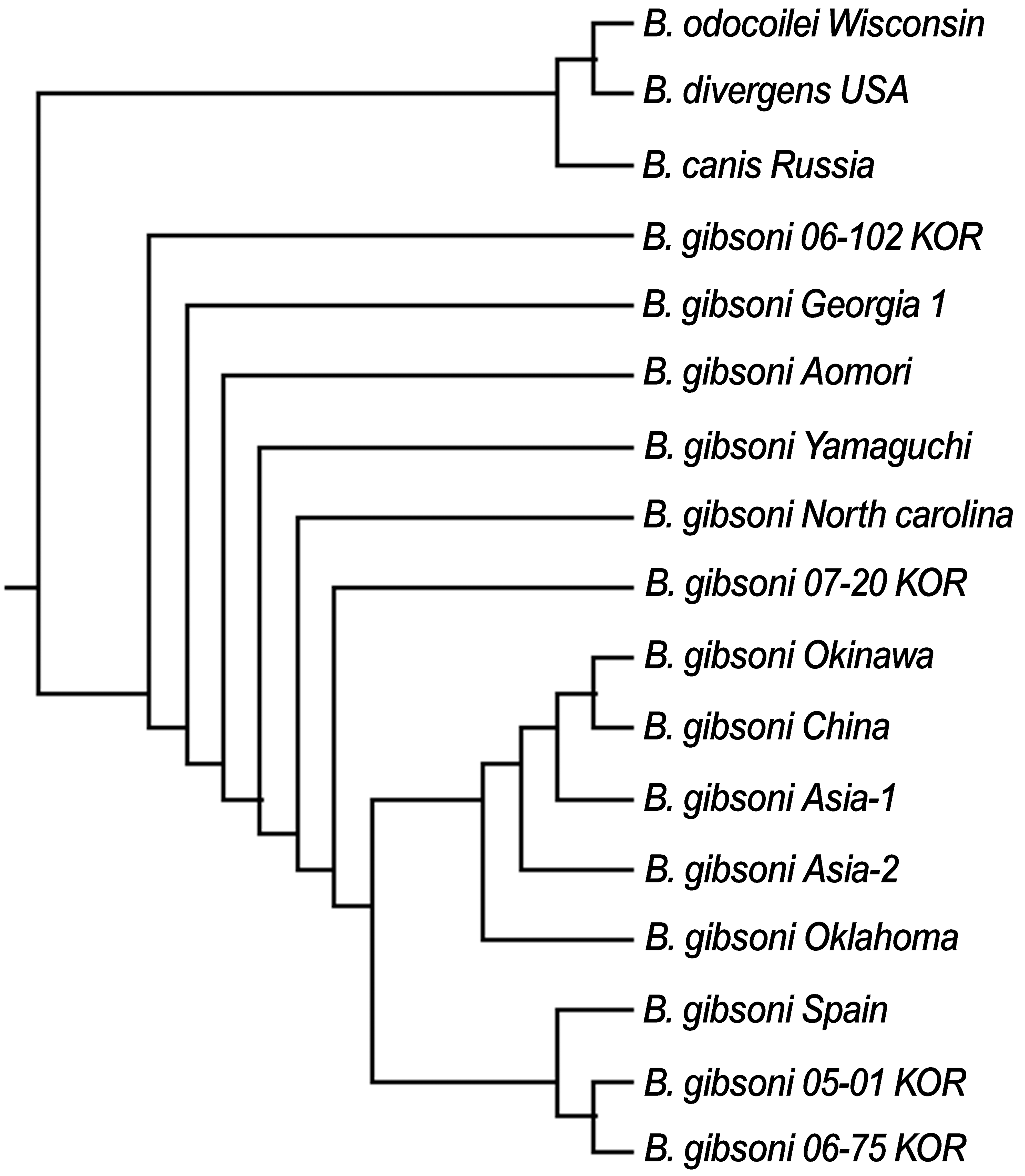
Phylogenetic analysis based on the nucleotide sequences of the18S rRNA gene obtained from Babesia gibsoni infected dogs in South Korea. Our KOR strains were found to be highly homologous to B. gibsoni strains obtained from other countries. The nucleotide sequences for 4 (06-102, 07-20, 05-01, and 06-75) of the 14 KOR strains were compared with various strains of B. gibsoni using the NCBI BLAST network service and MultAlin software. Phylogenetic trees were generated using Multiple Sequence Alignment by CLUSTALW.
Discussion
In eastern Japan, 26 of the 35 PCR-positive dogs had a history of bite wounds from other dogs and the rate of bites was significantly higher (Miyama et al. 2005). In addition, infection with B. gibsoni was observed in 41 of 42 tosa dogs with a history of fighting in Aomori prefecture, northeastern Japan (Matsuu et al. 2004). In the United States, B. gibsoni primarily affects pit bull terriers, and its symptoms are often subclinical. When other breeds of dogs are infected, they often have a history of fighting with a pit bull terrier (Birkenheuer et al. 2005, Macintire et al. 2002). In a previous study in South Korea, B. gibsoni infection was detected in nine German shepherds dogs (1.8%, n = 501) (Song et al. 2004). However, B. gibsoni infection was observed in 29 dogs (24.8%, n = 117), even though the number of samples evaluated in this study was much lower, and it is more common in pit bull terriers than German shepherds in this study. Pit bull terriers infected with B. gibsoni accounted for 15 (51.7%) of the 29 dogs that were infected, and all 15 of these dogs had suffered from mild to severe injury due to fighting with other dogs in this study. In addition, the proportion of male dogs infected with B. gibsoni was rather high (23 of 29 dogs), which is similar to the results of a study conducted by Matsuu et al. (2004). This finding may have occurred because male dogs tend to be more aggressive than female dogs and are more likely to be used as fighting dogs in South Korea and Japan. And these results indicate that B. gibsoni is primarily disseminated through bite wounds in pit bull terriers in South Korea and the spreading of B. gibsoni through bite wounds between fighting dogs has important implications in the dissemination of this organism in South Korea.
Though it has been not cleared the transmission of B. gibsoni in dogs in South Korea, the major part of the B. gibsoni infection could be established in Haemaphysalis longicornis ticks, because H. longicornis tick, known to transmit B. gibsoni in Japan, is distributed nationwide in South Korea (Lee et al. 2005). Indeed, three pit bull terriers had a history of tick exposure and three indoor dogs revealed clinical features of canine babesiosis after being walked and found to have been exposed to ticks. Moreover, all 11 indoor dogs that were found to be infected in this study had a history of being walked three to four times per week, which indicates that indoor dogs can be easily exposed to ticks due to frequent walking. Care should be taken to ensure that indoor dogs and pit bull terriers are not exposed to ticks. Future studies should be conducted to evaluate the infection of B. gibsoni in ticks in South Korea.
The largest number of samples infected with B. gibsoni was collected from Gyunggi province in this study. However, this likely occurred because most of the blood samples were referred from animal hospitals located in Gyunggi province. In addition, Gyunggi province is a populous area in which many dogs live; therefore, this area has the largest number of dogs referred to animal hospitals in South Korea. To obtain a better understanding of the distribution of B. gibsoni in South Korea, an additional survey that includes a larger number of dogs from other provinces should be conducted.
Thrombocytopenia is a prominent feature of B. gibsoni infection and may occur before and after parasitemia (Meinkoth et al. 2002). Tosa dogs in Aomori prefecture, Japan that had parasitemia were found to have lower mean platelet counts (137.5 × 10³/μL) than dogs that were not infected with B. gibsoni (Matsuu et al. 2004). Inokuma et al. (2005) reported that the mean platelet count of dogs with low parasitemia for B. gibsoni was 70 × 103/μL, whereas those with moderate to severe parasitemia and in dogs with IMHA for B. gibsoni infection were 49 × 103/μL and 155 × 103/μL, respectively. In the present study, 14 of 23 positive dogs were found to have thrombocytopenia, and all 14 of these dogs showed low parasitemia. The mean platelet count in dogs with low parasitemia and in dogs with IMHA in this study was 102.8 × 10³/μL and 217.5 × 10³/μL, respectively. Similar to other studies, we confirmed that B. gibsoni infection can induce more severe thrombocytopenia in dogs with parasitemia than in dogs with IMHA. Furthermore, Anaplasma or Ehrlichia infection is uncommon in dogs in South Korea, and thrombocytopenia is more likely to indicate B. gibsoni infection than other protozoal infection. Therefore, clinicians must use a test to determine if B. gibsoni infection is present in dogs that show thrombocytopenia.
B. gibsoni infections can easily be misdiagnosed as idiopathic or autoimmune hemolytic anemia because merozoites of B. gibsoni are not only always observed in blood smears (Farwell et al. 1982) but also clinically affected dogs often have very low levels of parasitemia (Inokuma et al. 2005). In the present study, 11 of 29 positive dogs were found to have IMHA based on other clinical signs. However, these 11 dogs were diagnosed with B. gibsoni infections using the PCR assay and subsequently treated with the antibiotics rather than immunosuppressive drugs. Therefore, B. gibsoni should be included as a differential category for the cause of hemolytic anemia by clinicians, and the PCR assay should be used to confirm or rule out B. gibsoni infection in dogs.
In this study, phylogenetic analysis revealed that our KOR strains were very closely related to other B. gibsoni strains from Spain, the United States, and Japan. In previous reports, the identity of the Miyazaki strain in Japan was closely related to the Asia 1 and 2 strains of B. gibsoni, but differed from B. gibsoni isolated in Europe and the United States (Ano et al. 2001). A previous study conducted by Zahler et al. (2000) also revealed that B. gibsoni strains from the United States and Asia vary enough genetically to be considered different species. However, the results of the present study suggest that B. gibsoni strains in South Korea have the highest homology to the B. gibsoni strains from the United States, Japan, and Europe. In addition, B. gibsoni strains obtained from Oklahoma, North Carolina, and Okinawa were found to be 92% identical with the B. canis strain obtained from South Africa (Kjemtrup et al. 2000). In the present study, the nucleotide sequences of the KOR strains were also 96% identical with the nucleotide sequence of B. canis obtained from Russia.
In conclusion, most of the dogs infected with B. gibsoni in South Korea were found to have anemia and thrombocytopenia, especially those that revealed icterus and splenomegaly upon physical examination. In addition, all of the infected dogs revealed low levels of parasitemia, which prevents the detection of merozoites for B. gibsoni in blood smears. In the present study, the PCR assay was shown to be a useful tool for detecting of B. gibsoni infection in dogs with acute and chronic infection. Although the results of this study indicate that B. gibsoni infection occurs nationwide, few clinical and epidemiologic surveys for B. gibsoni have been conducted in South Korea. Therefore, clinicians should be trained to notice the occurrence of B. gibsoni infection, which may cause anemia and thrombocytopenia in dogs, including indoor dogs. In addition, being bitten by a pit bull terrier was identified as a risk factor for the transmission of B. gibsoni in South Korea. Furthermore, because B. gibsoni infection in dogs may be an endemic disease that can be spread by ticks, an additional survey is necessary to clarify the relationship between the host and the vector. Overall, these results indicate that clinicians and dog owners must take care to prevent B. gibsoni infection because it may have epidemiologic implications in South Korea.