




Abstract
Sampling was conducted at The Ohio State University's Veterinary Teaching Hospital (OSU-VTH) to evaluate the extent of environmental contamination with Salmonella enterica, at 1-week intervals beginning March 19, 2007, through May 21, 2007. Environmental samples were collected from various surface and floor locations in the equine and food animal areas using sterile moistened gauze sponges. All samples were processed using standard bacteriologic culture to identify the presence of Salmonella spp. Genetic relatedness of isolates was assessed using amplified fragment length polymorphism (AFLP) procedures, and minimum inhibitory concentrations to a panel of antimicrobial drugs were determined using microbroth dilution. A total of 16 Salmonella isolates were recovered from 270 (5.9%) cultured environmental and animal samples, with prevalence ranging from 0% to 32% on individual sampling dates. A total of 9% of the samples from the food animal section and 2.5% of the samples from the equine section tested positive for Salmonella (p = 0.03). The 16 isolates represented seven different clonal strains and four different serotypes (Paratyphi B var. L-tartate n = 3, Kentucky n = 5, Cerro n = 7, Montevideo n = 1), most of which were pansusceptible to a panel of antimicrobial drugs. Our results indicate that animal treatment areas with a high population of animals or cases within the veterinary teaching hospital can become contaminated with Salmonella, especially in high traffic areas that may facilitate horizontal dissemination. The most common occurrence appears to be infected agricultural animals that contaminate the hospital environment, but normal cleaning and disinfection appears to effectively prevent long-term contamination.
Introduction
With diverse populations of ailing food and companion animals, veterinary teaching hospitals without appropriate biosecurity measures could provide the opportunity for the rapid spread of infectious agents among their patients. Thus, the potential for nosocomial infections and the zoonotic transmission of infectious agents are important concerns in veterinary medicine. Salmonella infections can cause serious illness in both humans and other mammals, with immunocompromised populations at highest risk (Blaser and Newman 1982). Outbreaks of salmonellosis among patients at veterinary teaching hospitals have been reported primarily in large animals (Tillotson et al. 1997, Schott et al. 2001). Not only can animals be infected, they can also serve as the reservoir for transmitting this agent to other animals, faculty, students, and hospital visitors. The direct zoonotic transmission of Salmonella in veterinary facilities resulting in human outbreaks has been reported (CDC 2001).
Although animals can be infected clinically or subclinically, Salmonella most often persists in the subclinical form, making the detection of infected animals difficult (Gay et al. 1993, Huston et al. 2002). One factor that can initiate fecal Salmonella shedding by infected animals is stress (Wray et al. 1991, House et al. 1999). Salmonella-infected animals, suffering from stresses such as transportation and environmental change, often arrive at the teaching hospital for treatment along with other immunocompromised animals. These stress-affected animals provide adequate potential for the spread of Salmonella infections to susceptible animals with weakened immune systems
Therefore, an emphasis on preventing and controlling potential nosocomial Salmonella infections is an important biosecurity concern for veterinary teaching hospitals (Kim et al. 2001). Environmental and animal samples can be easily collected in the veterinary teaching hospital on a routine basis as part of an ongoing surveillance program (Burgess et al. 2004). The purpose of this study was to determine the extent of environmental contamination with Salmonella in The Ohio State Veterinary Teaching Hospital. Monitoring and controlling Salmonella infections in veterinary facilities will have important future public health implications.
Materials and Methods
Sampling
Environmental and animal fecal samples were collected from The Ohio State University Veterinary Teaching Hospital (OSU-VTH) at 1-week intervals beginning March 19 through May 21, 2007. Prior to the start of regular sampling, an initial preliminarily sampling was conducted the week of February 20, 2007. Environmental samples were collected using a 2 × 2 inch sterile gauze sponge moistened with sterile phosphate-buffered saline solution (PBS, VWR, West Chester, PA). The sterile gauze was moistened for approximately 5 sec, and the surface was then swabbed for approximately 20 sec. The gauze was then placed into a sterile 50 mL plastic centrifuge tube. When fecal samples were obtained, approximately 10 g of fresh feces was collected from the floor and placed into a 50 mL plastic centrifuge tube.
Each week of sample collection, a single investigator walked systematically through the equine and food animal areas of the OSU-VTH and subjectively identified floors, drains, contact surfaces, and animal feces for sampling. A target number of 24 to 26 samples per week approximately balanced between the equine and food animal areas was maintained. Emphasis was placed on identifying drains and contact surfaces for sampling.
When floors were sampled, an area of approximately 24 × 24 inches was swabbed. Floors sampled in the equine area included floors in patient stalls and the floor of the hay storage area. The floors that were sampled in the food animal area included holding pen floors, the floor near the entrance and exit of the building, the floor of the primary treatment area, and the floor of the recovery stalls. Floor drains were sampled by swabbing the entire top surface of the drain and inside the drain as much as possible. Contact surfaces that were sampled included doors, tables, phones, sinks, railing, shovels, and manure storage bins. The contact surfaces were sampled by swabbing the entire surface of the object. These different contact surfaces were sampled in different combinations each week. Animal fecal samples were only collected when an obviously fresh fecal sample was readily and easily available for sampling. The samples that were collected were intended to be generally representative of the food animal and equine areas in OSU-VTH. Clinical history of the patients associated with these fecal samples was not obtained, but all fecal samples were of normal consistency and were not considered to be diarrhea.
Culture procedures
All samples were cultured for the presence of Salmonella using a standard protocol. The 2 × 2 inch sterile gauze sponges moistened with sterile PBS (VWR) were enriched in tetrathionate broth (TTB, VWR) using a 10:1 ratio immediately after sampling and incubated at 37°C overnight. Fecal samples were also enriched in TTB, with 4 g of feces in 36 mL of TTB. A 100 μL aliquot of the TTB mixture was then transferred to Rappaport-Vassiliadis R10 Broth (RV, VWR) and incubated at 42°C overnight. On the following day, samples from the RV were inoculated onto xylose lysine tergitol 4 (XLT4, VWR) agar plates, streaked for isolation, and incubated at 37°C overnight. Presumptive Salmonella-positive specimens with black colonies were confirmed using standard biochemical tests, including triple sugar iron (TSI), Urea, MacConkey's agar, and polyvalent antisera.
Isolated colonies from the XLT4 plates were then struck onto MacConkey Agar (MAC, VWR) plates and incubated at 37°C overnight. The appearance of clear, colorless, lactose-negative colonies confirmed the presence of Salmonella. Isolated colonies were also inoculated on to Triple Sugar Iron slants (TSI, VWR) and incubated at 37°C overnight. The tubes were observed for color changes that indicate acid production in the slant and butt, hydrogen sulfide precipitate production in the butt, and gas production in the form of an air bubble or crack in the media. Isolated colonies were also inoculated in urea broth at 37°C overnight. The absence of color change indicated positive Salmonella samples. Last, the results were confirmed by conducting an antisera agglutination test. A colony from the MAC agar was mixed with polyvalent antisera. Agglutination indicated a positive reaction.
Amplified fragment length polymorphism genotyping
DNA fingerprinting of Salmonella was done using the method previously described (Gebreyes et al. 2006). Briefly, genomic DNA was purified using the Qiagen DNAeasy tissue kit (Qiagen, Valencia, CA), and 100 ng of genomic DNA was digested with restriction enzymes, EcoRI and MseI (New England BioLabs, Beverly, MA), in NEB 2 buffer (New England BioLabs) containing 50 μg/μL bovine serum albumin (New England BioLabs) at 37°C for 2 h followed by 15-min inactivation at 70°C. Adapter oligosequences unique to each end were ligated to the restriction fragments using T4 DNA ligase (New England BioLabs) at 25°C overnight. Fragments were then amplified using EcoRI (5′-GACTGCGTACCAAATC) and MseI (5′-GATGAGTCCTGAGTAA) (Integrated DNA Technologies, Coralville, IA) primers. The amplification protocol was 20 cycles at 94°C for 30 seconds, 56°C for 1 min, and 72°C for 1 min. The amplified products, diluted at a 1:10 ratio using molecular-grade water, underwent a selective amplification using MseI primer (5′-GATGAGTCCTGAGTAA) and wellRED dye labeled EcoRI primer with an additional adenine at the 3′-end EcoRI-A (5′-GACTGCGTACCAAATCA) (Integrated DNA Technologies). The conditions for final amplification were one cycle of 94°C for 30 sec, 65°C for 30 sec, and 72°C for 1 min, followed by 12 cycles subsequently lowering the annealing temperature (65°C) by 0.7°C per cycle while keeping the denaturation temperature at 94°C for 30 sec and extension temperature at 72°C for 1 min. This was followed by 23 cycles of 94°C for 30 sec, 56°C for 30 sec, and 72°C for 1 min.
The amplified fragments were separated by capillary electrophoresis using the CEQ 8000 genetic analyzer (Beckman Coulter, Fullerton, CA, USA). Thirty-five microliters of sample loading solution (SLS) (Beckman Coulter) and 0.66 μL of DNA size standard 600 (Beckman Coulter) were mixed with 1 μL of the final amplification and overlayed with a drop of mineral oil. A fragment amplication method with denaturation at 90°C for 120 sec, injection for 30 sec at 1 Kv, and separation at 5 Kv for 55 minutes was used. Amplified fragments between 50 bp and 500 bp were scored and analyzed using BioNumerics v4.5 software (Applied Maths, Kortrijk, Belgium).
Minimum inhibitory concentration procedure
Minimum inhibitory concentrations (MIC) of specific drugs were determined using the semiautomated Sensititre broth microdilution system (Trek Diagnostics, Westlake, OH, USA) following manufacturer's guidelines. Isolates were inoculated into sterile water and turbidity adjusted to a 0.5 McFarland standard. Ten microliters of the suspension were then inoculated into 10 mL of cation-adjusted Mueller-Hinton broth. The Mueller-Hinton broth was then used to inoculate standard 96-well Sensititre plates containing defined concentrations of antimicrobial drugs. Plates were incubated 18 to 24 h at 35°C and MICs to specific antimicrobial drugs determined based on observed bacterial growth in individual wells. Quality control organisms included Escherichia coli 25922, Enterococcus faecalis 29212, and Pseudomonas aeruginosa 27853.
Data analysis
A table of descriptive statistics is generated to show the prevalence of Salmonella recovery by the different sections sampled, sampling sites, and by sampling date. Maps of the veterinary teaching hospital were developed to describe the spatial distribution of Salmonella recovery. The temporal distribution of the recovery of Salmonella positive samples was described graphically. The prevalence between groups was compared using the Pearson chi-square analysis or Fisher exact test when appropriate.
Results
A total of 270 samples were collected from OSU-VTH over a 3-month period. Salmonella were identified in 16 (5.9 %) of the collected samples. The sampling took place primarily in two distinct areas of the hospital: the food animal section and the equine section. Of the samples taken, 9% of the samples from the food animal section and 2.5% of the samples from the equine section tested positive for Salmonella (p = 0.03). The floor drain surfaces were the most common site for Salmonella recovery at 7.3%, and the floor surface samples were similar at 6.7%. Other various surface areas, such as manure barrels, hay grate covers, shovels, mats, and rails that were sampled were 5.4% positive. Salmonella was not isolated from any of 17 animal fecal samples collected. We could not detect a statistical difference in the prevalence of Salmonella recovery among the different sampling sites. However, drain samples collected in the food animal section (8/64) were more likely (p = 0.02) to contain Salmonella than drain samples from the equine section (0/45). The overall Salmonella results are displayed in Table 1. Differences were observed in the prevalence of Salmonella recovery among the sampling dates (p < .001), with the highest prevalence of Salmonella recovery in mid-April (32%), although commonly all samples collected on a single day were negative. The prevalence of Salmonella recovery for samples from different sources and over time is summarized in Table 1.
Total % column was calculated by dividing the total number of positive samples by the number sampled.
Hospital Salmonella isolates were most commonly resistant to streptomycin (12.5%), sulfamethoxazole (31.2%), and tetracycline (37.5%). All of the isolates were susceptible to amikacin, amoxicillin/clavulanic acid, ampicillin, cefoxitin, ceftiofur, ceftriaxone, chloramphenicol, ciprofloxacin, gentamicin, kanamycin, nalidixic acid, and trimethoprim/sulfamethoxazole. Some isolates exhibited a multiresistant phenotype, with 25% of isolates resistant to both sulfamethoxazole and tetracycline. Also, 6.25% of the isolates exhibited a multiresistant phenotype to streptomycin, sulfamethozole, and tetracycline. Table 2 depicts the resistance patterns for all isolates.
The 16 Salmonella isolates were identified as representing seven different clonal strains and four different serotypes. The four different serotypes were identified as Paratyphi B var. L-tartate (n = 3), Kentucky (n = 5), Cerro (n = 7), and Montevideo (n = 1). Clonal strains were defined as having ≥78% genetic similarity based on AFLP results and a similar resistance phenotype. Clonal strain 1 was recovered from four different sites in the food animal section on April 16, 2007. A second clonal strain (clone 3) was also recovered on April 16, 2007, in three different samples from the equine section. Clone 4 was recovered on 3 different weeks, and clone 5 was recovered on 2 different weeks. The AFLP results are shown in Figure 1, and a summary of the clonal Salmonella strains is presented in Table 3.
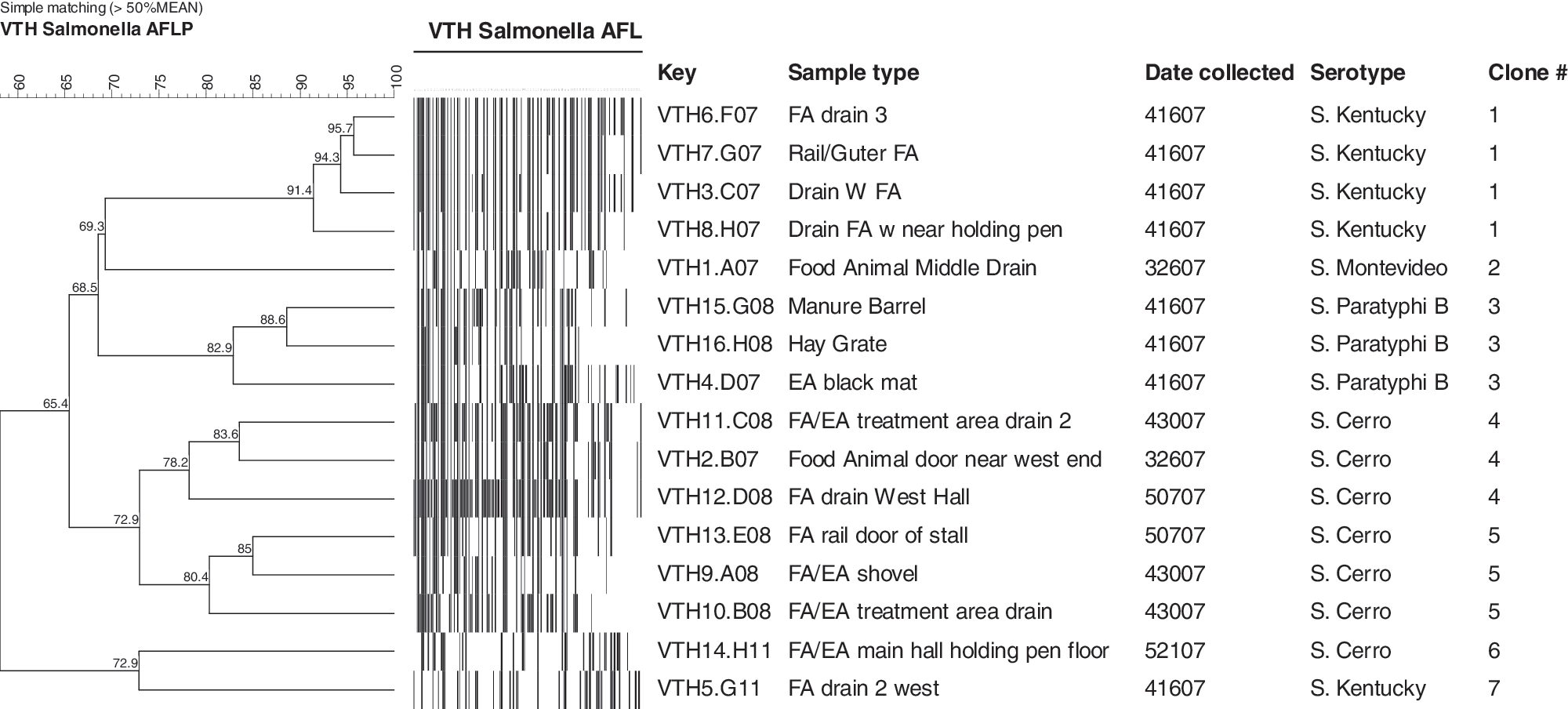
Amplified fragment length polymorphism analysis of 16 Salmonella isolates recovered from environmental samples from The Ohio State University Veterinary Teaching Hospital.
Discussion
Our results indicate that areas of the OSU-VTH can be intermittently contaminated with Salmonella. Our overall observed prevalence of 5.9% is lower than the 11.9% of environmental samples reported positive at the Colorado State University veterinary teaching hospital (Burgess et al. 2004), but higher than the 0.1% prevalence of culture-positive environmental swabs reported for the Michigan State University veterinary teaching hospital (Ewart et al. 2001). However, differences in sampling strategies and microbiologic techniques make direct comparison of environmental Salmonella prevalence rates between hospitals difficult.
We observed that the prevalence of environmental Salmonella in the food animal section of the OSU-VTH was 9%, compared to a prevalence of only 2.5% found in the equine section. This result may reflect the fact that populations of agricultural animals frequently maintain Salmonella infections and contain animals shedding subclinically. These animals may enter the veterinary hospital and contaminate the environment without the knowledge of the owner or clinicians involved. Environmental Salmonella contamination of veterinary hospitals poses a risk of nosocomial infection to patients and the risk of zoonotic infections to clients, clinicians, and students. This result suggests that students and clinicians in the food animal section should be aware of the potential risks. In addition, appropriate biosecurity practices should be in place to prevent nosocomial infections or zoonotic transmission.
Salmonella was most frequently recovered from floor drains and other hospital surface areas but was not found in any individual fecal specimens that were cultured. Others have also reported that drains are a common site for the recovery of Salmonella (Alinovi et al. 2003) or Salmonella DNA (Ewart et al. 2001, Schott et al. 2001) in veterinary hospitals. Drains are expected to be a high prevalence area for environmental Salmonella sampling because waste from a variety of sources can accumulate there and have been reported to be a source of nosocomial infections (Schott et al. 2001). Thus, these may be useful sites for Salmonella monitoring and surveillance in hospitals. However, with higher prevalence rates, drains may not reflect true prevalence of environmental contamination of the hospital. Our ability to recover Salmonella from surfaces suggests that a variety of human and animal contact surfaces can easily become contaminated, posing a risk of nosocomial or zoonotic transmission.
These results suggest that the routine cleaning and disinfection procedures used in the OSU-VTH are not completely effective in preventing maintenance of the Salmonella organisms for extended periods of time after they contaminate the environment. Table 1 indicates that the prevalence of Salmonella varied greatly from week to week. This suggests that infected animals were likely entering the hospital on a regular basis, contaminating the environment with Salmonella, and that the most severe contamination was likely cleared by the regular hospital cleaning and disinfection protocol. Thus, we detected no Salmonella in environmental samples collected in the majority of the weekly sampling periods. However, the results also indicated that the OSU-VTH can suffer from frequent environmental contamination, and individuals employed by or visiting the facility should take appropriate protective measures.
A total of seven unique clonal strains of Salmonella were identified from among the 16 isolates based on at least 78% genetic similarity using the AFLP results and similar resistance phenotype. The three isolates recovered in the equine section on April 16, 2007 were clonal, but different than any of the clonal strains recovered in the food animal section. Therefore, we found no evidence of Salmonella cross-contamination between the food animal and equine sections of OSU-VTH. Clonal strains 4 and 5 were each recovered repeatedly over time, with strain 4 recovered on three different occasions over a 10-week period. This result suggests that clonal strains of Salmonella can persist in the OSU-VTH environment despite the cleaning and disinfection protocols. These persistent strains can serve as a reservoir for nosocomial infection of patients and zoonotic infections of students, clinicians, and clients. While current efforts to prevent persistent environmental Salmonella contamination in the OSU-VTH are appropriate, total prevention in a large-animal hospital setting may be difficult or impossible. Repeated recovery of clonal strains over time might also represent repeat environmental contamination when a patient who was the source of the original contamination returns to the hospital, or the widespread dissemination of a specific clonal strain in the larger population that is the source of hospital patients.
A general infectious disease control program for preventing nosocomial infections in large-animal veterinary hospitals has been described (Smith et al. 2004). However, based on these results, we can make certain recommendations for the prevention and control of Salmonella that are specific to the OSU-VTH. First, it should be standard procedure to isolate all known Salmonella-positive patients. However, these can be difficult to identify because subclinical shedding is common. The mandatory culturing of fecal samples from all hospitalized animals in the food animal and equine sections could potentially identify these. However, the length of time between sample collection and confirmed result is several days, limiting the usefulness of this practice in a hospital setting. The use of foot baths and plastic disposal boot covers when working with high-risk patients may be an effective control method. The use of disposal boot covers has been reported to be effective in decreasing new nosocomial infections (Schott et al. 2001) in hospital settings. Regular and extensive surveillance for environmental Salmonella contamination will provide useful information for a better understanding of the need for intervention. Standard protocols for the OSU-VTH food animal and equine sections include cleaning and disinfecting of each stall after each animal. In addition, the treatment areas are washed at least twice a day, once in the morning and again at night, also in between each animal examination. The area is usually cleaned with a mixture of water, bleach, and detergent. The surface is scrubbed and then hosed down the drain, which has been deemed an effective means of controlling Salmonella. The institution of stricter monitoring and cleaning practices could be beneficial in containing the organism and preventing the spread of Salmonella throughout the hospital.
Footnotes
Disclosure Statement
No competing financial interests exist.