
Abstract
The aim of this study is to present molecular, serologic, and clinical findings for dogs that were naturally infected with Anaplasma phagocytophilum or Borrelia burgdorferi sensu lato (s. l.) in the Czech Republic. This data can provide information relevant to human infection. In total, blood samples from 296 dogs and 118 engorged ticks were examined. Samples were tested for A. phagocytophilum using polymerase chain reaction (PCR) amplification, nested PCR, and direct sequencing of the 16S rDNA, and for B. burgdorferi s. l. using PCR amplification of the 16S rDNA and restriction fragment length polymorphism analysis of the 5S-23S rDNA intergenic spacer. In addition, blood samples were screened for antibodies to these bacteria. Ten (3.4%) dogs were PCR-positive for A. phagocytophilum. Morulae of A. phagocytophilum in granulocytes were found in two of these dogs. Nine of the PCR-positive dogs had clinical signs related to anaplasmosis. Statistically significant differences in the PCR detection rates were found between breeds and between symptomatic and asymptomatic dogs. Infection with Borrelia garinii was detected by PCR in a dog with meningoencephalitis. DNA of A. phagocytophilum and B. burgdorferi s. l. (B. garinii or Borrelia afzelii) was detected in 8.5% and 6.8% of ticks, respectively. Immunoglobulin (Ig) G seropositivity to A. phagocytophilum was 26%. Significant differences were found with respect to breed and gender. IgM and IgG antibodies to B. burgdorferi s. l. were detected in 2.4% and 10.3% of dogs, respectively. Our findings suggest that the exposure to B. burgdorferi s. l. exists in dogs in the Czech Republic, and exposure to A. phagocytophilum is common.
Introduction
A. phagocytophilum is a Gram-negative obligate intercellular rickettsial bacterium found in neutrophil granulocytes (Parola and Raoult 2001). A. phagocytophilum is known to cause granulocytic anaplasmosis in humans and domestic animals such as dogs, horses, cattle, sheep, goats, llamas, and cats (Engvall et al. 1996, Barlough et al. 1997, Bjöersdorff et al. 1999, Engvall et al. 2002, Skotarczak 2003, Lester et al. 2005, Poitout et al. 2005, Lillini et al. 2006). Anaplasmosis signs include fever, fatigue, inappetence, lethargy, lameness, and gastrointestinal and central nervous system signs (Rikihisa et al. 1991, Greig et al. 1996, Egenvall et al. 1997, Engvall et al. 2002). The related hematologic and biochemical abnormalities are anemia, thrombocytopenia, lymphopenia, and elevated serum alkaline phosphatase activity (Greig et al. 1996, Goldman et al. 1998). Epidemiologic studies of A. phagocytophilum in dogs using polymerase chain reaction (PCR) and serologic methods are available in the literature for various countries. Only case reports are available in the Czech Republic. Two cases of granulocytic anaplasmosis in dogs in the Czech Republic were described by Huml et al. (1996), one case by Melter et al. (2007), and four cases by Spejchalova et al. (2008). A. phagocytophilum was identified also in horses and cows by Hulinská et al. (2004).
Lyme disease (borreliosis) is a zoonotic, tick-borne disease caused by a spirochete B. burgdorferi s. l., which actively migrates in body tissues. The clinical form of borreliosis occurs in humans and domestic animals, especially dogs, horses, and cattle (Burgess et al. 1987, Greene et al. 1991, Cohen et al. 1992). Canine borreliosis most commonly affects the limb joints (Skotarczak and Wodecka 2003, Skotarczak et al. 2005), with clinical manifestations such as arthritis and arthralgia (Jacobson et al. 1996). Other associated signs are malaise, fever, inappetence, fatigue, and lameness. Even though the signs are the same as in humans, they are more difficult to detect in dogs and develop in relatively few of them (Levy and Magnarelli 1992). Serologic reactivity to B. burgdorferi s. l. in dogs was analyzed in the Czech Republic (Pejchalová et al. 2006). B. burgdorferi s. l. infection prevalence in questing ticks from Czech Republic were found by Pejchalová et al. (2007) to be 12.1%. A. phagocytophilum prevalence in ticks (14.7%) was suggested by Sikutová et al. (2007); however, less specific primers EHR521/747 were used.
Data on the prevalence of infection with A. phagocytophilum and B. burgdorferi s. l. in dogs in the Czech Republic is lacking, except for the above-mentioned serologic study. In the present study, we attempt to gather molecular, serologic, and clinical findings for dogs that were naturally infected with A. phagocytophilum or B. burgdorferi s. l. in the Czech Republic.
Material and Methods
Study sites and animals
All dogs examined in the Faculty of Veterinary Medicine, University of Veterinary and Pharmaceutical Sciences, Brno, Czech Republic and in the Veterinary Clinic in Jablonec nad Nisou, Czech Republic between November 2005 and October 2007 were included in the study. We also included dogs of the clinic students and employees and 39 hunting dogs examined for a different study. The 292 dogs came from various locations in the Czech Republic, principally from South (202) and North Moravia (32) and from North Bohemia (62). None had traveled outside of the country during the 6 months prior to presentation. Data on the age, gender, breed, geographical origin, health status, and purpose (working dog or pet dog) were recorded.
The dogs were divided into two groups. Group A consisted of dogs showing clinical signs attributable to infection with A. phagocytophilum or B. burgdorferi s. l. The inclusion criteria for group A was the presence of any the following signs: apathy, fever, lameness, lethargy, inappetence, or gastrointestinal and central nervous system disorders. Dogs of group B were healthy or with a diagnosis or signs not attributable to the studied infections such as trauma or epilepsy. Samples from dogs were collected on the day of presentation to the clinic. Blood was drawn from the jugular or cephalic vein. Buffy-coat blood smears were made and immediately stained by the Giemsa method. Ethylenediaminetetraacetic acid-anticoagulated whole blood and serum samples were taken from each dog. Attached engorged ticks were removed using forceps, identified by species and life stage, and stored individually in microcentrifuge tubes at 2 to 8°C prior to DNA extraction.
DNA extraction
Total DNA was isolated from blood samples with a High Pure PCR Template Preparation Kit (Roche, Mannheim, Germany): 200 μL of blood was transferred to a tube containing 200 μL of lysis buffer and 40 μL of proteinase K, mixed, lysed at 56°C for 1 h, and continued according to manufacturer's protocol. Purified DNA was stored at −20°C before using it as a template for PCR amplification.
Ticks were washed in phosphate-buffered saline solution before DNA extraction using a DNeasy Tissue Kit (Qiagen, Hilden, Germany). Ticks were transferred to a tube containing 180 μL of lysis buffer and 20 μL of proteinase K, crushed with a sterile scalpel, mixed, and incubated at 56°C for at least 2 h.
PCR amplification
All samples were analyzed by standard PCR with the Ehr 521-790 primer set (for Anaplasma) (Pancholi et al. 1995) and the LD primer set (for Borrelia) (Marconi and Garon 1992), both targeting the 16S rDNA. PCR amplification was performed in a Peltier Cycler (MJ Research, Waltham, MA, USA). The reaction mixture consisted of 2.5 μL of DNA extract as a template and 1 μM solution of each primer (Generi Biotech, Hradec Kralove Czech Republic) in a total volume of 25 μL Hot Start Master Mix (Qiagen, Hilden, Germany). The DNA was amplified with the Ehr 521–790 primer set as follows: an initial 15-min denaturation at 95°C and then 40 cycles of 45 sec at 95°C, 45 sec at 60°C, and 45 sec at 72°C. A final extension was done for 7 min at 72°C. Cycling conditions for the LD primer set were described previously (Kybicová et al. 2008). The PCR products were separated by electrophoresis in 1% agarose gel and stained with ethidium bromide. Previously extracted DNA of B. garinii strain 310M was used as a positive control. Purified water served as a negative control.
Nested PCR for Anaplasma detection
The samples found PCR positive for Anaplasma were retested by nested PCR with two sets of primers targeting also the 16S rDNA gene (ge3a, ge10r and ge9f, ge2) (Massung et al. 1998). It has been demonstrated that nested PCR with these primers and standard PCR with the Ehr 521–790 primers have the same detection limits (Massung and Slater 2003). The primary reaction mixture used 2.5 μL of DNA extract and 0.5 μM solution of each primer in a total volume of 25 μL Hot Start Master Mix. Cycling conditions involved an initial 15 min denaturation at 95°C, 40 cycles of 30 sec at 94°C, 30 sec at 55°C, and 60 sec at 72°C and a final extension of 5 min at 72°C. The reaction mixture for the nested amplifications used 0.5 μL of the primary PCR product as the template and 0.2 μM solution of each primer in a total volume of 25 μL. The nested cycling conditions were the same as those for the primary amplification, except that only 30 cycles were run. DNA of A. phagocytophilum of an infected dog was used as a positive control.
DNA sequencing
To prove that the positive PCR results were truly due to A. phagocytophilum, all positive PCR products from nested PCR amplification were directly analyzed on a CEQ 2000XL sequencer (Beckman Coulter, Buckinghamshire, UK). Size of targeted DNA sequence was 497 bp without primer-annealing areas.
Restriction fragment length polymorphism analysis for Borrelia detection
The Borelia-positive DNA samples were tested by restriction fragment length polymorphism (RFLP) analysis with 5S (rrfA)-23S (rrlB) rDNA intergenic spacer primers (222–255 bp) (Derdakova et al. 2003) using the same procedure as in Kybicová et al. (2008).
Serology, indirect immunofluorescence assay, and enzyme-linked immunosorbent assay
Indirect immunofluorescence assay (IFA) for the detection of canine immunoglobulin (Ig) G antibodies against A. phagocytophilum (Fuller Laboratories, Fullerton, CA) was used according to the manufacturer's instructions. Examination of the slides was performed using a fluorescence microscope at 400-fold magnification. The fluorescence intensity at a dilution of 1:640 was used as the cutoff level. A positive reaction was manifested by apple green fluorescence of inclusion bodies (morulae).
The sera were also examined by an enzyme-linked immunosorbent assay (ELISA) (Test-line, Brno, Czech Republic) for detection of specific antibodies against B. burgdorferi s. l. in dogs in the IgG and IgM class (against antigens: OspA, OspC, p41, p100) according to the manufacturer's instructions, with serum dilution 1:400.
Buffy coat smears
Buffy coat smears were stained with Giemsa and observed under 1000-fold magnification. For each Anaplasma-positive dog found by PCR, one buffy coat smear was examined for the presence of morulae.
Statistical analysis
All statistical tests were performed with respect to breed, age group, purpose (working dog or family dog), gender, and group A/B (symptomatic/asymptomatic). For statistical analysis, the dogs were divided into five age groups (≤1, 2–4, 5–7, 8–10, ≥11 years) according to their age after the onset of clinical signs. Breeds were separated according to FCI groups (Federation cynologique internationale,
Results
Dogs
The study group included 296 dogs, 131 females and 165 males, mean age 6.5 years, age range 2 months to 16 years. Their distribution by age, gender, breed, and purpose is shown in Table 1. The group was divided into two subgroups: group A, 141 symptomatic dogs, and group B, 155 asymptomatic dogs. Most frequent hematologic abnormalities in group A (symptomatic) were anemia, thrombocytopenia, and leukocytosis. In group B, 55 dogs were clinically healthy and 100 had a neurologic (epilepsy) or oncologic diagnosis, unrelated to infection. None of the dogs of group B showed hematologic or biochemical abnormalities.
Group A is symptomatic dogs and group B is asymptomatic dogs. FCI, Federation cynologique internationale.
Molecular detection of A. phagocytophilum
DNA of A. phagocytophilum was detected by PCR in 10 (3.4%) of 296 dogs. Of these dogs, nine belonged to group A (9/141, 6.4%) and one to group B (1/155, 0.6%) (Table 2); the difference between groups was statistically significant (odds ratio 11.8, confidence interval 1.44-96.9, p = 0.02 by logistic regression). Six infected dogs were terriers and four were retrievers (FCI groups III and VIII), with the difference between breeds being significant (p = 0.001). Eight of the 10 PCR-positive dogs were diagnosed between April and July, one dog was diagnosed in August, and one in September.
PCR, IFA,
PCR, polymerase chain reaction; IFA, immunofluorescence assay; ELISA, enzyme-linked immunosorbent assay; FCI, FCI, Federation cynologique internationale.
Clinical sings and diagnosis of the nine PCR positive dogs in group A were fever (3), apathy (2), inappetence (1), pyometra (1), gastroenteritis (2), and seizure (2). Hematologic abnormalities included anemia (4), trombocytopenia (2), leukocytosis (2), neutropenia (2), neutrofilia (3), lymphopenia (2), monocytosis (1), eosinopenia (1), and leukopenia (1). Biochemical abnormalities such as elevated alkaline phosphatase (3) or alanine aminotransferase (1), hyperbilirubinemia (1), hypoproteinemia (1), hyperglycemia (1) were observed.
Sequencing of the 10 positive PCR products revealed the gene sequence of A. phagocytophilum. The sequences of the isolates were submitted to the NCBI database with the GenBank accession numbers: EU847526 to EU847535.
Molecular detection of B. burgdorferi s. l.
DNA of B. burgdorferi s. l. was only found in one dog of group A, an 8-year-old labrador retriever. The dog was diagnosed with lymphocytic meningoencephalitis in April 2006. We found no biochemical and hematologic abnormalities in blood, except for a slight anemia. Pleocytosis with small and activated lymphocytes was detected in the cerebrospinal fluid. Using RFLP, the agent was identified as Borrelia garinii.
Ticks
A total of 118 engorged I. ricinus adult ticks (106 females, 12 males) were collected from 67 dogs of groups A and B (42 and 76 ticks, respectively) and individually screened for the presence of DNAs of B. burgdorferi s. l. and A. phagocytophilum. Ten ticks (8.5%), all females, were infected with A. phagocytophilum. Three of these 10 positive ticks were collected from two Anaplasma-positive dogs. Borrelial DNA was found in eight ticks (6.8%), seven females and one male. All infected ticks originated from Borrelia-negative dogs. B. garinii was detected in five ticks and Borrelia afzelii in three ticks, using RFLP.
Serologic findings
Using IFA, IgG antibodies to A. phagocytophilum were detected in 77 dogs, which corresponds to an overall seroprevalence rate of 25.9%. There was no significant difference between groups A and B with 39 (27.5%) and 38 (24.7%) seropositive dogs, respectively. Similarly, there was no significant difference between groups A and B with respect to antibody titers. Very high antibody titers (≥1280) were similarly frequent in both groups, found in 17 of 141 group A dogs and in 17 of 155 group B dogs. Five of the 10 PCR-positive dogs were seropositive, with antibody titers of 1:2560 (n = 1), 1:1280 (n = 2), and 1:640 (n = 2). There was a statistically significant difference in seropositivity between breeds (p = 0.002). The highest seropositivity rates were found in FCI groups II and VIII; males showed higher seropositivity rates than females (odds ratio 2.8, confidence interval 1.52-5.13, p = 0.001 by logistic regression, Table 2).
Using ELISA, IgM and IgG antibodies to B. burgdorferi s. l. were detected in 7 and 30 dogs, respectively (Table 2). The one PCR-positive dog was found negative in both IgM and IgG. Thus the overall seroprevalence rates were 2.4% and 10.3%, respectively. There was no significant difference in seroprevalence between groups A and B or with respect to other studied parameters. The number of dogs with IgG antibodies to both A. phagocytophilum and B. burgdorferi s. l. was 13 (4.4%).
Buffy coat smears
By examination of buffy coats, morulae of A. phagocytophilum (Fig. 1) were detected in peripheral blood neutrophil granulocytes of 2 of the 10 PCR-positive dogs of group A, mostly at a rate of one morula per infected neutrophil.
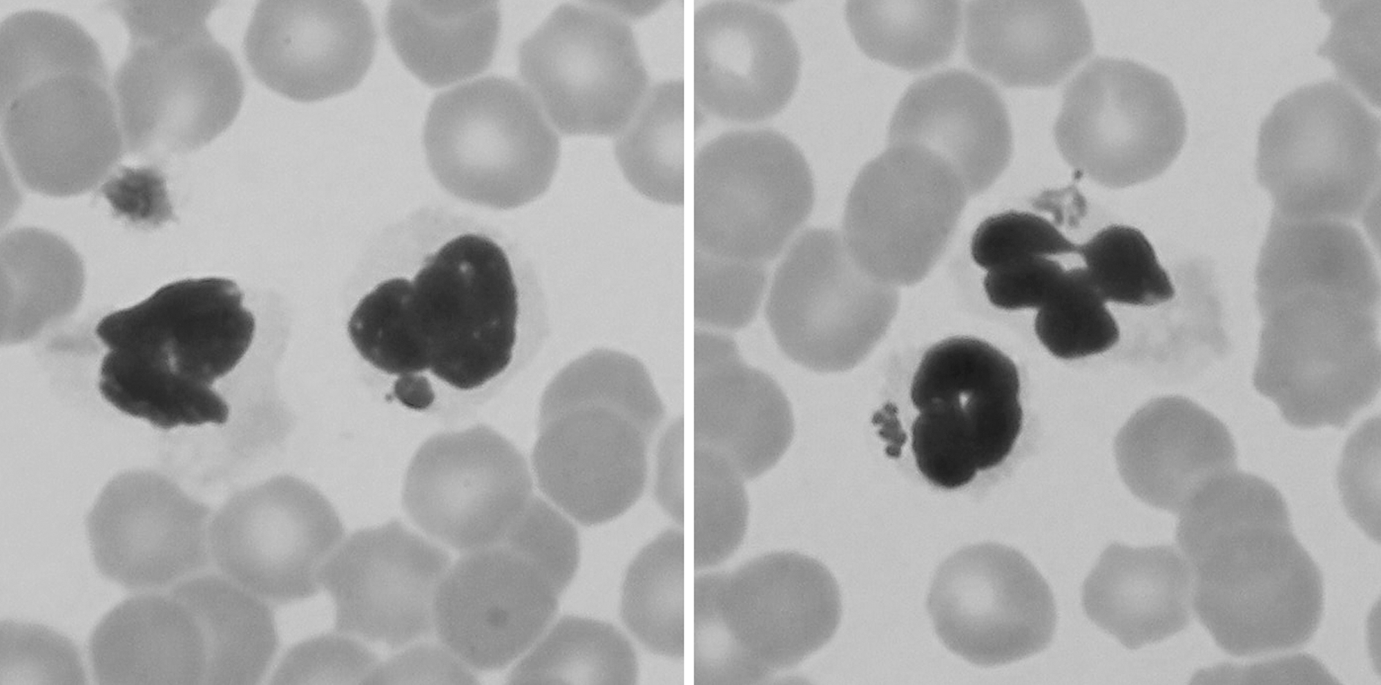
Morulae of A. phagocytophilum in neutrophil granulocytes of a dog.
Discussion
The seropositivity rates in the examined dogs indicate natural exposure to A. phagocytophilum and B. burgdorferi s. l. We found a 25.9% positivity of IgG antibodies against A. phagocytophilum. For comparison, the reported canine seroprevalence rates were 17.7% for granulocytic Ehrlichia in Sweden (Egenvall et al. 2000b) and 7.5% for A. phagocytophilum in Switzerland (Pusterla et al. 1998). In Germany, antibodies to A. phagocytophilum were found in 43.2% of examined dogs (Jensen et al. 2007), in Israel in 9% (Levi et al. 2006), and in the United States, the canine seroprevalence rates were between 9.4% (Magnarelli et al. 1997) and 29% (Beall et al. 2008). The canine seroprevalence was reported to be 11.5% and 15.5% in Spain (Solano-Gallego et al. 2006, Amusategui et al. 2008) and 34.4% in Italy (Torina and Caracappa 2006). All studies used IFA, except Beall et al. (2008), who used ELISA. The above differences may have resulted from the use of different selection criteria for examined dogs. In the present study, no difference in seropositivity was found between symptomatic (27.5%) and asymptomatic (24.5%) dogs. Similar findings have been reported in Germany and the United States (Jensen et al. 2007, Beall et al. 2008). In our study, seropositivity in dogs without clinical signs can be explained by persistent antibodies as reported in chronically infected animals (Egenvall et al. 2000a). Seropositivity in many healthy dogs indicates that antibodies to A. phagocytophilum in dogs can persist (Madigan et al. 1990, Magnarelli et al. 1997, Engvall et al. 2002).
In the present study, DNA of A. phagocytophilum was detected in 3.4% (10/296) of dogs. Similar PCR positivity rates (i.e., 6.3%, 5.5%, and 9.5%) have been reported in Germany (Jensen et al. 2007), Italy (Torina and Caracappa 2006), and the United States (Beall et al. 2008), respectively. One out of 155 healthy dogs was PCR positive for A. phagocytophilum, which is similar to the study of Beall et al. (2008) in the United States (7/222 healthy dogs). Morulae in peripheral blood granulocytes were observed in only two of 10 PCR positive dogs, which corresponds to the findings of Jensen et al. (2007) (2/6). The inclusion bodies produced by A. phagocytophilum appear in granulocytes and can be demonstrated by microscopy only at the peak of the acute infection, which usually lasts only a few days (Engvall et al. 2002).
We found 2.4% IgM and 10.3% IgG canine seropositivity rates for B. burgdorferi s. l. Serologic detection of B. burgdorferi s. l. in dogs in the Czech Republic performed in 2006 (Pejchalova et al. 2006) showed a seroprevalence rate of 6.5%. The reported figures vary widely from 0.6% and 6.9% in Spain (Solano-Gallego et al. 2006, Amusategui et al. 2008*), 3.9% in Sweden (Egenvall et al. 2000b*), and 1.85% in Canada (Gary et al. 2006) to 11% in the United States (Beall et al. 2008), approximately 20% in France, the United Kingdom, and Denmark (Doby et al. 1988*, May et al. 1991, Hansen and Dietz 1989*), 40.2% in Poland (Skotarczak et al. 2005), and 50% in Slovakia (Stefanciková et al. 1998). Most of the studies were based on ELISA, except for those marked by an asterisk (*), which were based on IFA. As for A. phagocytophilum, the differences may have resulted from different dog selection criteria. Differences in canine seroprevalence rates for tick-borne diseases can arise from variability in tick densities or proportion of infected ticks.
In the present study, we detected DNA of B. burgdorferi s. l. in peripheral blood of only one PCR-positive dog. This low yield can be explained by the transient nature of the presence of the spirochetes in the blood (Straubinger et al. 1997, Chang et al. 2001). Using PCR, borrelial DNA can only be detected in blood in the early stage of infection (Skotarczak and Wodecka 2003). Once the microorganism is disseminated in the body, a variety of organs can be affected, especially the skin, joints, heart, and central and peripheral nervous systems (Straubinger et al. 1997).
We found DNA of A. phagocytophilum and B. burgdorferi s. l. in 8.5% and 6.8% of ticks, respectively. Anaplasma-positive ticks on positive dogs might have become positive during feeding. Similar findings have been reported in Poland (Zygner et al. 2008). We found no coinfection of Anaplasma and Borrelia in dogs or in ticks.
In conclusion, our findings suggest that exposure to A. phagocytophilum is common in dogs in the Czech Republic. This study demonstrated that symptomatic dogs from North and South Moravia and from North Bohemia had higher chance to be infected with A. phagocytophilum that asymptomatic dogs. On the other hand, no correlation was found between clinical signs of anaplasmosis or borreliosis and positive antibody titers to A. phagocytophilum or B. burgdorferi s. l., respectively.
Footnotes
Acknowledgments
The authors thank M. Maly for statistical analysis, M. Musílek for sequencing, A. Nemec and J. Kybic for helpful comments, and E. Kodytková for reviewing the manuscript.
Disclosure Statement
No competing financial interests exist.