
Abstract
Bartonella spp. are fastidious, gram-negative, rod-shaped bacteria and are usually vector-borne. However, the vector has not been definitively identified for many recently described species. In northern California, gray foxes (Urocyon cinereoargenteus) are infected with two zoonotic Bartonella species, B. rochalimae and B. vinsonii subsp. berkhoffii. Fleas (range 1–8 fleas per fox) were collected from 22 (41.5%) of 54 gray foxes from urban and backcountry zones near Hoopa, California. The flea species were determined, and DNA was individually extracted to establish the Bartonella species harbored by these fleas. Of the 108 fleas collected, 99 (92%) were identified as Pulex simulans. Overall, 39% (42/108) of the fleas were polymerase chain reaction (PCR)-positive for Bartonella, with B. rochalimae and B. vinsonii subsp. berkhoffii identified in 34 (81%) and 8 (19%) of the PCR-positive fleas, respectively. There was no difference between the prevalence of Bartonella spp. in P. simulans for the urban and backcountry zones. Fourteen (64%) of the 22 foxes were Bartonella bacteremic at one or more of the capture dates. In 10 instances, both the foxes and the fleas collected from them at the same blood collection were Bartonella-positive. B. rochalimae was the predominant species identified in both foxes and fleas. The competency of Pulex fleas as a vector of B. rochalimae has not been confirmed and will need to be demonstrated experimentally. Pulex spp. fleas readily feed on humans and may represent a source of human exposure to zoonotic species of Bartonella.
Introduction
Bartonella spp. are usually vector-borne bacteria, transmitted mainly by fleas, lice, or sandflies (Boulouis et al. 2005). Ctenocephalides felis, the cat flea, is the main vector for B. henselae and presumably for B. clarridgeiae (Chomel et al. 1996, Kordick et al. 1997, Finkelstein et al. 2002, Rolain et al. 2003a, Kelly 2005). DNA from several Bartonella species has been detected in cat fleas, including B. henselae, B. koehlerae, B. clarridgeiae, and B. quintana (Rolain et al. 2003a). Bartonella DNA has also been detected in recent years in Pulex spp. fleas. Bartonella quintana DNA was detected in three P. irritans fleas taken from a pet moustached monkey (Cercopithecus cephus) in Gabon (Rolain et al. 2005). In addition, DNA from three unidentified species of Bartonella has been detected in Pulex spp. fleas collected from the bedding and clothes of schoolchildren and adults in Peru (Parola et al. 2002). A partial sequence from the 16S–23S intergenic spacer (ITS) region from one of these fleas was most similar to B. clarridgeiae. The ITS sequence from this flea was later discovered to be 100% identical to the ITS sequence from a Bartonella sp. isolated from an American woman with fever, rash, splenomegaly, and a recent history of travel in Peru. This person was also reported to have many insect bites. This human isolate, recently described as a new species of Bartonella, has been designated Bartonella rochalimae (Eremeeva et al. 2007).
Bartonella vinsonii subsp. berkhoffii, a pathogen of dogs and humans, has been suspected to be carried by ticks, based both on seroepidemiological studies and DNA detection in different tick species, but transmission by ticks has not been confirmed (Chang et al. 1999, 2001, Chomel et al. 2006). Detection of B. vinsonii subsp. berkhoffii DNA has not been reported to date from cat fleas. However, DNA extracted from a Pulex flea collected from a human in Peru showed a sequence closely related to B. vinsonii subsp. berkhoffii (Parola et al. 2002). In domestic dogs (Canis familaris), B. vinsonii subsp. berkhoffii infection has been associated with endocarditis, myocarditis, arrhythmia, and other diverse clinical outcomes (Breitschwerdt and Kordick 2000, Chomel et al. 2006). At least one human case of endocarditis-associated B. vinsonii subsp. berkhoffii has also been reported (Roux et al. 2000).
Wildlife may function as natural reservoirs or maintenance hosts for Bartonella species, and peridomestic wildlife may bring arthropod vectors in close contact with domestic animals and humans. Coyotes (Canis latrans) are suggested to be the main sylvatic reservoir of B. vinsonii subsp. berkhoffii in California due to the high percentage (28%) of bacteremic animals reported in one study (Chang et al. 1999). In northwestern California, gray foxes (Urocyon cinereoargenteus) may serve as a reservoir for the recently described B. rochalimae and B. vinsonii subsp. berkhoffii (Henn et al. 2006, 2007). Among 53 gray foxes from Humboldt county, 26 (49%) were Bartonella bacteremic, 22 (42%) with B. rochalimae (formerly B. clarridgeiae-like), and 5 (9.4%) with B. vinsonii subsp. berkhoffii (Henn et al. 2006, 2007). Bartonella rochalimae, previously designated B. clarridgeiae-like, has also been associated with a case of endocarditis in a dog (MacDonald et al. 2004).
Gray foxes are a cosmopolitan species, which can often occur in high densities (Trapp and Hallberg 1975, Fritzell and Haroldson, 1982, Cypher 2003), and commonly harbor ectoparasites that could be vector-competent for Bartonella species. Moreover, gray foxes are frequently observed near human dwellings, can interact with pets, and eat unattended outdoor pet food, garden crops, or refuse left outside of these dwellings (Harrison 1993, Cypher 2003). The purpose of the present study was to identify fleas collected from gray foxes in a rural area of northern California to determine if Bartonella spp. DNA could be amplified from these fleas. The gray foxes had been previously tested for Bartonella (Henn et al. 2007), allowing for comparison of Bartonella spp. DNA identified in the fleas with the results of blood culture performed on their hosts.
Materials and Methods
Sampling was conducted on the Hoopa Valley Indian Reservation in northeastern Humboldt County, California (UTM 10 04 43624 E, 45 44 450 N). Gray foxes were live-trapped from June 2003 to mid-October 2004 in 81 × 25 × 31–cm wire mesh traps (Model 108 Tomahawk Live Trap Company, Tomahawk, WI) and the trapping protocol followed the methods described previously (Gabriel 2006, Henn et al. 2007). A total of 54 foxes were trapped 70 times, as several foxes were trapped more than once (Henn et al. 2007).
A thorough systematic examination (≥5 minutes) of the fur on each individual fox was conducted. All fleas observed were collected in 70% ethanol for later identification. Foxes were designated as an “urban fox capture” or a “backcountry fox capture” (Gabriel 2006). The urban zone included the valley floor as it follows the Trinity River over 16.4 km in a south–north direction with elevations varying between 76 m and 152 m above sea level (Singer and Begg 1975), where the majority of the people live (Fig. 1). The backcountry zone included the remaining area of the Hoopa Valley Indian Reservation with elevations ranging from 152–1170 m. This area is managed for its natural resources and cultural preservation. Fleas that were removed from the foxes were also assigned the same code number and locality designation than the fox from which it was removed.
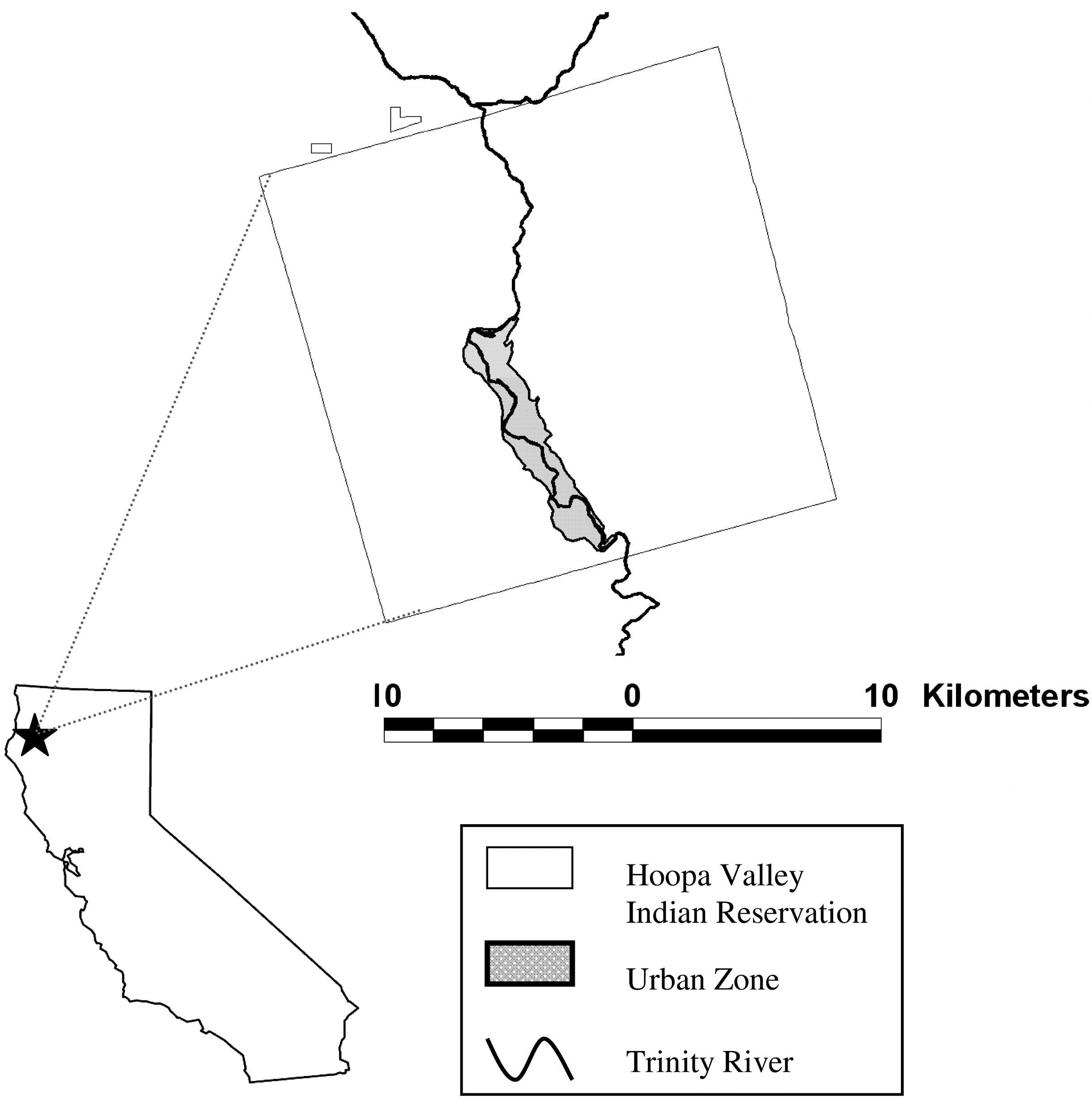
Map of the Hoopa Valley Indian Reservation, Humboldt County, California.
Flea identification
All fleas in vials that had five or fewer fleas per fox capture were identified to species. In the vials that had more than five fleas (up to eight fleas maximum), only five individual fleas that showed superficially visible morphological differences (i.e., pronotol combs, genal combs) were identified from each capture and were processed. Fleas were maintained in 70% ethanol, removed from ethanol individually onto a clean glass slide and allowed to sit until the ethanol had evaporated. An incision was made with a sterile scalpel blade across the tergites to release body contents, then the entire flea was incubated in a 1.5 mL Eppendorf tube with 180 μL of ATL buffer (QuiAmp, Valencia, CA) and 20 μL of proteinase K at 55°C for 6 hours. Subsequently, the exoskeleton was removed for mounting for identification by immersion in potassium hydroxide for 24 hours and then transfer through dehydration series (70%, 80%, 95%, and 100%) of ethanol for 30 minutes per step. Specimens were then transferred to methyl salicylate for 30 minutes, then xylene for 1 hour, and were mounted in Canada balsam. Each flea was identified to species using western North American taxonomic keys (Hubbard 1968, Lewis et al. 1988). All remaining material in the tube was processed for DNA extraction as per the Qiagen kit tissue extraction protocol (QuiAmp, Valencia, CA).
DNA extraction and polymerase chain reaction–restriction fragment length polymorphism procedures
DNA was extracted from the material remaining after the flea was removed from incubation in proteinase K using a commercial DNA extraction kit (QuiAmp). Flea DNA samples were analyzed by polymerase chain reaction (PCR) of the 16S–23S ITS region (Rolain et al. 2003b) and the ftsZ (Zeaiter et al. 2002) gene. PCR reaction vials were set up as previously described (Chang et al. 2000). Briefly, DNA was diluted 1:10 in buffer (10 mM Tris, 1 mM ethylenediaminetetraacetic acid) and 0.25 μM of each primer was used. The primers used for the ITS region were 5′-CTTCGTTTCTCTTTCTTCA-3′ and 5′-CTTCTCTTCACAA-TTTCAAT-3′ and the primers for the ftsZ gene were 5′-ATTAATCTGCAYCGGCCAGA-3′ and 5′-ACVGADACACG-AATAACACC-3′. Amplified PCR products were identified by ethidium bromide fluorescence after electrophoresis in 2% agarose gels (SeaKem LE agarose, Cambrex Bio Science Rockland Inc., Rockland, ME). The amplified product of the ITS region was digested with HaeIII restriction endonuclease (Promega, Madison, WI). Banding patterns were compared with the patterns observed for B. rochalimae and B. vinsonii subsp. berkhoffii isolates from the gray foxes from which the fleas were collected.
DNA sequencing
PCR products used for DNA sequencing were purified with QIAquick PCR purification kit (QIAGEN Sciences, Germantown, MD), and sequencing was done using a fluorescence-based automated sequencing system (Davis Sequencing, Davis, CA).
Statistical analysis
All statistical analyses (chi-square for trend) were conducted using statistical software from Number Cruncher Statistical Software (NCSS 2001). Results were considered significant if p values were ≤0.05.
Results
Of the 53 foxes for which a blood culture was performed, 22 (41.5%) had fleas and 108 fleas were removed for further analysis. On average, there were five fleas collected per infested fox (range 1–8 fleas). Ninety-nine (94%) fleas were identified as P. simulans, three (2.8%) as Ctenocephalides felis, and one each as Ctenocephalides canis, Cediopsylla inequalis interrupta, and Orchopeas laens. Seventy-five (76%) P. simulans were female and 24 (24%) were males. Two of the three C. felis were females and the single C. canis was a female. Three fleas could not be identified to species due to damage from mounting of the specimen leading to absence of distinctive morphological characters.
Forty-two (39%) of the 108 flea DNA samples tested were PCR-positive for Bartonella spp. based on the ITS region. Thirty-nine (93%) of the 42 Bartonella PCR-positive fleas were identified as P. simulans, with 28 (37%) positive females and 11 (46%) positive males. Despite a higher prevalence of infection in male P. simulans fleas, the difference was not statistically significant (χ 2 = 0.55, df = 1, p = 0.45). One C. felis, one C. inequalis, and one unidentified flea species were PCR-positive for Bartonella. Thirty-three (78.6%) of the 42 Bartonella PCR-positive samples had an ITS RFLP profile characteristic of B. rochalimae, and 8 (19%) had a banding pattern similar to that observed for the B. vinsonii subsp. berkhoffii strains isolated from gray foxes in Humboldt county. Banding patterns could not be determined for the remaining two Bartonella PCR-positive fleas.
Six flea samples with RFLP profiles from HaeIII digests of ITS PCR products matching B. rochalimae and four flea samples with RFLP profiles matching B. vinsonii subsp. berkhoffii were selected for nucleic acid sequencing. Whenever enough DNA was available, sequencing was performed for both the ITS region and ftsZ gene. For the six fleas with RFLP profiles matching B. rochalimae, partial sequences from the ITS region and ftsZ gene were ≥99.6% identical to B. rochalimae previously isolated from the gray foxes. For two of the four flea products sequenced with RFLP profiles similar to B. vinsonii subsp. berkhoffii, ITS and ftsZ DNA sequences were 100% identical to B. vinsonii subsp. berkhoffii previously isolated from the gray foxes. For the third flea, the ITS sequence was identical to B. vinsonii subsp. berkhoffii, but the partial sequence from the ftsZ gene amplicon was identical to B. rochalimae. For the fourth flea, the RFLP profile for the initially amplified PCR product had suggested B. vinsonii subsp. berkhoffii, but when DNA was amplified a second time and sent for sequencing, the ITS sequence was identical to B. rochalimae; there was not enough DNA available for partial sequencing of the ftsZ gene. Finally, for the two Bartonella-positive flea samples that did not have clear RFLP profiles due to low amounts of DNA in the ITS PCR product, sequences from the ftsZ PCR product for these samples were identical to B. rochalimae. Overall, 39% (42/108) of the fleas were PCR-positive for Bartonella, with B. rochalimae and B. vinsonii subsp. berkhoffii identified in 33 (78.5%) and 8 (19%) of the PCR-positive fleas, including two fleas that appeared to be coinfected.
Sixty-six P. simulans were removed from foxes in the urban zone, and 33 P. simulans were removed from foxes in the backcountry zone. Two C. felis, the C. inequalis, and C. canis were removed from a fox in the urban zone. One C. felis and the O. laens flea were removed from a fox in the backcountry zone. Twenty-three (34.8%) of the 66 urban P. simulans were PCR-positive, and 16 (48.5%) of the 33 backcountry P. simulans were positive. The difference between the prevalence of Bartonella spp. in P. simulans between the urban area and the backcountry was not statistically significant (χ 2 = 1.71, df = 1, p = 0.19).
Seven (32%) of the 22 foxes that had fleas removed from them were trapped more than once during the course of the study (Table 1). Fourteen (64%) of the 22 foxes were found to be infected with Bartonella spp. on one or more capture dates when tested as part of a previous study (Henn et al. 2007). There were 10 (71.4%) instances in which both the foxes and the fleas collected from them on the same capture date were positive for Bartonella DNA. In 7 (72.7%) of these 11 pairs (foxes #6, 8, 12, 13, 14, 18, 21), both the foxes and fleas were positive for B. rochalimae. For two pairs, the foxes (#1 and #3) were bacteremic with B. vinsonii subsp. berkhoffii and had fleas on them that were PCR-positive for both B. vinsonii subsp. berkhoffii and B. rochalimae. The last pair was composed of a fox (#22) infected with B. rochalimae, but with one out of five fleas infected with B. vinsonii subsp. berkhoffii. There were three foxes (#2, 9, 17) infected with B. rochalimae that had no Bartonella-positive fleas at the time of the positive blood culture. One fox (#11) was sequentially bacteremic with B. vinsonii subsp. berkhoffii and B. rochalimae, but the only PCR-positive fleas (infected with B. rochalimae) were collected on the last recapture, when the animal was already abacteremic. Finally, there were eight foxes (#4, 5, 7, 10, 15, 16, 19, 20) that were Bartonella culture-negative that had 32 fleas collected from them, 12 (37.5%) of which were Bartonella PCR-positive. Eleven (91.7%) of these fleas were positive for B. rochalimae.
The Flea PCR+column describes the number of fleas that were Bartonella PCR-positive out of the total number of fleas tested from that fox. PCR, polymerase chain reaction; NA, not applicable; ND, not done; Neg, negative; Br, Bartonella rochalimae; Bvb, Bartonella vinsonii berkhoffii.
Discussion
This is the first report of the presence of two zoonotic bartonellae, B. rochalimae and B. vinsonii subsp. berkhoffii, in fleas collected from gray foxes. Most of these fleas were identified as P. simulans, which are commonly found on gray foxes (Lewis et al. 1988, Hubbard 1968). Overall, 42 (39%) of the 108 fleas were Bartonella PCR-positive. B. rochalimae was the predominant (81%) species identified in these fleas. Two of the eight samples that were initially identified as B. vinsonii subsp. berkhoffii based on the restriction fragment length polymorphism profile also appeared to have B. rochalimae DNA present when PCR amplification was performed a second time before sequencing. This unexpected finding suggests that these fleas were harboring both B. vinsonii subsp. berkhoffii and B. rochalimae.
The prevalence of B. rochalimae and B. vinsonii subsp. berkhoffii identified in fleas was similar to the prevalence of the two species identified in foxes by blood culture. Bartonella rochalimae was isolated from 22 (42%) of the 53 gray foxes tested, and B. vinsonii subsp. berkhoffii was isolated from only 5 (9.4%) gray foxes (Henn et al. 2007). Fleas from six of the eight nonbacteremic foxes were PCR-positive for B. rochalimae, and a flea on one of those six foxes was PCR-positive for B. vinsonii subsp. berkhoffii. This suggests that the amplification of Bartonella DNA from these fleas could reflect more than just a recent blood meal on a bacteremic host and that the organism may be multiplying in the flea gut, as has been demonstrated with B. henselae (Higgins et al. 1996). Furthermore, it may also reflect earlier infection of the fleas when feeding on another bacteremic fox or our lack of detecting a very low level of bacteremia in the culture-negative foxes.
Bartonella clarridgeiae-like and B. rochalimae DNA has also been detected in Pulex fleas from other parts of the world. Bartonella DNA was detected in Pulex fleas collected from people in Peru, including a sample described as similar to B. clarridgeiae (Parola et al. 2002) and later found to be identical to a human B. rochalimae isolate based on partial sequence of the 16S–23S ITS region (Ereemeva et al. 2007). In Chile, 5 (15%) of 33 P. irritans fleas collected on dogs were found to be Bartonella PCR-positive, and partial sequencing of the rpoB gene identified a Bartonella similar to B. clarridgeiae (95% identity; Gonzales-Acuna et al. 2006). Furthermore, Bartonella DNA was detected in 4 of 19 pools (total 95 P. irritans) collected from four different red foxes (Vulpes vulpes) in Hungary (Sreter-Lancz et al. 2006). The groEL sequences of all four positive pools were identical and similar to the sequence of a Bartonella detected in rat fleas (Xenopsylla cheopis) from Egypt and similar to B. clarridgeiae in the maximum parsimony tree published by Loftis et al. (2006). These data are suggestive that DNA from a B. clarridgeiae-like group, possibly B. rochalimae, could be identified in P. irritans fleas collected on red foxes in central Europe and in rat fleas in Egypt, two flea species that can infest humans. Furthermore, strains closed to B. clarridgeiae were recently identified in Ctenophthalmus lushuiensis fleas collected in vole nests from Yunnan, China (Li et al. 2007), and strains close to B. clarridgeiae and B. rochalimae were described from two fleas (Polygenis gwyni) collected from cotton rats (Sigmodon hispidus; Abbot et al. 2007).
There were no differences between the Bartonella prevalence in fleas collected from foxes in the urban zone and in the backcountry, suggesting that this pathogen is well distributed among the fox population in the Hoopa Valley. Gray foxes are likely to represent one of the main reservoirs of B. rochalimae in this area of northern California. The competency of P. simulans as a vector of B. rochalimae and B. vinsonii subsp. berkhoffii has not been established, and experimental studies will be needed to demonstrate transmission of these organisms by P. simulans to pathogen-free hosts. As Pulex spp. will readily feed on humans, these fleas may represent a source of human exposure to zoonotic species of Bartonella.
Footnotes
Acknowledgments
We would like to thank the Hoopa Tribe and the Hupa people for providing access to this study area. We also would like to acknowledge Mark Higley at Hoopa Tribal Forestry, Nikki Drazenovich at the Center for Vector-Borne Diseases, University of California, Davis, and Mark Early at Humboldt State University for assistance in the construction of traps.
Disclosure Statement
Financial support was provided by MGW Biological, Integral Ecology Research Center, Stockton Sportsman’s Club, Stanley W. Harris Scholarship, Humboldt State University Graduate Equity Fellowship, and the Center for Vector-Borne Diseases, University of California, Davis.