




Abstract
A pilot study was conducted to determine whether existing human or canine strains of Anaplasma phagocytophilum would reproduce clinical disease in experimentally inoculated dogs similar to dogs with naturally acquired granulocytic anaplasmosis. Six hounds were inoculated intravenously with one human and two canine strains of A. phagocytophilum that were propagated in vitro in HL-60 cells or in infected autologous neutrophils. Infected dogs were monitored for lethargy, anorexia, petechiae, lymphadenopathy, and fever. Dogs were assessed for complete blood count (CBC), serum chemistry, and serology (IFA and SNAP® 4Dx®); for A. phagocytophilum blood load by quantitative polymerase chain reaction; and for cytokine production. Prominent clinical signs were generalized lymphadenopathy and scleral injection; only one dog developed fever lasting 4 days. Notable laboratory alterations included sustained leukopenia and thrombocytopenia in all dogs. A. phagocytophilum morulae were noted in blood between days 10 and 11, although all dogs retained A. phagocytophilum DNA in blood through day 60. All dogs seroconverted by days 10–15 by IFA, and by days 17–30 by SNAP 4Dx; cytokine analyses revealed 10-fold increases in interleukin-2 and interleukin-18 in the neutrophil-propagated 98E4 strain-infected dog. All A. phagocytophilum strains produced infection, although canine 98E4 strain reproduced clinical signs, hematologic changes, and inflammatory cytokine elevations most consistent with granulocytic anaplasmosis when recognized clinically. Therefore, this strain should be considered for use in future studies of A. phagocytophilum canine infection models.
Introduction
A pilot study was conducted to determine which strain and delivery method of A. phagocytophilum would be most suitable to reproduce clinical disease in dogs. For these studies, we compared two inoculation methods and three different A. phagocytophilum strains. The inoculation methods chosen are advantageous because they are used extensively in the laboratory, bypass tick bite delivery by intravenous inoculation, and utilize cell-free bacterial preparations.
Materials and Methods
Inoculum and culture of A. phagocytophilum
A. phagocytophilum was propagated in HL-60 cells in vitro, and all strains were less than eight in vitro passages when used. Infected and uninfected HL-60 cells, a promyelocytic cell line, were maintained in the RPMI 1640 culture medium supplemented with 5% fetal bovine serum and 2 mM L-glutamine at 37°C in a 5% CO2 environment until sufficient quantities of bacteria or infected cells were obtained for infection. Prior experiments in the laboratory demonstrated that 1 × 10⁶ infected cells or 1 × 10⁷ cell-free bacteria are sufficient to reproduce detectable infection in dogs, mice, and horses (Choi et al. 2003 and unpublished data).
Because inoculation with infected HL-60 cells introduces the potential confounding factor of immune stimulation by human cells, we used infected autologous neutrophils for half of the studies. Neutrophils from ∼5 to 10 mL of ethylenediaminetetraacetic-acid-anticoagulated blood of each dog were isolated by Ficoll-Hypaque density gradient centrifugation from dextran-sedimented leukocyte-rich plasma. Hypotonic erythrocyte lysis resulted in >95% neutrophil purity and >90% viability by trypan blue exclusion. Isolated neutrophils were then infected with cell-free A. phagocytophilum as follows. A quantity of A. phagocytophilum–infected HL-60 cells (106–107 cells) equal to that of neutrophils to be infected was centrifuged at 450 g for 10 min, and the cell pellet was suspended in 2 mL of the medium and sonicated just until complete HL-60 cell lysis was achieved. Thereafter, the sonicate was centrifuged at 200 g for 5 min to remove cellular debris and nuclei. The cell-free bacterium-enriched supernatant was washed in 5–10 volumes of the tissue culture medium and centrifuged at 15,000 g to harvest bacteria. Bacteria were then resuspended in a volume of RPMI 1640 medium sufficient for infection of isolated neutrophils. These cells were incubated for 18 h at 37°C in 5% CO2.
Dogs
Six specific pathogen free (SPF) large hounds were purchased from a Class A USDA vendor and housed in our canine facilities according to the Guide for the Care and Use of Laboratory Animals (National Research Council) and the Animal Welfare Act regulations. Two dogs each were infected with one of three different A. phagocytophilum strains (Table 1). One group received the A. phagocytophilum Webster strain, originally isolated from a human patient, and the two other groups received one of two A. phagocytophilum strains isolated from the peripheral blood of canine clinical cases in Minnesota (98E4 strain, courtesy of Barbara Grieg, DVM) and California (E06 strain, courtesy of Jane Sykes, DVM). The Webster strain was chosen since it is the A. phagocytophilum type strain and the standard strain we used for many murine studies. Each dog was infected intravenously (cephalic vein) with 106 infected HL-60 cells or neutrophils (Table 1).
Clinical assessment
All dogs were observed for baseline parameters and behaviors before infection, assessing activity levels, food consumption, and water intake. Once infected, dogs were monitored daily and clinical findings recorded. Rectal temperature was also performed once daily, and pulse and respiration were noted. Food consumption, water intake, fecal production, urine output, changes in ambulation, and occurrence of lameness were also recorded. Particular attention was paid to differentiating arthralgia versus arthritis. All animals were followed for 30 days postinfection. All clinical observations were scored using a numerical grading system from 0 to 3, with 0 being absent clinical signs, and 3 being a severe manifestation of the clinical sign. The total clinical score represents the sum of grades for lethargy, anorexia, lameness, lymphadenopathy, weight loss, petechiae, scleral injection, and icteric mucous membranes.
Bloodwork and laboratory analysis
Blood was collected on days 0, 2, 4, 7, 10, 14, 17, 21, 25, 30, 45, 54, and 60 (13 time points). CBC with blood smear review was performed on all samples, as was complete serum chemistry analysis, paying particular attention to hepatic transaminases (IDEXX Laboratories).
DNA extraction and A. phagocytophilum quantitative polymerase chain reaction
Quantitative A. phagocytophilum polymerase chain reaction (PCR) was performed on blood at both Johns Hopkins and IDEXX. Briefly, nucleic acids were prepared from ethylenediaminetetraacetic-acid-anticoagulated blood using the Qiagen or Promega Wizard DNA mini extraction kit. Bacterial DNA was quantified at Johns Hopkins using the BioRad IQ5 multicolor Real-Time PCR detection system, targeting A. phagocytophilum major surface protein-2 (msp2), as previously described (Scorpio et al. 2004). Quantitative PCR (qPCR) at IDEXX Laboratories was performed using the Lightcycler 480 with proprietary forward and reverse primers and donor/acceptor probes. Preliminary experiments demonstrated the ability for these qPCR methods to detect as few as one infected HL-60 cell/μL (1–2 bacteria/μL) of whole-blood DNA (data not shown). It has been determined that based on the known 105 copies of msp2 in the A phagocytophilum HZ strain genome and the known number of infected cells put into the reaction that the msp2 qPCR detects ∼1–2 bacteria/infected cell, whether an HL-60 cell or neutrophil. Samples were tested in duplicate with appropriate positive and negative controls, and quantitation was achieved using a standard curve with serial dilutions of 106 infected HL-60 cells down to 10−2 infected cells.
Serology
Antibody detection was performed on dog sera collected over the 60-day period by in-house IFA and the SNAP® 4Dx® Test Kit (IDEXX Laboratories). In the IFA test, A. phagocytophilum–infected HL-60 cells were used, and a titer of 80 or greater was considered positive. The SNAP 4Dx Test is a rapid, in-clinic enzyme-linked immunosorbent assay that uses a synthetic peptide from the major outer surface protein (P44) for the detection of A. phagocytophilum antibodies. For the SNAP 4Dx® Test, a visual +/− result is observed and demonstrates the presence of antibodies without quantification.
Cytokine analysis
Inflammatory cytokines in infected animals at baseline and through day 21 of infection were evaluated in serum samples using the Luminex multianalyte platform (canine interferon gamma [IFNγ], KC, interleukin-2 [IL-2], IL-6, IL-8, IL-10, IL-18, and tumor necrosis factor alpha [TNFα]; Canine Cytokine/Chemokine LINCOplex Kit; R&D Biosystems) per manufacturer's instructions. The kit has been validated for canine cytokines and chemokines by the manufacturer, and samples were run in duplicate. Quality control was assured by using canine quantitation standards for each cytokine. Quality control was further evaluated using Bioplex (BioRad) computer software for assessment of appropriate, reliable, and accurate performance with each replicate.
Statistical analysis
Mann–Whitney tests were used to compare the effects of infection methods on clinical, cytokine, and molecular parameters. Nonparametric statistics were chosen due to the assumption that variances would not be equal across groups due to small sample size. Means were calculated rather than medians to accurately calculate standard errors. Data were also analyzed with median values to verify similar statistical results when compared to mean values. Specific variables measured included clinical scores for lymphadenopathy, scleral injection, qPCR, cytokines (IL-2, IL-6, IL-8, IL-18, KC, and TNFα), and blood cell counts, including platelets, white blood cells, lymphocytes, monocytes, eosinophils, and neutrophils. Analyses were performed for each independent variable at each time point comparing autologous infected neutrophil versus infected HL-60 cell inocula. Overall means for both inoculum methods were plotted as change that occurred over time. SPSS software was used for Mann–Whitney analysis. Nonparametric results were verified using David Howell's re-sampling freeware (DC Howell, University of Vermont, 2007).
Results
Clinical assessment
All baseline scores were 0 for all dogs before infection. The most prominent clinical signs observed among dogs included generalized lymphadenopathy and bilateral scleral injection. Only one dog (98E4 in neutrophils) developed fever that lasted 4 days (103.9°F–104.1°F), with the highest temperature occurring between days 7 and 10 postinfection. No other dogs developed fever in the study. All dogs maintained appetite, while voiding functions and baseline activity levels remained unchanged. Lameness and petechiae were not observed. Total clinical scores were highest for dogs infected by either the California strain E06 (score = 70) in neutrophils or by HL-60 cells (score = 45). Clinical scores were similar for Webster and 98E4 strain-infected dogs (scores between 22 and 32), regardless of inoculation with bacteria grown in neutrophils or HL-60 cells.
Bloodwork and laboratory analysis
Biochemistry parameters remained normal throughout the experiment, with no changes noted in hepatic or renal function tests. The most prominent changes occurred in the CBC, predominantly involving leukocyte counts, differential leukocyte counts, and platelet counts. Most dogs showed thrombocytopenia beginning on day 4 and resolving by day 30, although none returned to baseline levels. Changes in leukocyte count were less dramatic and most dogs remained within normal limits. Dog 98E4-N (inoculated with infected autologous neutrophils) exhibited leukopenia of 4900 and 3400 leukocytes/μL (normal range 5700–16,300 leukocytes/μL) at days 7 and 10, respectively, corresponding to the febrile period and the time of thrombocytopenia (normal range 164,000–510,000 platelets/μL) (Figs. 1 and 2). This was also the time of peak A. phagocytophilum load and when morulae were noted in blood smear preparations. A. phagocytophilum morulae were also noted in blood smear preparations in dog E06-HL (inoculated with infected HL-60 cells) on day 10, when there was also a peak in bacterial load and lowest thrombocytopenia.

(

qPCR data plotted against platelet counts demonstrating the difference between inoculum delivery by infected autologous neutrophils versus infected HL-60 cells for three strains. All dogs developed infection (positive through and including day 60) with A. phagocytophilum as detected by qPCR using the msp2 gene target. Platelet counts reached a nadir in and around time of peak A. phagocytophilum bacterial load. msp2, major surface protein-2; qPCR, quantitative polymerase chain reaction. N, neutrophils; HL, HL60 cells.
PCR results
All dogs developed evidence of sustained A. phagocytophilum infection through day 60 (Figs. 1 and 2), and as early as day 2 postinoculation. For the Webster and 98E4 strains, peak bacterial load occurred between days 10 and 17 and coincided with peak thrombocytopenia. With the California E06 strain, one dog (E06-N) developed A. phagocytophilum peak load at day 54 and remained infected through day 60, the last time point. In the other dog (E06-HL), the bacterial load peaked twice, first around day 7–10 and again at day 45, and corresponded to platelet count nadirs each time (Fig. 2). qPCR results from both laboratories were similar 82% of the time. The remaining 18% of results were dissimilar due to differing qPCR amplification cut-off values between the two laboratories, although peak bacterial loads and infection kinetics were comparable throughout the study.
Serology
Using the SNAP 4Dx system, seroconversion occurred in four dogs by days 17–21 and in two dogs by day 30, and all remained sero-reactive through day 60. Using IFA, seroconversion occurred by days 10–14 for all dogs, and they remained sero-reactive for A. phagocytophilum immunoglobulin G at titers ≥2560 through day 60.
Cytokine levels
Cytokines were of low level or not detected for IFNγ, TNFα, IL-6, and IL-10. There was detection of cytokines IL-2, IL-8, IL-18, and KC, although mean levels across all six dogs at each time point exhibited large standard deviations and wide confidence intervals. This finding suggests significant individual variation in subjects that can depend upon genetic makeup, host immune response, inoculum type, or other unknown factors. For the detected cytokines, mean cytokine levels did generally follow infection status across time, particularly when cytokine elevations were detected between days 7 and 10, when clinical and hematologic findings were most abnormal and when bacterial loads peaked (data not shown). IL-8 levels remained stable across all time points. These cytokine patterns were largely due to levels noted in dog 98E4-N, since response to infection was less predictable and less dramatic when this dog was excluded from the mean levels described above. For dog 98E4-N on day 10, IL-2 and IL-18 levels were elevated to 10 and 7 times baseline values, respectively (Fig. 1C). Both levels declined by day 15 and returned to baseline by day 21.
Comparison of delivery methods
To determine the effects that heterologous versus autologous host cells used during inoculation have on clinical and molecular parameters in this canine model, dogs administered infected HL-60 cells and dogs administered infected autologous neutrophils were compared (Fig. 3). Temperatures were significantly higher (p < 0.05) for dogs given infected neutrophils than infected HL-60 cells at day 7, primarily due to one dog (98E4-N) with a temperature that reached 104.1°F. However, dogs given infected HL-60 cells exhibited higher overall temperatures from day 10 through 24 compared to dogs given infected neutrophils, although this was not statistically significant.
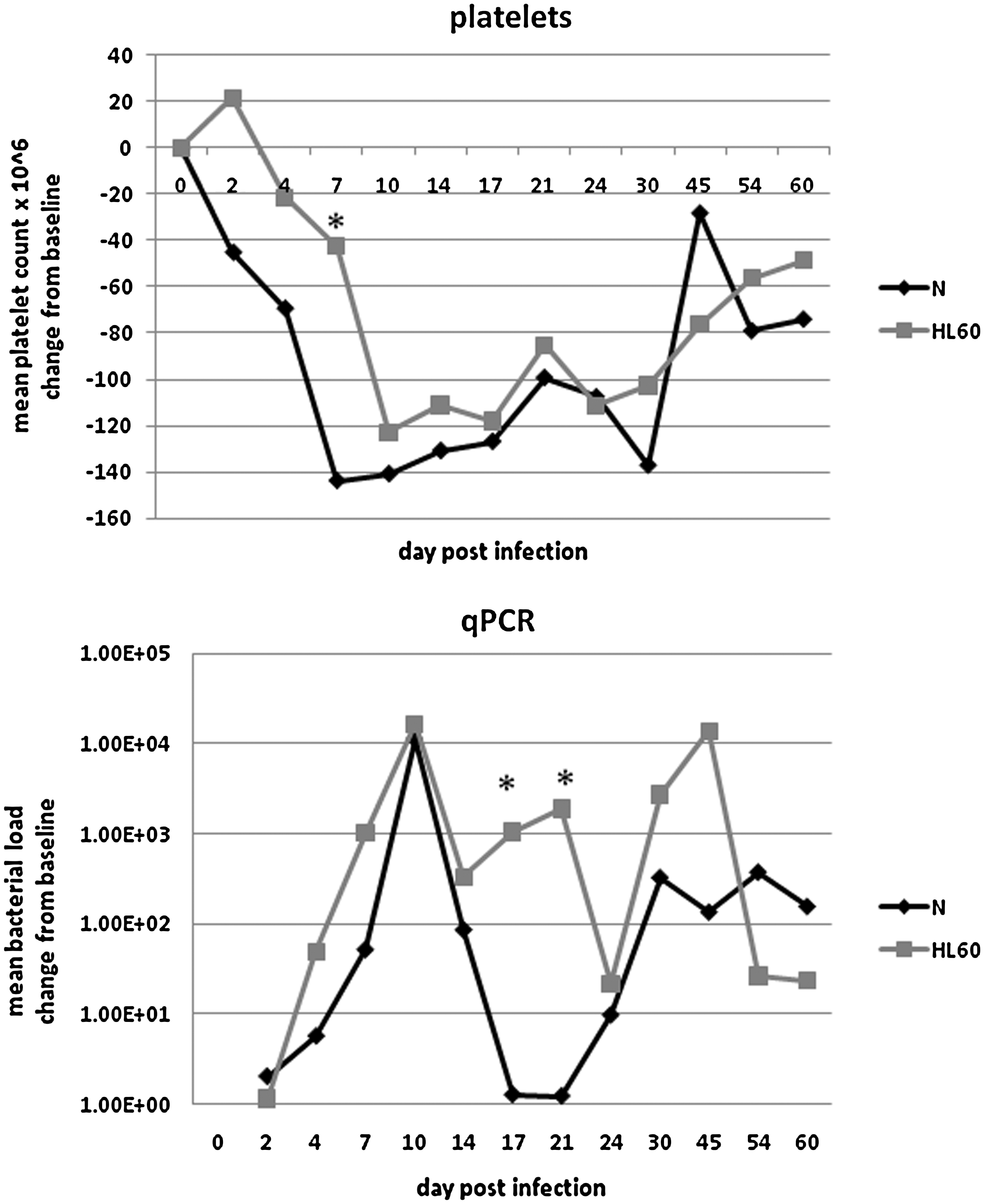
Comparison of mean changes in quantitative load and platelet counts between delivery of A. phagocytophilum by HL-60 cells (HL60) versus autologous neutrophils (N). In dogs given infected autologous neutrophils, there were lower bacterial loads (p < 0.05) on days 17 and 21 compared to those who received the heterologous inocula. A single peak was also noted at day 10 for neutrophil delivery compared to double peaks note at days 10 and 45 for the HL-60 inocula. Platelet counts declined sooner in dogs administered infected neutrophils compared to infected HL-60 cells; thrombocytopenia in dogs given infected HL-60 cells lagged kinetically, especially at day 7 (p < 0.05). Of note is that neither group of infected dogs returned to baseline platelet levels by the end of the study (day 60). Significant differences are noted by an asterisk.
Platelet counts declined sooner in dogs administered infected neutrophils compared to infected HL-60 cells; thrombocytopenia in dogs given infected HL-60 cells lagged, especially at day 7 (p < 0.05; Fig. 3). Dogs administered autologous neutrophils on average experienced a single peak in bacterial load at day 10 compared to a double peak phenomenon that occurred at days 10 and 45 in dogs administered infected HL-60 cells (Fig. 3). In dogs administered infected autologous neutrophils, bacterial loads were lower (p < 0.05) on days 17 and 21 compared to those that received the heterologous inocula (Fig. 3).
Among cytokines/chemokines, only KC exhibited any difference between inoculation groups, with dogs administered infected HL-60 cells demonstrating higher levels at days 4 and 21 compared to dogs given infected autologous neutrophils, although this finding was not statistically significant (p = 0.08 for both days).
Discussion
This study provides evidence that the human and canine strains of A. phagocytophilum tested produce infection in dogs. The canine A. phagocytophilum 98E4 strain administered in autologous neutrophils produced clinical signs and hematologic changes most consistent with human and canine granulocytic anaplasmosis, including thrombocytopenia, leukopenia, fever, lymphadenopathy, and an inflammatory response. This strain is a good candidate for use in future studies of tick-borne infections and coinfections in dogs. Animal numbers in this study were small; thus, additional studies are needed. However, it is important to highlight that this report is the first to demonstrate consistent ability to reproduce clinical signs in dogs after experimental infections with three different geographic strains of A. phagocytophilum.
In addition to clinical response, an important consideration when developing models of canine granulocytic anaplasmosis is the simultaneous occurrence of key infection and disease features, including the peak bacterial load at day 10, core body temperature, and cytokine response, and nadirs in thrombocytopenia and leukopenia. The time course of findings—early PCR detection of organisms, with expected seroconversion at 2 weeks and subsequent positive SNAP 4Dx Test—correlated well with laboratory abnormalities such as thrombocytopenia and presence of morulae. Coupled with consistent clinical signs, the disease presentation gives confidence for using the A. phagocytophilum 98E4 strain administered as infected autologous neutrophils in establishing a canine infection model. The other A. phagocytophilum strains and delivery methods established infection but with variable success in simultaneously reproducing the various elements of disease.
Increased levels of IL-2, KC, and IL-18 concentrations were observed in dogs at time of peak bacterial infection and clinical manifestations. Although CD4+ T lymphocytes are a main source of IL-2, CD8+ T cells and NK cells also can secrete it under certain conditions (Hoyer et al. 2008). Several signaling pathways, including the NF-κB pathway, regulate IL-2 expression (Hoyer et al. 2008). High concentrations of IL-2 can activate neutrophils, the host cell for A. phagocytophilum (Hoyer et al. 2008). IL-18 was originally discovered because of its ability to induce release of IFNγ and other pro-inflammatory mediators from macrophages and has since been shown to influence expression of other cytokines as well (Lapaque et al. 2009). IL-18 is now widely recognized as a cytokine stimulated with inflammasome activation (Pedra et al. 2007). It also synergizes with IL-2 to induce the production of other cytokines by T lymphocytes and NK cells (Lapaque et al. 2009).
In our canine studies, cytokines IFNγ and IL-10 were not detected, although their levels are routinely detected in other infected animals such as mice and horses (Martin et al. 2001, Kim et al. 2002), and humans (Dumler et al. 2005). While not anticipated, this finding could be explained since dogs can serve as reservoirs for tick-borne pathogens, and this and other studies demonstrate persistent infection with A. phagocytophilum. Thus, over time, the canine immune response has evolved to only partially control infection with these organisms to allow timely transmission to the vector. The lack of detection of other pro-inflammatory cytokines was anticipated since these are often absent in humans and particular animal models of A. phagocytophilum infection (Dumler et al. 2000, Kim et al. 2002).
Infected heterologous and homologous cells differed in their impact on clinical and hematologic parameters (Fig. 3). In general, inoculation of infected heterologous cells (human HL-60 cells) was associated with features of immune and inflammatory stimulation, as anticipated. The effect that this early stimulation has upon A. phagocytophilum propagation and subsequent disease manifestations is not entirely clear, but is likely to confound the model. Thus, these data support the use of autologous infected cells or cell-free bacteria as inocula for human granulocytic anaplasmosis (HGA) animal models.
Of interest, the strain E06, isolated from a canine clinical case in California, exhibited a bimodal pattern of infection, with bacterial load first peaking at days 10–14 postinfection, and then again between days 45 and 54, regardless of whether administered as infected autologous neutrophils or heterologous infected HL-60 cells. It is unclear what the bimodal infection phenotype suggests, but its occurrence is consistent with the re-emergence of antigenically diverse clones that would be predicted by the presence of the >105 paralogs of msp2 found present in the HZ strain genome sequence (Barbet et al. 2003, Lin et al. 2004, Scorpio et al. 2004, Hotopp et al. 2006). Further investigation of this particular strain in both canine and mouse models is warranted.
In light of published evidence demonstrating persistence of A. phagocytophilum in horses without prolonged clinical signs (Franzén et al. 2009), the topic of persistence in dogs is timely and relevant. This study also demonstrates that dogs given diverse strains via different delivery methods can remain PCR positive without clinical manifestations, as also observed in the equine study, although this is likely strain dependent (Franzén et al. 2009). Persistence of A. phagocytophilum organisms in dogs through day 60 did reveal continued abnormalities in platelet counts that did not return to baseline normal values at the conclusion of the study. One difference between this study and the equine investigation is that there was no stressor or dexamethasone used in our dogs. The persistence of A. phagocytophilum DNA was evident without immunosuppression. Since these dogs were not euthanized at the end of the study, we were not able to ascertain the level of bacterial burden in tissues, but suspect that the findings would be similar to the published results of Franzén et al. (2009). The data published in this investigation do point to the possibility that dogs with persistence of A. phagocytophilum in blood could serve as reservoirs for infection. This conclusion requires additional investigation, and should prompt consideration for more extensive evaluation in domestic pets for improved control and reduced public health threat.
Footnotes
Acknowledgments
The authors acknowledge the staff of Research Animal Resources at Johns Hopkins University for the daily care of the animals. Funding for this study was provided by IDEXX Laboratories Research and Development.
Disclosure Statement
There are no commercial associations for authors D.G.S., J.S.D., N.C.B., J.A.C., or C.B. For B.S., K.C.D., M.J.B., and R.C. of IDEXX Laboratories, there is no commercial conflict of interest since the information generated here is solely for scientific dissemination. There are no conflicts of interest to report by any author.