




Abstract
Wild deer are one of the important natural reservoir hosts of Anaplasma species, which cause granulocytic anaplasmosis in equines, canines, and humans. The objective of the present study was to determine whether and what species of Anaplasma naturally infect Korean water deer (KWD) in the Republic of Korea. A total of 66 spleens from KWD carcasses were collected by the Conservation Genome Resource Bank for Korean Wildlife in Korea between March 2008 and May 2009. Polymerase chain reaction (PCR) was performed using 16S ribosomal (r)RNA, with ankA, groEL, and msp2 gene primers to amplify the genes of Anaplasma and Ehrlichia. Using 16S rRNA-based nested PCR, Anaplasma phagocytophilum and Anaplasma bovis were detected in 42 (63.6%) and 23 (34.8%) of 66 KWD spleens, respectively. The 42 A. phagocytophilum were classified into five genotypes and the 23 A. bovis were classified into two genotypes by sequence analysis. By ankA-, groEL-, and msp2-based nested PCR, A. phagocytophilum was detected in 1 (1.5%), 7 (10.6%), and 3 (4.6%) of 66 samples, respectively. These gene sequences had only one genotype. Five of seven obtained 16S rRNA gene sequences have never been identified. The ankA, groEL, and msp2 obtained gene sequences represented new genotypes. This is the first report of A. phagocytophilum and A. bovis in KWD, suggesting that they may act as reservoirs for anaplasmosis zoonotic pathogens.
Introduction
A. phagocytophilum (formerly Ehrlichia phagocytophila) is the causative agent of human granulocytic anaplasmosis (Chen et al., 1994). Anaplasmosis is a febrile disease and an acute infection in ruminants, equines, and canines (Dumler et al. 2001), which is transmitted by Ixodes ticks. A. phagocytophilum was found in a variety of animal species including bears (Drazenovich et al. 2006, Víchová et al. 2010), cats (Bjöersdorff et al. 1999), dogs (Madewell and Gribble 1982, Dumler et al. 2001), gray squirrels (Nieto and Foley 2008), horses (Madigan and Gribble 1987), mice (Tyzzer 1938, Telford et al. 1996, Liz et al. 2000), mountain lions (Foley et al. 1999), raccoons (Levin et al. 2002), voles (Walls et al. 1997, Liz et al. 2000), skunks (Levin et al. 2002), white tailed-deer (Little et al. 1998), and woodrats (Nicholson et al. 1999). In the Republic of Korea (ROK), Heo et al. (2002) and Chae et al. (2009) demonstrated antibodies against Ehrlichia chaffeensis and A. phagocytophilum among serum samples from humans with febrile illnesses and cats, horses, and cattle of otherwise unknown etiology by an indirect fluorescent antibody test and Western blotting. Moreover, A. phagocytophilum was found in wild rodents and ticks by polymerase chain reaction (PCR) (Kim et al. 2003, 2006, Oh et al. 2009).
By sequencing analysis, A. bovis is more closely related to A. phagocytophilum than to A. centrale. Infection with A. bovis has been mainly reported in African cattle; however, A. bovis was also detected in Ixodes and Haemaphysalis species ticks and sika deer (Cervus nippon yesoensis) in Japan and the ROK (Kawahara et al. 2006, Kim et al. 2006, Lee et al. 2009, Oh et al. 2009).
Wild deer are suspected reservoirs of Anaplasma and Ehrlichia species infection. In the United States, A. phagocytophilum was detected in white-tailed deer (Odocoileus virginianus), roe deer (Capreolus capreolus), and mule deer (Odocoileus hemionus) (Belongia et al. 1997, Little et al. 1998, Liz et al. 2002, Yabsley et al. 2005). In Europe, Anaplasma spp. was identified in Iberian red deer (Cervus elaphus hispanicus) and roe deer (C. capreolus) (Petrovec et al. 2002, Oporto et al. 2003, De La Fuente et al. 2004). Further, in Japan and the ROK, Anaplasma and Ehrlichia spp. have been detected in sika deer (C. nippon yesoensis) (Kawahara et al. 2006, Lee et al. 2009). These findings indicated that wild deer could be reservoirs for Anaplasma spp. infection. However, Anaplasma spp. have not been evaluated in water deer and other wild deer in Asia. Water deer (Hydropotes inermis) are indigenous to the lower reaches of rivers in China and Korea. They have two subspecies: the Korean water deer (KWD, Hydropotes inermis argyropus) and the Chinese water deer (Hydropotes inermis inermis).
The aim of the present study was to investigate the prevalence of A. phagocytophilum in KWD and determine what genotypes of Anaplasma species could infect KWD in the ROK based on multiple PCR gene sequencing. Here, we report the first molecular evidence of Anaplasma spp. among naturally infected KWD in northeastern Asia.
Materials and Methods
Sample collections
KWD carcasses (n = 66) were collected in Chungcheongbuk-do, Gangwon-do, Gyeonggi-do, and Jeollanam-do provinces and Ulsan City in the ROK between March 2008 and May 2009 by the Conservation Genome Resource Bank for Korean Wildlife. All of the carcasses were stored at −20°C until necropsy at which time the spleens were sterilely harvested. Portions of the splenic tissues (100 mg) were frozen at −80°C until DNA extraction.
DNA extraction, PCR amplification, and PCR–restriction fragment length polymorphism
DNAs were extracted from spleens using DNeasy Blood and Tissue Kits (Qiagen) according to the manufacturer's instructions. For DNA extraction, 10 mg of spleen was used. PCR and nested PCR were performed using specific primers for A. phagocytophilum and A. bovis (Table 1). The A. phagocytophilum genomic DNA provided by J. Stephen Dumler (Johns Hopkins University School of Medicine, Baltimore, MD) was used as a positive control for detecting tick-borne pathogenic Anaplasma species. The first and nested PCRs were performed in a total volume of 25 μL. Each PCR mixture consisted of 10 pmol of primers, 1 U recombinant Taq DNA polymerase (Takara Bio, Inc.), 10× PCR buffer (Takara Bio, Inc.), 2.5 mM dNTP mixture (Takara Bio, Inc.), and 10–100 ng samples of genomic DNA for the first PCR and 1 μL of the first PCR product for the second PCR. The amplification was carried out in a PTC-200 thermal cycler (MJ Research, Inc.). Table 2 shows the conditions of the PCRs. Amplified products were separated by electrophoresis on 1.5% agarose gel and visualized by ethidium bromide. PCR products were digested with restriction endonucleases HinfI and HaeIII according to the instructions of the manufacturer (ELPIS-Biotech., Inc.). The HinfI and HaeIII restriction enzymes cut the sequences 5′-G/ANTC-3′ and 5′-GG/CC-3′, respectively. The digestion products were visualized by electrophoresis on an ethidium bromide-stained 1.5% agarose gel.
PCR, polymerase chain reaction.
ROK, Republic of Korea.
Cloning, nucleotide sequencing, and phylogenetic analysis
The PCR products were purified with QIAquick Gel Extraction kits (Qiagen). After purification, the amplicons were cloned with pGEM®-T Easy Vectors (Promega Corporation), followed by transformation into Escherichia coli JM109, and then plated onto Luria-Bertani (LB) agar containing 50 μg/mL of ampicillin. Plasmid DNA for sequencing was purified using the Wizard® Plus SV Minipreps DNA Purification System (Promega Corporation) according to the manufacturer's instructions. Purified recombinant plasmid DNA was sequenced using a T7 and SP6 promoter primer set by dideoxy termination with an automatic sequencer (ABI 3730xl capillary DNA sequencer). The obtained sequences were evaluated with Chromas software (Ver 2.33;
Nucleotide sequence accession numbers
The GenBank accession numbers of 16S ribosomal (r)RNA, groEL, msp2, and ankA gene sequences and specific genospecies sequences related to Anaplasmataceae pathogens for sequence comparisons are listed in Table 2.
Results
Anaplasma 16S rRNA genes were detected by conventional PCR and species-specific nested PCR assays with DNA from KWD spleens collected in the ROK (Table 2). Anaplasma spp. were detected in 50 of 66 KWD spleens (75.8%). A. phagocytophilum was identified in 42 (63.6%) specimens: 4 (80%) from Chungcheongbuk-do, 11 (73.3%) from Gangwon-do, 3 (60%) from Gyeonggi-do, 13 (72.2%) from Jeollanam-do, 8 (57.1%) from Ulsan City, and 3 (37.5%) from unknown areas. A. bovis was identified in 23 (34.8%) specimens: 4 (26.7%) from Gangwon-do, 1 (20%) from Gyeonggi-do, 8 (44.4%) from Jeollanam-do, 7 (50.5%) from Ulsan City, and 3 (37.5%) from unknown areas. In addition, 15 (22.7%) KWD were identified as being coinfected with both A. phagocytophilum and A. bovis: 4 (26.7%) from Gangwon-do, 1 (20%) from Gyeonggi-do, 7 (38.9%) from Jeollanam-do, and 3 (21.4%) from Ulsan City (Table 3).
Infected with A. phagocytophilum and A. bovis.
To investigate the genetic relationship between the Anaplasma species detected in the deer spleens, 65 genome sequences were analyzed and compared with the fragments of 16S rRNA gene sequences. A total of 42 variants of A. phagocytophilum 16S rRNA gene sequences were divided into five genotypes by sequence similarity. Pairwise comparison revealed the presence of KWDAP1 (n = 33), KWDAP2 (n = 4), KWDAP3 (n = 3), KWDAP4 (n = 1), and KWDAP5 (n = 1) genotypes. Phylogenetic analysis indicated that four new genotypes were identified in this study and one genotype (KWDAP1) corresponded to the previously sequenced A. phagocytophilum (AY527214) (Fig. 1). The 16S rRNA gene sequences from all 42 infected deer were 98.7%–100% identical to A. phagocytophilum (AY527214).
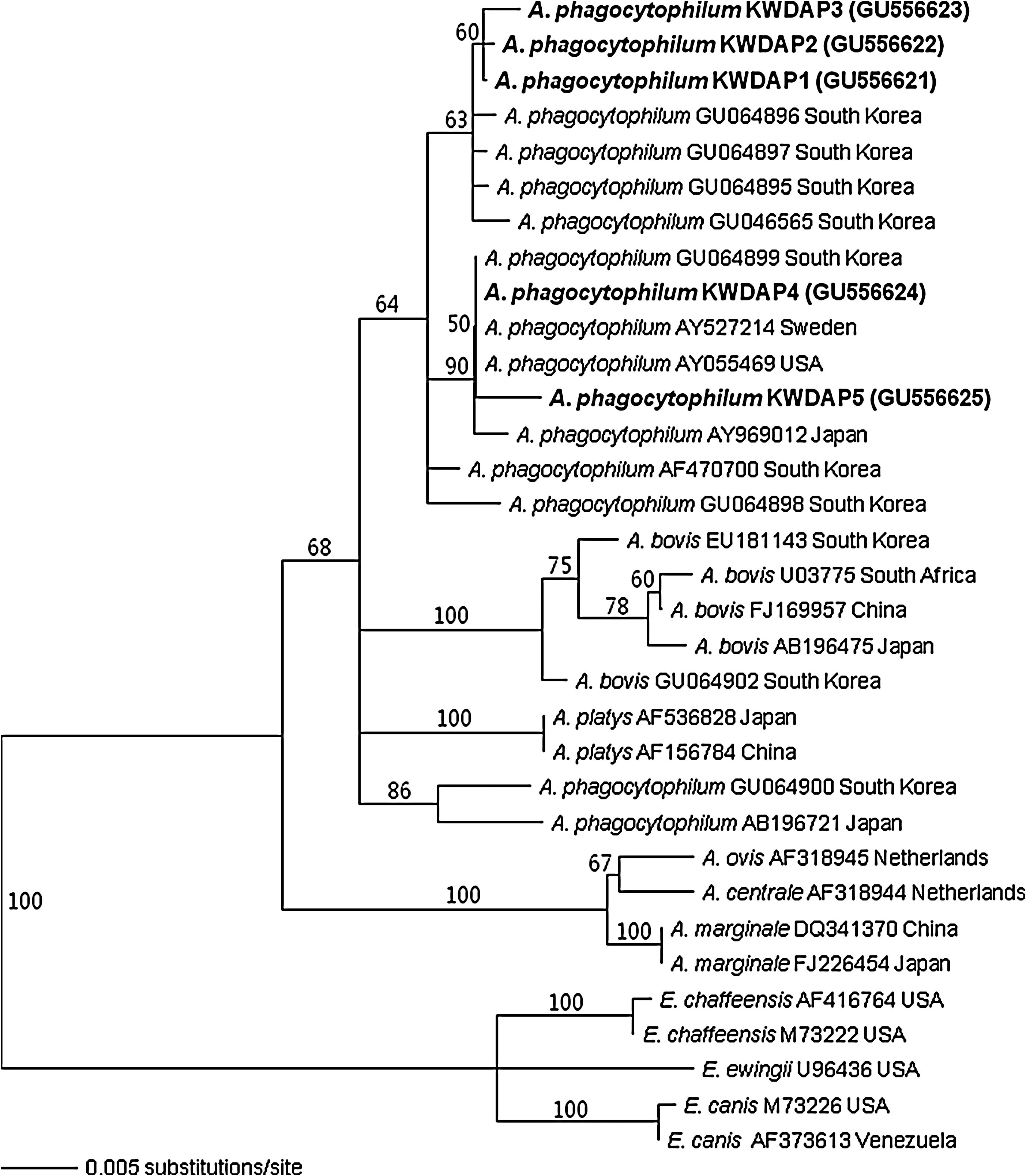
Phylogenetic relationships among Anaplasma phagocytophilum (bold letter) detected from the Korean water deer (KWD) and Anaplasma and Ehrlichia species based on partial nucleotide sequences of the 926-bp 16S rRNA gene. The neighbor-joining method was used for constructing a phylogenetic tree. The numbers at nodes are the proportions of 100 bootstrap resamplings that support the topology shown.
Using the groEL gene-specific primer pairs shown in Table 2 for PCR, seven A. phagocytophilum groEL gene sequences were obtained from 66 KWD spleens. The product had a size of 1297 base pairs (bp) and translated 409 amino acids of groEL. The seven groEL of amino acid sequences (KWDAPg) were identical to each other and 99.7% similar to A. phagocytophilum (AF172163) from a human in the United States (Fig. 2A). The KWDAPg genotype showed 86.3% and 84.6% homology to E. chaffeensis (L10917) and Ehrlichia canis (U96731), respectively.
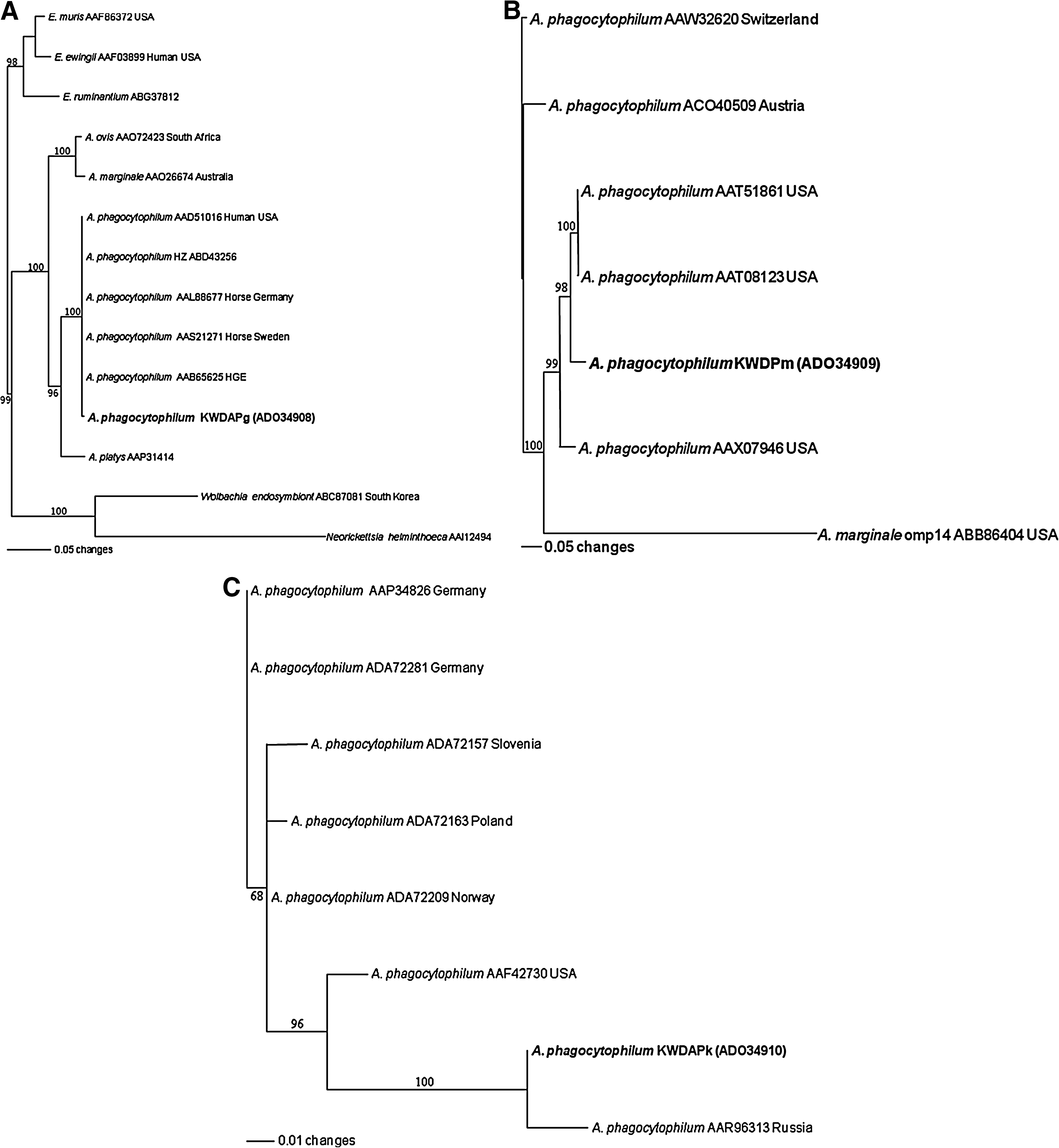
Phylogenetic relationships among A. phagocytophilum (bold face) detected in the KWD and Anaplasma and Ehrlichia species based on partial protein sequences of the 409-amino acid groEL
By msp2-specific nested PCR (Table 2), three A. phagocytophilum msp2 gene sequences were detected in the 66 KWD spleens. The product had a size of 457 bp and contained coding sequence for 151 amino acids of msp2. The three msp2 sequences of the amino acids (KWDAPm) were 100% identical to each other and were 89.4%–94.0% identical to A. phagocytophilum sequences from the United States (AAX07946, AAT08123, and AAT51861) (Fig. 2B).
By ankA-specific nested PCR (Table 2), only one sample of A. phagocytophilum ankA gene sequence was obtained from the 66 samples. The sequenced product was 460 bp long and included 137 amino acids of ankA (KWDAPk). The KWDAPk genotype was most similar to A. phagocytophilum from a tick (AAR96313), with 97.8% homology (Fig. 2C).
The HinfI restriction pattern of KWDAP1, KWDAP2, and KWDAP3 genotype amplified regions (three fragments: 661, 195, and 70 bp) was different from the restriction pattern produced after digestion of amplicons of KWDAP4, KWDAP5, and A. phagocytophilum Webster strain 16S rRNA gene (four fragments: 369, 292, 195, and 70 bp) (Fig. 3A). The restriction patterns using HaeIII from A. phagocytophilum groEL gene fragments showed differences between KWD samples (two fragments: 1126 and 171 bp) and A. phagocytophilum Webster strain (three fragments: 705, 421, and 171 bp) (Fig. 3B).

Polymerase chain reaction–restriction fragment length polymorphism pattern for identification of A. phagocytophilum genotypes.
A total of 23 samples of A. bovis 16S rRNA gene sequences were divided into two genotypes. The pairwise comparison showed the presence of KWDAB1 (n = 21) and KWDAB2 (n = 2) genotypes. The 16S rRNA sequences from all 23 infected deer were 99.8%–100% identical. A phylogenetic analysis revealed that the new KWDAB2 and KWDAB1 genotypes determined in this study corresponded to previously sequenced A. bovis (EU181143). The KWDAB2 genotype was 99.8% identical to the A. bovis (EU181143) from a tick in Korea (Fig. 4). A. bovis was not detected by ankA-, groEL-, and msp2-specific nested PCR.

Phylogenetic relationships among A. bovis (bold letter) detected in the KWD and Anaplasma and Ehrlichia species based on partial nucleotide sequences of the 547-bp 16S rRNA gene. The neighbor-joining method was used to construct a phylogenetic tree. The numbers at nodes are the proportion of 100 bootstrap resamplings that support the topology shown.
Discussion
Our results indicate that the KWD in the ROK were naturally infected with A. phagocytophilum and A. bovis. These findings suggest that KWD may play an important role in the enzootic maintenance of Anaplasma spp. in the ROK. White-tailed deer (O. virginianus) in the United States have been known to be infected with A. phagocytophilum (Belongia et al. 1997, Little et al. 1998, Arens et al. 2003). Anaplasma species have also been detected in roe deer (C. capreolus) and mule deer (O. hemionus) in the United States (Liz et al. 2002, Yabsley et al. 2005). In Europe, the prevalence of A. phagocytophilum infection in blood and spleens from roe deer (C. capreolus) and red deer (C. elaphus) was detected by serology and PCR (Alberdi et al. 2000, Liz et al. 2002, Oporto et al. 2003, De La Fuente et al. 2004). In northeast Asia including the ROK and Japan, Anaplasma species infections have been mainly reported in sika deer (C. nippon yesoensis) (Kawahara et al. 2006, Lee et al. 2009). Thus, the ROK might be in a geographic location where a high prevalence of Anaplasma species in wild deer as reservoir animals may be found.
The 16S rRNA gene sequences of A. phagocytophilum from KWD were divergent from previously reported A. phagocytophilum sequences from deer, other mammals, or ticks in the United States, Europe, and Asia (Belongia et al. 1997, Little et al. 1998, Alberdi et al. 2000, Liz et al. 2002, Arens et al. 2003, Oporto et al. 2003, De La Fuente et al. 2004, Yabsley et al. 2005) (Fig. 1). Five genotypes of A. phagocytophilum 16S rRNA gene sequences from KWD were 99.8%, 99.7%, 99.6%, 99.2%, and 98.8% similar to the genotype of the Korean tick (GU064897), 99.7%, 99.3%, 99.1%, 99.0%, and 98.9% identical to the genotype from Japanese ticks (AY969012), and 100.0%, 99.5%, 99.3%, 99.2%, and 99.1% identical to genotypes from Sweden (AY527214) and the United States (AY055469). In a previous study, we conducted PCR to test for infection by Anaplasma species from ticks from Jeju Islands, ROK. The obtained sequences (GU064895, GU064896, and GU064897) (Oh et al. 2009) were characterized as being in the same subgroup with KWDAP1, KWDAP2, and KWDAP3 genotypes (Fig. 1).
The 16S rRNA, ankA, groEL, and msp2 genes are considered to be useful molecular tools for phylogenic studies (Liz et al. 2000, Massung et al. 2000, Lin et al. 2004, Shukla et al. 2007). Therefore, we characterized three genes (ankA, msp2, and groEL) of A. phagocytophilum to distinguish other regional genotypes. The ankA, groEL, and msp2 gene sequences of A. phagocytophilum from KWD were one (1.6%), seven (10.6%), and three (4.6%), respectively. The groEL and msp2 gene sequences were identical to each other with regard to nucleotide. None of the obtained sequences had been previously reported. Three different genotypes (KWDAP1, KWDAP2, and KWDAP4) of 16S rRNA gene were shown from seven amplified groEL gene sequences in this study (data not shown). This means that genotypes of the 16S rRNA gene were not related to the groEL gene in this study. Also, the obtained sequences were easily distinguished from previously reported sequences by analysis of PCR–restriction fragment length polymorphism patterns (Fig. 3). Therefore, this may point to the existence of new major variants of A. phagocytophilum in Korean wild animals.
The prevalence of A. phagocytophilum infection in KWD determined with the use of HS43/HSVR, msp2F/msp2R, and B8/1R7 primer pairs were nearly 40, 14, and 6 times lower, respectively, than when using EE3/EE4 primer pairs. These results indicate that the detection of A. phagocytophilum infection in wild animal spleens by PCR using 16S rRNA gene primers is significantly more sensitive than when using primers that detect other genes. Our results are not consistent with the view that application of primers detecting the ankA gene increases the sensitivity of PCR for detection of A. phagocytophilum (Walls et al. 2000, Rymaszewska 2004) but are consistent with those reported by Chmielewska-Badora et al. (2007) and Fingerle et al. (1999), which indicate that the detected prevalence of A phagocytophilum can vary depending on the primers used.
A. bovis infection is not a zoonosis and can be isolated from cattle with subclinical signs such as fever, lymphadenopathy, depression, and abnormal conditions (Ooshiro et al. 2008). In the ROK and Japan, infection by A. bovis in sika deer has been reported (Kawahara et al. 2006, Lee et al. 2009). In this study, the 16S rRNA sequence homology of A. bovis retrieved from 23 KWD was 99.8%–100.0% identical and divided into two subgroups, KWDAB1 and KWDAB2 (Fig. 4). The KWDAB1 genotype was identical to A. bovis from sika deer in Japan (AB196475) and Haemaphysalis longicornis ticks in the ROK (EU181143). The KWDAB2 genotype also showed the highest sequence homology to A. bovis obtained from sika deer in Japan and H. longicornis ticks in the ROK. The KWDAB1 and KWDAB2 genotypes were 99.5% and 99.3% identical to the sequences of A. bovis from sika deer in the ROK (EU682762).
Table 3 shows that A. phagocytophilum and A. bovis were commonly detected in KWD in the ROK and coinfection by A. phagocytophilum and A. bovis was considerably higher (22.7%) than that previously reported (Kawahara et al. 2006). The KWDAP1 genotype showed three different major points with high degrees of heterogeneity observed in positions 192, 206, and 207 bp (Table 4) from previously reported sequences of A. phagocytophilum (AY527214). Also, the features of KWDAP2 and KWDAP3 genotypes were similar to the KWDAP1 genotype (Fig. 1).
Bold letters indicate variable sites that are different from A. phagocytophilum partial gene, but correspond with A. bovis partial gene (192, 206, and 207 positions).
GenBank accession no. AY055469.
GenBank accession no. EU181143.
−, deletion; +, consensus sequence.
This study is the first report of molecular detection of A. phagocytophilum and A. bovis in KWD, which suggests that they may act as reservoirs for Anaplasma species pathogens. Also, seven genotypes of A. phagocytophilum and one genotype of A. bovis are new genetic variants from KWD. Our results suggest that A. phagocytophilum and A. bovis may have more than two different strains existing in the ROK based on 16S rRNA, groEL, msp2, and ankA gene sequences. Therefore, further study will be necessary to determine the relationship between public health, domestic and wild animals, and vector-borne diseases.
Footnotes
Acknowledgments
This work was supported by a National Research Foundation of Korea Grant funded by the Korean Government (2009-0071622) and through the BK21 Program for Veterinary Science.
Disclosure Statement
No competing financial interests exist.