
Abstract
We report natural infection by Leishmania (Viannia) braziliensis in Mus musculus and Necromys lasiurus using molecular analyses (PCR-RFLP) of femoral bone marrow and skin fragments. The aim of this study was to detect infection by pathogenic species of Leishmania in small mammals in the state of Mato Grosso, Brazil. The animals were captured in Peixoto de Azevedo, a cutaneous leishmaniasis-endemic region located in the north of the state, from October 30 to November 3, 2008. Natural infection by Leishmania in synanthropic rodents may be a threat to humans due to cohabitation of human domiciles in this area.
Introduction
Materials and Methods
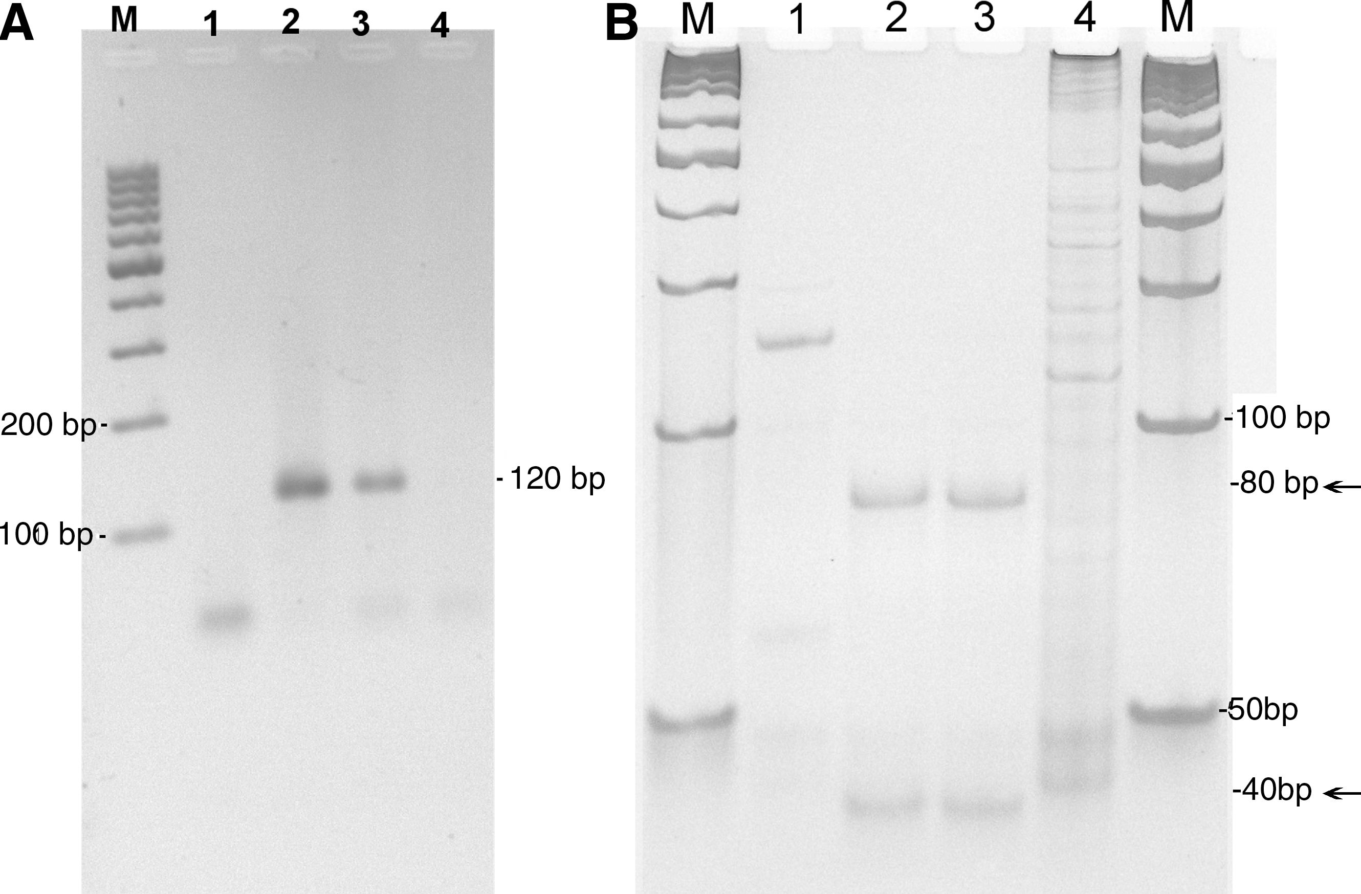
Eleven rodents were captured in the municipal district of Peixoto de Azevedo, Mato Grosso State, from October 30 to November 3, 2008: four wild rodents (Necromys lasiurus) and seven synanthropic specimens of two different species (four M. musculus and three Rattus rattus). We set 120 baited traps per night (Shermann®), distributed throughout an old abandoned mining area (10°15.092′S; 054°59.005′W), currently utilized for cattle raising. Despite its location in the Amazon biome, the study area has suffered serious degradation by humans, currently an open field with sparse trees and shrubs, creeping plants over poor mineral soil and a silty river. Rodent captures were licensed by the Brazilian Institute for the Environment and Renewable Natural Resources (ICMBio no. 13373-1) according to the standards for small mammals collection and handling procedures in compliance with biosecurity protocols. Following biometric data registration, the rodents were killed by anesthetic overdose for extration of femoral bone marrow and ear skin fragments for Leishmania sp. infection detection in accordance with the Ethics Committee of Federal University of Mato Grosso (23108.018419/08-0 CEPA). The cryopreserved samples were submitted to a DNA extraction protocol (Gomes et al. 2007), and then PCR kDNA amplification (Passos et al. 1999). For each PCR, positive and negative controls were supplemented. The positive amplified products (120 base pairs [bp]) were digested by HaeIII restriction endonucleases–PCR-RFLP (Volpini et al. 2004), resulting in 40 and 80 bp L. braziliensis fragments. L. braziliensis reference DNA (MHOM/BR/75/M2903) was adopted as a positive control (Fig. 1). The rodents were preserved in formaldehyde 37% and conserved in ethanol 70% as voucher specimens for morphological taxonomic identification.
Results
All four adult specimens of M. musculus (100%), two males and two females, were L. braziliensis positive by molecular analysis of ear skin samples and the bone marrow of one female. The bone marrow of two of the four adult N. lasiurus specimens (50%), one female and one male adults, also proved positive. Leishmania DNA was detected in neither of these tissues from the adult R. rattus (two female and one male) nor in the other two N. lasiurus (adult females). No suggestive lesions were apparent in the animals during clinical examination.
Discussion
N. lasiurus is representative of the sylvatic fauna and, together with R. rattus, was considered as L. braziliensis reservoirs in an endemic area of the Brazilian Northeast based on in vitro parasite culture isolation (zymodeme IOC/Z74) from humans, rodents, and L. whimani samples (Brandão-Filho et al. 2003). Heretofore, in another Northeast area, L. braziliensis was isolated from R. rattus and from Lutzomyia whitmani (Vasconcelos et al. 1994). Both M. musculus and Rattus spp. are worldwide species and were introduced in the Americas during European colonization, Rattus spp. being a potential reservoir of several species of Leishmania (Vasconcelos et al. 1994, Brandão-Filho et al. 2003, Di Bella et al. 2003, Ferreira et al. 2010, Motazedian et al. 2010). In an L. infantum visceral leishmaniasis–endemic region of Italy, M. musculus proved seropositive (Di Bella et al. 2003). More recently in the southeast of Brazil, M. musculus, N. lasiurus, and R. rattus were detected positive but without identification of the Leishmania species (Ferreira et al. 2010).
In the study area, Peixoto de Azevedo, an outbreak of CL was reported in the mid-1980s, related to drastic environmental changes (Azevedo et al. 2002), and since then cases in humans have been reported, totaling 26 in 2008 (unpublished data of State Secretary of Health of Mato Grosso). The detection of Leishmania infection in M. musculus in this locality has great importance as this species adopted the human domiciles as its main habitat. Since the study area has proximity with a region of recent human occupation, there is increased possibility of human infection.
A previous study undertaken in this region demonstrated a diverse phlebotomine fauna, with predominance of several species of Lutzomyia related to the CL transmission, with L. whitmani being the culprit in peridomiciles (Azevedo et al. 2002). This association between infection in synanthropic rodents and the presence of phlebotomines in the peridomicile has been reported in other endemic regions (Vasconcelos et al. 1994, Brandão-Filho et al. 2003).
Despite the L. braziliensis infection in this synanthropic rodent species having been detected in ear tegument, an accessible area to perpetuade vector infection, isolation of the parasite in both M. musculus and phlebotomines from this locality will be essential to incriminate this rodent species as a potential Leishmania reservoir. Nevertheless, these preliminary results suggest the participation of M. musculus in the zoonotic or enzootic transmission cycle of L. braziliensis in this endemic area.
Footnotes
Acknowledgments
Peixoto de Azevedo, Municipal Health Secretary, Mato Grosso State Health Secretary, and Secretary of Health Surveillance for logistical support. Oswaldo Cruz Foundation (Fiocruz/RJ) for providing the Leishmania DNA control. Dr. Reginaldo Peçanha Brazil (Oswaldo Cruz Institute/Fiocruz) for text revision.
Disclosure Statement
No competing financial interests exist.