
Abstract
Leishmaniasis has been considered endemic in Sinaloa, Mexico, since 1994. Despite that Leishmania mexicana is the main etiological agent of cutaneous leishmaniasis (CL) in other regions of Mexico, the species causing CL in patients from Sinaloa state has not been previously established, although Leishmania braziliensis has been found in the neighboring southern state, Nayarit. L. braziliensis is also associated with mucocutaneous leishmaniasis, which is a more complicated clinical variant. Due to the implications on individual and public health, the objective of this report was to identify the Leishmania species present in Sinaloa, Mexico. Using the first internal transcribed spacer (ITS-1) polymerase chain reaction-restriction fragment length polymorphism, we identified L. mexicana in a CL patient from Sinaloa and confirmed the extended distribution of this parasite in Mexico.
Introduction
CL is the major endemic type and has spread over 17 states of Mexico, including Campeche, Chiapas, Coahuila, Jalisco, Michoacán, Nayarit, Nuevo León, Oaxaca, Tamaulipas, Quintana Roo, Tabasco, Veracruz, Yucatán, Durango, and Sinaloa. VL has been found in Guerrero and Morelos, and MCL has been found in Tabasco and Chiapas (Cordova-Uscanga et al. 1993, Rodríguez Domínguez 2002, Pérez-Vega et al. 2009, Salazar-Mejía et al. 2010). In Sinaloa state, CL has been reported since 1996 (Rodríguez Domínguez 2002); however, the etiological species has not been identified at the molecular level. Here, we report for the first time the molecular confirmation of the presence of L. mexicana in a 17-year-old boy with CL in Sinaloa, Mexico.
Case Report
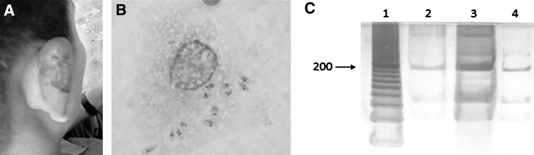
A 17-year-old school boy from Coco village, municipality of Concordia, Sinaloa, Mexico, was referred to our group at the Urban Health Center of Mazatlan, Sinaloa (SSA jurisdiction 5), on December 9, 2009. He had been living in his town during the previous year and was first seen at the Concordia Medical Services without success in diagnosis and treatment. Upon admission, the physical examination showed an amorphous, ulcerated nodule of 20×30 mm in diameter at the back of the right ear with a 6-month evolution (Fig. 1A). A pruritic ulcer began as a papular lesion that grew until it converted into an ulcer. The patient answered an epidemiological questionnaire in the presence of an adult parent and submitted a written and signed informed consent form according to the guidelines of the ethics committee of the Ministry of Health of Sinaloa. An impression of the lesion was then taken using standard methods. The ulcer impression was adsorbed on filter paper (Whatman; #3) for species identification by polymerase chain reaction–restriction fragment length polymorphism (PCR-RFLP) according to the method previously described by Schönian et al. (2003) and modified by Pérez-Vega et al. (2009). Briefly, genomic DNA was extracted using a DNA extraction kit (Dneasy; Qiagen) following the manufacturer's instructions. The DNA obtained was used as a template for PCR amplification with the following primers: LITSR (5′-CTGGATCATTTTCCGATG-3′) and L5.8S (5′-TGATACCACTTATCGCACTT-3′). The internal transcribed spacer (ITS-1) PCR products were digested with HaeIII (New England Biolabs) as described by Bensoussan et al. (2006) and Pérez-Vega et al. (2009). Restriction fragments were then subjected to gel electrophoresis on a 12% polyacrylamide gel at 100 V and observed by silver stain using a commercial kit (Silver stain; Sigma) based on the manufacturer's instructions. To identify the species of Leishmania, the ITS-1 RFLP pattern was compared to DNA from a culture of promastigotes of L. mexicana MHOM/MX/92/UAY68 isolated from a patient with LCL (kindly donated by Dr. Patricia Talamas Rohana from CINVESTAV-IPN).
Cutaneous leishmaniasis (CL) caused by Leishmania mexicana.
Results and Discussion
L. mexicana is the major species associated with CL in Mexico. Recently, we reported the first clinical case in Durango, Mexico, which confirmed the dissemination of this parasite in Mexico (Pérez-Vega et al. 2009). However, in other Mexican states, such as Nayarit, CL has been reported to be caused by L. braziliensis (Sanchez-Tejeda et al. 2001). Leishmania species identification is critical for the optimal treatment of patients, clinical course prediction, and epidemiological registration and contribution to strategy design in public health. Sinaloa was considered as an endemic for leishmaniasis since 1994; however, the etiological species of this parasitic disease has not been reported to date.
In the stained histological sample of the ulcerative lesion from this patient (Fig. 1A), several macrophages parasitized with Leishmania sp amastigotes were observed (Fig. 1B), confirming the parasitological diagnosis of CL. In addition, the Leishmania species was identified by PCR-RFLP using the HaeIII restriction enzyme. As shown in Figure 1C, the band pattern was similar to the reference strain of L. mexicana MHOM/MX/92/UAY68, where 60, 80, and 200 bp digested fragments were observed. Importantly, the band pattern of L. braziliensis previously reported using the same primers and restriction enzyme (Schönian et al. 2003) was different than the pattern from our result.
In conclusion, the results presented in this report confirm the presence of L. mexicana as the etiological agent of CL in Sinaloa state. However, it is critical to continue studies of the species of the sandfly vector and the reservoirs of this parasite. In addition, epidemiological studies are needed to determine the prevalence of CL in the state of Sinaloa, Mexico.
Footnotes
Acknowledgments
The authors would like to thank Dr. Celia Rosa Tejeda Aguirre for the clinical diagnosis and treatment of the patient. Moreover, they thank CECYT and PROFAPI-UAS for financial support. A special thanks to Dr. Dolores Correa for the critical review of this report.
Disclosure Statement
No competing financial interests exist.