
Abstract
Animal models for Ehrlichia chaffeensis have been unsuccessful in recapitulating the natural disease cycle. We have developed an animal model for tick feeding and transmission using white-tailed deer (Odocoileus virgianus), the intracellular bacterium (Ehrlichia chaffeensis), and the lone star tick vector (Amblyomma americanum). Here, we report the acquisition and transmission of E. chaffeensis infections by refeeding male ticks in this experimental model. This finding is important because techniques for gene silencing are most successful for unfed adult ticks. Males are able to refeed several days after acquiring a tick-borne pathogen. Using refeeding male lone star ticks and RNA interference technology, we plan to decipher underlying molecular mechanisms involved in transmitting E. chaffeensis to a host via a lone star tick bite.
Introduction
Materials and Methods
E. chaffeensis infection using refeeding male ticks
All unfed adult A. americanum ticks used in these studies were purchased from the National Tick Research and Education Resource (NTRER) at Oklahoma State University. Briefly, laboratory-reared ticks were propagated following the published protocols of Patrick and Hair (1975). These ticks were pathogen-free and have been in continuous culture for many years. Off-host ticks were held in the laboratory in desiccator cabinets that were modified to maintain high humidity (96%) with a saturated salt solution (K2SO4).
Two- to 3-day-old deer were purchased from local deer breeders and hand-fed until they were 3–4 months old. E. chaffeensis bacteria were grown in canine macrophage cell lines (DH82). One milliliter of 108 organisms was injected intravenously in each of four fawns. To verify infections, blood was collected from each fawn before infection and then weekly. These animals were positive for E. chaffeensis infection at day 21 and were not followed beyond the tick infestation. An aliquot of blood was used for DNA isolation. Blood genomic DNA extraction was done using the Illustra™ blood genomicPrep Mini Spin Kit (GE Healthcare, Piscataway, NJ) following the manufacturer's guidelines. E. chaffeensis genomic DNA was identified using specific PCR primers (Table 1) and a nested PCR protocol (Table 1; Dawson et al. 1996). We used GoTaq Master Mix (Promega, Madison) for all of our reactions. Amplifications were performed using the PTC-100™ Thermocycler (MJ Research Inc.) with the following protocol: First reaction, 95°C for 5 min, and 30 cycles at 94°C for 1 min, 55°C for 1 min, 72°C for 1 min; and second reaction, 95°C for 5 min, and then 40 cycles at 94°C for 1 min, 55°C for 1 min, 72°C for 1 min with one cycle of 72°C for 5 min. Amplicons were purified using the GENECLEAN II protocol, and E. chaffeensis products were verified by sequencing.
After verifying infection in the deer, tick infestations were established. Thirty pairs of adult A. americanum were placed in modified Nalgene containers that were glued to the shorn backs of the 4 infected deer. At the end of 7 days, all ticks were removed. Due to limitations in the number of ticks that could be placed on each animal, none of the males was tested for E. chaffeensis in the acquisition stage. All male ticks were placed in high humidity for 3 days before using them for transmission experiments. Significant mortality occurred if male lone star ticks were held off the host longer than 3 days (D.C. Jaworski, unpublished).
After 3 days, we infested 2 naïve deer with acquisition-fed male ticks and uninfected unfed female ticks and permitted these ticks to feed for 7 days. All ticks were removed at 7 days and dissected. We stored salivary glands and midguts from each tick individually in TRI Reagent (Molecular Research Center [MRC], Inc., Cincinnati) at −70°C. Tick DNA was extracted using the published protocol from MRC. Tick DNA was tested for E. chaffeensis infection using the PCR primers and protocol described above.
Results
Male lone star ticks acquired E. chaffeensis and subsequently were able to transmit the infection to naïve deer. One of 2 naïve deer subjected to tick bite inoculation had a positive PCR for E. chaffeensis infection 19 days after ticks were removed from the deer (Fig. 1A, lane 2). In other studies, the prepatent periods for tick-bite infections were anywhere from 15 to 36 days after infestation (Ewing et al. 1995). We verified that ticks feeding on the other deer were PCR positive for E. chaffeensis, and this may reflect a positive infection state for this deer as suggested by other authors (see de los Santos et al. 2007).
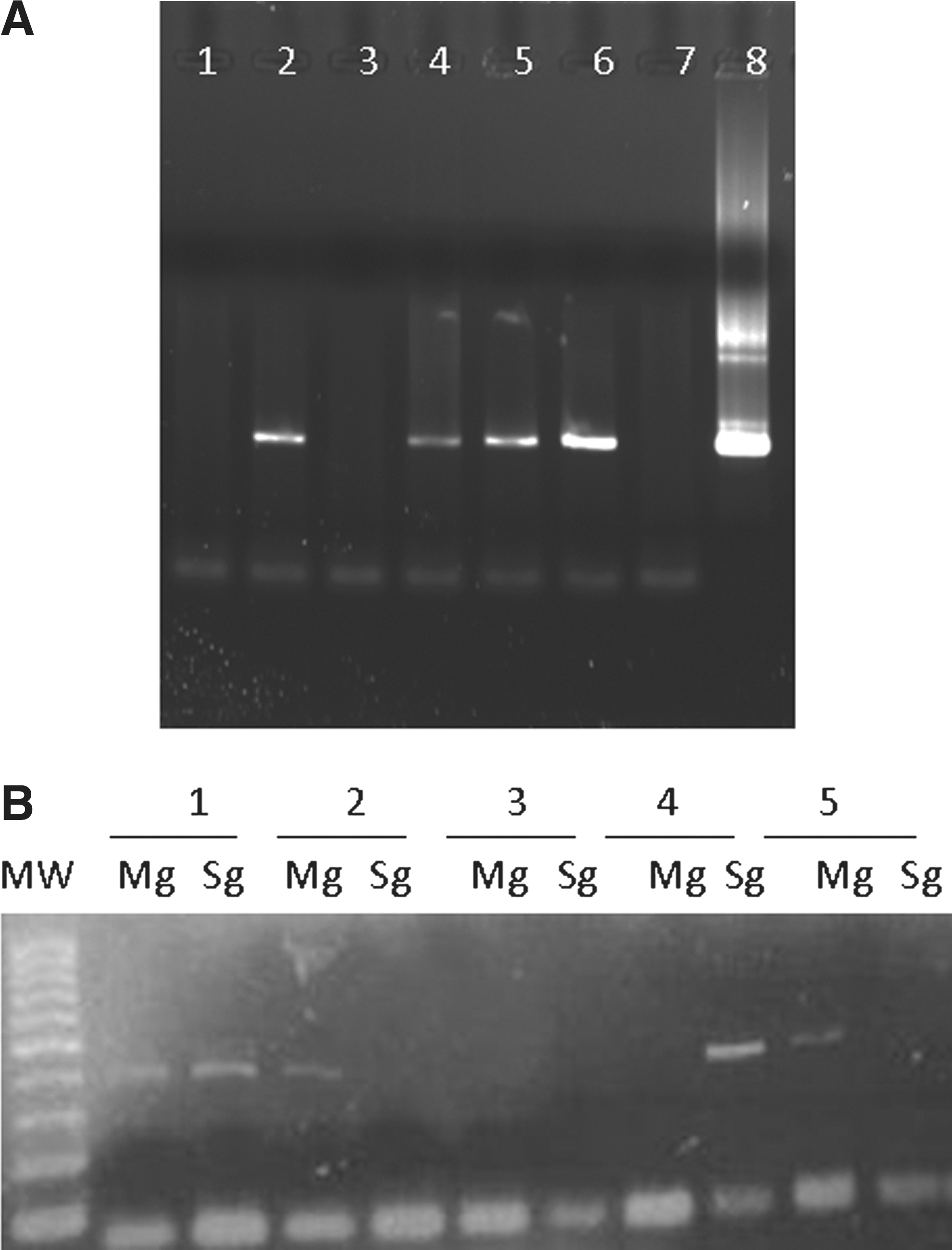
Acquisition and transmission of Ehrlichia chaffeensis using white-tailed deer and refeeding male lone star ticks (Amblyomma americanum). (
However, in our model, xenodiagnosis may not be an appropriate method for assessing the infection status of a host. For example, the male ticks acquired the bacteria in a previous feeding and are placed on a naïve host to assess transmissibility of E. chaffeensis. The ticks are likely already positive for E. chaffeensis when refeeding occurs. We did not assess co-feeding females, but then we could not discount the co-feeding environment as a source of infection (see Randolph 2011). Further studies would be required to clarify the value of xenodiagnosis in assessing host infections. Infection profiles varied from tick to tick. Infections that are seen in the salivary glands are assumed to reflect the activation of the bacteria to the salivary glands for injection into the host. Not all infections appeared to be activated to salivary glands from the midgut during transmission feeding (Fig. 1B). Bacteria could be found in both tissues (Fig.1B, tick 1), others did not activate to salivary glands (Fig. 1B, ticks 5 and 2), some were cleared from the midgut and only found in the salivary glands (Fig. 1B, tick 4) and some did not appear to have any bacterial infection (Fig. 1B, tick 3). We are continuing to evaluate our model using different strains of E. chaffeensis and nymphal tick feeding. Future experiments are planned to develop molecular information critical to the midgut to salivary gland activation phase for these intracellular bacteria.
Footnotes
Acknowledgments
The authors gratefully acknowledge Susan Little and Misti Spatz for preparing the bacteria for these experiments. In addition we acknowledge the helpful reviews by Susan Little, Michael Reiskind, and Astri Wayandande. This paper was approved for publication by the Director of the Oklahoma Agricultural Experiment Station and supported under project OKL02623 and Animal Health 1433 Funds.
Author Disclosure Statement
No competing financial interests exist.