
Abstract
The European subtype of tick-borne encephalitis virus (TBEV-Eu) and louping-ill virus (LIV) are two closely related tick-borne flaviviruses. However, whereas the first is the cause of one of Europe's most important zoonoses, the latter most often only causes disease in sheep and grouse. TBEV-Eu is typically found in the forests of central and northeastern Europe, and LIV typically is found in sheep pastures in the British Isles. In the 1980s, however, LIV was isolated from sheep with encephalomyelitis in Norway. In the 1990s, the first cases of human TBEV were also detected in this country, but while Louping-ill in sheep is very rare, the number of human TBEV cases is increasing. No larger investigations of TBEV and/or LIV seroprevalence and distribution in Norway have been published. However, before such studies are initiated, it is pertinent to know if LIV and TBEV are potentially co-circulating. In the current study, we examined if antibodies against LIV and TBEV were found in wild cervids in one location (Farsund) in southern and one location (Molde) in northwestern Norway using a commercially available enzyme-linked immunosorbent assay for detection of anti-TBEV immunoglobulin G (IgG) and a hemagglutination inhibition test for anti-LIV IgG. Positive results were confirmed by serum neutralization tests. In Farsund, 22 of 54 cervids had antibodies against TBEV and 8 antibodies against LIV. In Molde, 1 of 64 cervids was confirmed positive for TBEV, whereas none were positive for LIV. This shows that TBEV and LIV may co-circulate in southern Norway and that virus(es) antigenetically very similar to TBEV may be found in northwestern Norway. The latter is intriguing, because the climatic conditions typical of TBEV locations should not be expected this far north.
Introduction
TBEVs have often been divided into three major subtypes: (1) the European (TBEV-Eu), transmitted by Ixodes ricinus, and (2) Siberian (TBEV-Sib) and (3) Far Eastern (TBEV-FE), both transmitted by I. persulcatus. (Gritsun et al. 2003, Lindquist and Vapalahti 2008). It has previously been suggested that tick-borne flaviviruses have evolved in a cline across Asia and Europe, implying that new subtypes have evolved as a response to the local environment as the virus extended its distribution range from east to west, resulting in a virus species showing gradual genotypical and phenotypical changes, with the oldest virus lineages in the east and the more recent lineages in the west (Zanotto et al. 1995). In line with this theory, it has been thought that louping-ill virus (LIV), a virus found west of the main distribution area of TBEV-Eu, and genetically a closer relative to TBEV-Eu than TBEV-Eu is to the other two subtypes (Charrel et al. 2001), has represented the most recently evolved variant of the TBEV complex. Phylogenetic analyses based on the envelope glycoprotein gene have suggested that its origin was a single introduction of TBEV from Continental Europe to Ireland 330–800 years ago (McGuire et al. 1998). However, more recently published phylogenetic analyses based on sequencing of the complete protein-encoding genome of the virus strains indicate that both LIV, the closely related Spanish sheep encephalitis virus, and TBEV-Eu have evolved from a common sheep and goat encephalitis virus cluster, and that this evolution has occurred independently from and as long ago as the evolution of TBEV-Sib and TBEV-FE (Uzcátegui et al. 2012).
Today, LIV is predominantly distributed on the sheep-rearing hillsides of Scotland, England, Wales, and Ireland (McGuire et al. 1998). It causes encephalomyelitis in sheep and red grouse (Lagopus lagopus scotia), and is considered a major cause of red grouse mortality in endemic areas (Reid et al. 1978). Sheep and grouse are apparently the only species capable of developing LIV viremia of sufficient titer to infect ticks (Hudson et al. 1995). The mountain hare (Lepus timidus) can play a role in the maintenance of LIV cycles, probably via a nonviremic co-feeding mechanism, but rodents do not seem to be involved in the transmission of LIV in the United Kingdom (Gilbert et al. 2000, Norman et al. 2004). LIV is capable of infecting humans, but cases of naturally acquired human disease are few and have occurred mainly in individuals with occupational exposure, such as sheep farmers, veterinarians, or butchers, and verified tick-borne transmission to humans is not reported (Davidson et al. 1991). The clinical picture is very similar to that of the biphasic meningitis typical of European TBEV (Charrel et al. 2004).
In Norway between 1998 and 2010, only 54 cases of autochthonous human TBE have been reported (
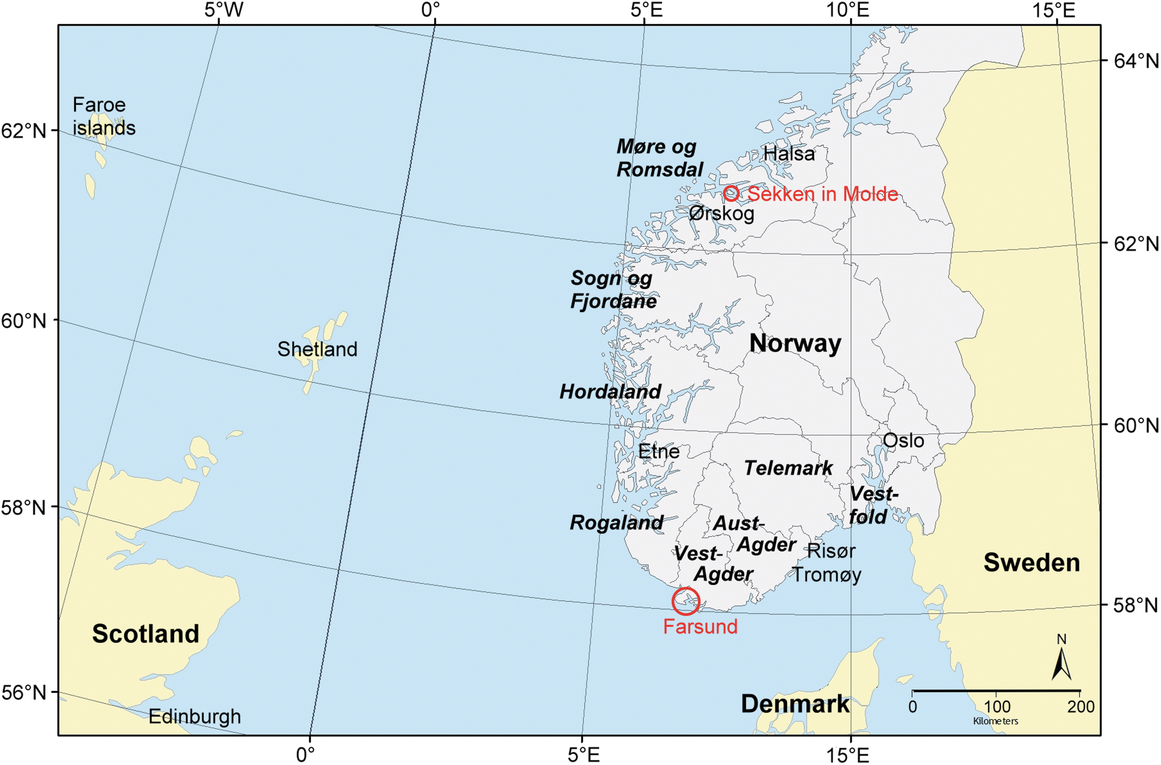
Map of southern Norway. Sekken in Molde and Farsund are marked with circles. (Color image available online at
A few cases of clinical and histological Louping-ill–like disease were reported in sheep in south and southwestern Norway (Farsund in Vest-Agder and Etne in Hordaland counties; Fig. 1) back in the 1980s (Ulvund et al. 1983, Ulvund 1987). Brain and spinal cord tissue from some of these cases was inoculated intracerebrally into sheep and mice and a virus was cultivated and isolated (J. Krogsrud, pers. comm., Ulvund et al. 1983). This isolate was later characterized as LIV indistinguishable from Scottish strains (Gao et al. 1993). Phylogenetic studies indicate that LIV quite recently (first part of 20th century) was introduced to Norway from Scotland (McGuire et al. 1998). Serological studies do indicate although that LIV or antigenetically very similar flaviviruses are relatively prevalent among sheep flocks along the coast from southern to northwestern Norway (Risør in Aust-Agder to Halsa in Møre og Romsdal county (J. Krogsrud et al., unpublished studies)(Fig. 1). The disease is, however, very rare in Norway, in spite of the fact that I. ricinus is very common, the sheep population is big, many sheep are exposed to the tick, and vaccination against LIV is not performed (Tore Tollersrud, pers. comm.).
Older studies have also indicated that viruses closely related to TBEV and LIV may be found relatively far north along the western coast of Norway. In 1973, Traavik published a serological investigation of 81 cattle from localities from Farsund in southern Norway to Ørskog in Møre og Romsdal (Fig. 1). According to his results, 14 animals, spread on all sampling localities, were found seropositive for TBE viruses. He also used a hemagglutination inhibition test (HI) and a closed hexagon immunodiffusion procedure (CHI) to examine sera from 341 human patients from Hordaland, Sogn, and Fjordane and Møre og Romsdal counties (Fig. 1). Traavik found a seroprevalence of 19.6% (Traavik 1979), but noted that sera strongly positive by HI not could be confirmed by CHI, and suggested that this could be attributed to infection with other flaviviruses than TBEV.
The aim of the current study was to investigate if wild cervids in two different localities, one on the coast of southern and another on the coast of western Norway, are exposed for TBEV and LIV. On the basis of current knowledge, we believed that both TBEV and LIV should be present in the location in southern Norway, whereas only LIV should be present in the location in western Norway.
Material and Methods
Study area and study populations
The study areas were Farsund municipality in southern Norway and the island Sekken in Molde municipality in northwestern Norway (Fig. 1). Farsund municipality covers an area of 252 km2 land, ranging from sea level to 506 meters above sea level (m.a.s.l.) and consists of rugged hills covered by oak and pine forest intermingled with agricultural land (10%) and peat bogs (3%) (
Sample collection
Serum from 25 and 34 roe deer, 4 and 28 red deer, and 25 and 2 moose was collected from carcasses of animals shot in Farsund and Molde, respectively, during ordinary hunting in the autumn of 2005. The hunters were instructed to collect blood from the thoracic cavity with a plastic Pasteur pipette and transfer it to a tube with clot activator and gel for serum separation. The blood samples, together with other samples from the same animals, were delivered as soon as possible and within the same day to a field collection station, where the blood was centrifuged at 3000 turns/min for 10 min, the supernatant transferred to 5-mL tubes, and the samples stored at −20°C until the end of the hunting season. The samples were then transferred to the Norwegian Veterinary Institute (NVI) with freeze transport, thawed and aliquoted (1-mL tubes), refrozen, and stored at −40°C or −80°C until use.
Anti-TBE IgG-antibody detection
Serum samples were analyzed for immunoglobulin G (IgG) antibodies against TBEV using a modified commercial enzyme-linked immunosorbent assay (ELISA) assay (Enzygnost® Anti-TBE virus IgG; Siemens, Eschborn, Germany). IgG to TBEV was detected by peroxidase-labeled affinity-purified antibody to deer IgG (H+L) produced in rabbit (KLP, Gaithersburg, MD, USA) in a dilution of 1:20,000. Except for the conjugate, the assay was performed as recommended by the manufacturer. The samples were interpreted qualitatively as positive or negative, and the cutoff used for qualitative evaluation of positivity was calculated as optical density (OD) values according to the manufacturer's instructions. Positive and negative roe deer sera, analyzed by serum neutralisation test (SNT) at the laboratory of Prof. F.X. Heinz, Department of Virology, Medical University of Vienna, Austria, were used as positive and negative controls in each run. All ELISA-positive samples were retested by SNT assay at the same laboratory.
Anti-LIV IgG-antibody detection
A HI test for antibody to LIV was performed at the Moredun Research Institute (MRI) using gander erythrocytes, as described by Clarke and Casals (1958), and modified to use tissue culture–grown virus and a microtiter plate. This assay format is validated in routine diagnostic use for many species, including deer. Samples were required to be heat inactivated at 56°C for 30 min prior to importation. Because heat inactivation can damage IgM, the remaining antibody is considered to be mostly IgG. Nonspecific inhibitors and goose erythrocyte agglutinins were removed by kaolin and goose erythrocyte absorption. The assay was controlled using known antibody-positive and -negative ovine serum samples. Samples giving a result of HI at a titer of greater than 1/20 were considered seropositive. Samples where the titer was 1/10 were considered inconclusive. All samples where a titer of ≥1/10 was detected were tested in the MRI laboratories by SNT using the constant virus varying serum method (Grist et al. 1966). The test is modified to be performed in 96-well plates and used baby hamster kidney (BHK) cells and the LIV strain L31 using 30–300 median tissue culture infective dose (TCID50) per well. Virus controls, known positive and negative titrated serum controls, toxicity controls, and uninfected control wells were run with each test.
Results
In Farsund, 9 of 25 roe deer, 14 of 25 moose, and 0 of 4 red deer were found positive for antibodies against TBEV using the ELISA assay. This result was confirmed by SNT in all but one positive roe deer and all positive moose (Table 1), resulting in a seroprevalence of 32% among tested roe deer and 56% among tested moose. The positive titers ranged from 1/10 to 1/640.
One of the 2 moose cows from Farsund that in this table is presented as positive for LIV was judged as uncertain both on HAI and neutralization test for LIV, while the other was judged as positive on HAI, but not tested by neutralization test due to lack of serum.
TBEV, tick-borne encephalitis virus; LIV, louping-ill virus.
In Molde, 4 of 34 roe deer, 0 of 2 moose, and 1 of 28 red deer were positive for TBEV on ELISA, but only one roe deer was confirmed positive by SNT (Table 1), having a titer of 1/15.
Antibody to LIV was detected at a titer of ≥1/20 in eight samples from Farsund, comprising 2 red deer, 1 roe deer, and 5 moose. An inconclusive result (titre 1/10) was obtained for a further five samples (3 roe deer and 2 moose). One moose sample had insufficient serum remaining to test by SNT. The remaining 7 HI-positive samples were tested by SNT and antibody to LIV was detected in 6, including both red deer. The discordant (HI positive, SNT negative) was a moose sample. Interestingly, while SNT titers in roe deer and moose were in the order of 1/20 and 1/40, both red deer had much higher SNT titers (1/220 and 1/450). Antibody to LIV was also detected by SNT in two inconclusive samples from roe deer at titers of 1/20 and 1/40, one moose sample remained inconclusive and no antibody to LIV was detected by SNT in two of the samples inconclusive in HAI (1 moose and 1 roe deer) (Table 1). The seroprevalences were hence ∼13% both among tested roe deer and moose. There were only four red deer samples from this sample area, but two of them were seropositive. In Molde, all samples were negative for antibodies against LIV.
Among the samples from Farsund, six of the sera that were positive with ELISA for TBEV did also show seroreactivity when tested with the HAI for LIV, whereas 15 of the TBEV-positive sera were negative for LIV (Table 2). Among the sera that were negative for TBEV, seven were positive and 23 were negative for LIV (Table 2). This gives us an odds ratio of 1, 31 (95% confidence interval 0.37–4.98) and a relative risk of 1.22 (0.48–3.01). These low measures of association may indicate that there is a low degree of serological cross-reaction between TBEV and LIV using these two tests, although the number of examined samples is too low to draw any firm conclusions. A recent study of humans vaccinated against TBEV, Japanese encephalitis virus, and/or yellow fever virus demonstrated plaque reduction neutralization test antibody to LIV at titers of ≥1:10 in 68 % of samples (Mansfield et al. 2011), supporting the possibility that cross-reactivity may be occurring in some samples. Analysis of individual samples demonstrated that while six samples with detectable TBEV SNT titers had low LIV SNT titers, which may well be due to cross-reactivity, TBEV antibodies were not detected for the remaining four samples in which LIV SNT antibody was detected (Table 3). In particular, the red deer samples, none of which were TBEV positive but two of which had high titers in the LIV SNT test, suggest that LIV is genuinely present in this region.
ELISA, enzyme-linked immunosorbent assay; TBEV, tick-borne encephalitis virus; HAI, haemagglutination inhibition test; LIV, louping-ill virus.
TVEV, tick-borne encephalitis virus; IgG, immunoglobulin G; SNT, serum neutralization test; LIV, louping-ill virus; ND, test not performed as TBEV IgG negative.
Discussion
The current study indicates that both TBEV and LIV are likely to be circulating in Farsund in southern Norway, whereas only TBEV or a virus serologically very similar to TBEV is found in Molde in northwestern Norway. The first result is as expected, because clinical cases of TBE in humans and LI in sheep have been observed in the area. Possible co-circulation of TBEV and LIV has also been reported from Bornholm in Denmark (Jensen et al. 2004). The latter result is somewhat surprising, because it has been believed that serological evidence for presence of TBEV-like viruses in western Norway was caused by infection with LIV rather than TBEV, as the climatic conditions presumed to be needed for maintenance of sylvatic TBEV cycles are not present in this part of the country (Randolph and Rogers 2000).
However, the prevalence is low, and only one out of the five sera that were positive on ELISA was confirmed positive on neutralization test, having a relatively low titer (1/15). It may be pertinent to ask if the serological reaction may indicate presence of an unknown flavivirus closely related to TBEV. The existence of such viruses has been suggested by Traavik (1979, 1984). Another explanation could be that the seropositive cervids have been infected by ticks carried by birds migrating to Molde from TBEV-endemic areas, because TBEV has been found both in larvae and nymphs of ticks attached to birds during long-distance migration (Waldenström et al. 2007). However, looking at the migration routes of birds ringed in northwestern Norway, most birds found in the study locality should be expected to use western migratory flyways, i.e., the British Isles and the countries surrounding the North Sea coasts (Bakken et al. 2006), which are not regarded as TBEV-endemic areas. One could also argue that this single roe deer fawn could have dispersed from a TBEV-endemic area in southern Norway. However, the roe deer fawn would have had to have crossed rugged mountain areas and traveled at least 350 km in a straight line to get from the known endemic areas to Sekken. Typical dispersion distances for roe deer fawns are not that far (Mysterud 1999), although dispersion up to 261–280 km has been reported in the relatively more flat taiga of northern Sweden (Wahlström and Liberg 1995).
Rather, the current finding of a TBEV-seropositive deer may actually mean that the virus, although the general conditions in the area are regarded as suboptimal, persists in small niches where microclimate and other factors support TBEV transmission. The public health implications of the current findings are that: (1) medical doctors should consider TBE as a possible diagnosis also in patients from western Norway even without a story of visiting known TBE-endemic areas and (2) louping-ill should be regarded as a differential diagnosis to tick-borne encephalitis in southern Norway. LIV infection in humans is of course uncommon, even in endemic areas, but TBE-like clinical signs without positive serology should raise suspicion, especially if the patient has some occupational risk (sheep farmer, abattoir worker, veterinarian, etc.) or is immunocompromised (Davidson et al. 1991). Differentiation between these two virus infections may be difficult serologically, but this is important knowledge for public health authorities responsible for prophylactic measurements such as public advice and vaccination policy. In clinically affected humans, virus genome detection by PCR of cerebrospinal fluid may be an alternative diagnostic method.
It has been demonstrated that intragenic recombination can occur between similar flaviviruses, especially in mosquito-transmitted viruses like dengue (Twiddy and Holmes 2003). During co-infection, recombination can theoretically result in gene shifts between viruses belonging to the same family and result in virus evolution. However, there is limited evidence that dual infection and/or superinfection with flaviviruses in mammalian hosts is highly unusual; there are only sparse individual case reports of co-infection with the related dengue viruses in humans (Wenming et al. 2005, Araujo et al. 2006, Terzian et al. 2011). In a persistently infected cell culture system, LIV-infected cell cultures have been demonstrated to resist superinfection with homologous viruses and a variety of heterologous viruses (Venugopal and Gould 1992), including TBEV-Eu, Negeshi virus, Langat virus, West Nile virus, Powassan virus, and two of three yellow fever strains (the exception was a neurotrophic yellow fever virus). In mosquito cells, competitive suppression occurs between dengue virus strains (Pepin et al. 2008). In a dual-infection tick study, infection of ticks with Skalica virus (a virus closely related to TBE) 14 days prior to tick infection with a mouse-lethal strain of TBE prevented transmission of TBE to mice (Weismann et al. 1990). Therefore, while co-circulation of LIV and TBE may pose diagnostic problems, the available evidence suggests that this co-circulation is unlikely to pose a significant threat to animals or man due to virus recombination.
In conclusion, TBEV and LIV are apparently co-circulating in southern Norway and a TBEV-like virus may circulate also in the northwestern part of the country. Using cervids as sentinel animals in serological surveys for flaviviruses seems to be a feasible way of getting an overview of the prevalence and distribution of these viruses, and thus further studies of flavivirus distribution in Norway should be encouraged.
Footnotes
Acknowledgments
We would like to thank the hunters of Farsund and Molde, Reidun Moseid, Anja Johannessen, Just Ove Westad, Nils-Bjørn Venås, and Jan-Fredrik Sundt for their invaluable help in collecting samples and Inger-Lise Larsen for sample preparation and providing institutional memory. We are also grateful to Prof. Franz X. Heinz, Vienna, and his laboratory for confirmatory testing of sera. This work was supported by the National Surveillance Program for Cervids (HOP) in Norway. The Virus Surveillance Unit at the Moredun Institute is funded by the Scottish Government.
Author Disclosure Statement
No competing financial interests exist.