




Abstract
Both Rickettsia helvetica and Anaplasma phagocytophilum are common in Ixodes ricinus ticks in Sweden. Knowledge is limited regarding different animal species' competence to act as reservoirs for these organism. For this reason, blood samples were collected from wild cervids (roe deer, moose) and domestic mammals (horse, cat, dog) in central Sweden, and sera were tested using immunofluorescence assay to detect antibodies against spotted fever rickettsiae using Rickettsia helvetica as antigen. Sera with a titer ≥1:64 were considered as positive, and 23.1% (104/450) of the animals scored positive. The prevalence of seropositivity was 21.5% (23/107) in roe deer, 23.3% (21/90) in moose, 36.5% (23/63) in horses, 22.1% (19/90) in cats, and 17.0% (17/100) in dogs. PCR analysis of 113 spleen samples from moose and sheep from the corresponding areas were all negative for rickettsial DNA. In roe deer, 85% (91/107) also tested seropositive for A. phagocytophilum with a titer cutoff of 1:128. The findings indicate that the surveyed animal species are commonly exposed to rickettsiae and roe deer also to A. phagocytophilum.
Introduction
R
Several studies have investigated the presence of Rickettsia spp. in their arthropod vectors, in which vertical transmission helps to maintain the infection in nature. To date, only limited data are available regarding the animal reservoirs of these bacteria. Rickettsia rickettsii and Rickettsia sibirica have been isolated from various wild mammals (Parola et al. 2005), and for Rickettsia conori subsp. israeliensis, and R. rickettsii, dogs are demonstrated to act as competent reservoirs (Shaw et al. 2011, Levin et al. 2012). The infection may cause different symptoms of illness in the dog and sometimes also cause serious disease in humans when transmission of an infectious agent involves ticks with a broad host range (I. ricinus), including humans (Shaw et al. 2011; Liedó et al. 2014).
Molecular studies on blood samples from dogs have also supported that dogs may have rickettsaemias and possibly be an important reservoir host for Rickettsia felis (Oteo et al. 2006, Giudice et al. 2014). A study conducted in Germany also suggests that rodents and other small mammals may act as reservoir hosts for Rickettsia helvetica and Rickettsia felis (Schex et al. 2011). R. helvetica nucleotide sequences have been detected in blood samples of sika deer (Cervus nippon) in Japan and roe deer in The Netherlands (Inokuma et al. 2008, Sprong et al. 2009). Molecular and serological studies have suggested cattle as reservoir for Rickettsia africa in Nigeria, and wild boars and domestic ruminants as reservoir for Rickettsia slovaca in northeastern Spain (Ortuno et al. 2007, 2012, Reye et al. 2012). Further studies are needed to confirm these assumptions as well as to examine whether conditions are comparable in Sweden.
Another by I. ricinus tick-transmitted bacterium within the order Rickettsiales, is Anaplasma phagocytophilum, which is the causative agent of human granulocytic anaplasmosis (HGA) (Bjöersdorff et al. 1999), tick-borne fever (TBF) in sheep, cattle, and goats, as well as equine granulocytic anaplasmosis (EGA) in horses, canine granulocytic ehrlichiosis (CGE) in dogs, and feline granulocytic ehrlichiosis (FGE) in cats (Woldehiwet 2006). In Sweden, moose (Alces alces) were found to be serologic and DNA-positive for A. phagocytophilum (Malmsten et al. 2014). Thus, testing moose and deer may be useful in monitoring of anaplasmosis and other tick-borne infections in given areas (Jenkins et al. 2001, Stuen et al. 2001, 2002, 2013. Malmsten et al. 2014).
The southern, central, and coastal parts of Sweden are known to be endemic areas for SFG rickettsiae in ticks, and the only species found in free-living ticks in the given area (besides a single reported finding of R. sibirica) is R. helvetica, occurring in approximately 1–15% of I. ricinus ticks (Severinsson et al. 2010, Wallménius et al. 2012). It is comparable to the R. helvetica prevalence reported from Denmark and Poland of 1.1–13% in I. ricinus (Svendsen et al. 2009). However, the role of animals, both rodents and larger wild and domestic mammals, in the life cycle of tick-borne rickettsiae has not been fully elucidated.
In their natural life cycle, Rickettsia spp. are characterized by their transovarial mode of transmission in the vector and its stages, and larvae, nymphs, and adults are infective for vertebrate hosts. It is assumed that the infections of mammals may result in a rickettsemia that allows noninfected ticks to become infected, thus enabling the life cycle to continue. In this perspective, the vector may be regarded as the actual reservoir and the animal as the intermittent reservoir in the course of the the transient rickettsemia, during which time it could be involved in further geographical dispersion of Rickettsia bacteria (Raoult et al. 1997). This assumption is also supported by a previous study in which feeding ticks picked from mammals were PCR positive at a higher rate than ticks gathered free-living in nature (Nilsson et al. 1999).
The aim of the present study was to conduct a survey to determine whether wild and/or domestic animals in Sweden are potential reservoirs for Rickettsia spp. by detecting DNA and analyzing the presence of antibodies in serum. In a limited part of the study, the seroprevalence of A. phagocytophilum for deer was analyzed for comparison with Rickettsia as another measure of exposure for tick-borne infections in the area.
Materials and Methods
Samples
Blood samples were collected either from domestic animals (cats, dogs, and horses) investigated at an animal hospital or from harvested game animals (moose and roe deer). Serum was stored at the Swedish National Veterinary Institute (SVA) for further analysis. In the present study, these samples were analyzed without specific identity, i.e., no information on the animal or the animal's owner's identity. The study included a total of 450 samples, of which 63 serum samples were from horses, 90 from cats, 100 from dogs, and, collected during the hunting season, 107 from roe deer (Capreolus capreolus), and 90 samples from moose (A. alces) (47 adult and 43 juveniles). Most of the domestic animals were free roaming, but besides that, there was no information of symptoms of disease, because the animal's identity was not known and access to medical records not possible. The majority of the serum samples were collected during the period 2010–2011, with the exception of the roe deer samples, which were collected during 1990–1993. Those latter serum samples were stored in a −20°C freezer from collection to analysis and were not thawed in the meantime. In addition, 113 spleen samples were analyzed by PCR for the presence of rickettsial DNA. These samples consisted of spleen tissue from 30 moose from the island of Öland (2007), 33 domestic (sheep) and wildlife animals (moose) from central and southern Sweden (2011–2013), and lysates from the spleen of 50 moose from the same areas collected during 2011. We have no information on the individual animals regarding tick infestation.
Serological analysis
R. helvetica serologic analyses were performed using an immunofluorescence assay (IFA). Blood sera were diluted 1:64 (or 1:32 for cervids) in phosphate-buffered saline (PBS) and were screened for immunoglobulin G (IgG) antibodies to SFG rickettsiae using R. helvetica–coated slides with antigen prepared from an aliquot of R. helvetica–infected Vero cells, applied to microscope slide wells, and incubated with serial dilutions of serum, as previously described (Elfving et al. 2008). As a secondary antibody, fluorescein isothiocyanate– (FITC) labeled goat anti-deer IgG (cat. no 02-31-06, for deer and moose samples)/anti-horse IgG (cat. no 02-21-06)/anti-dog IgG (cat. no 02-19-02), and anti-cat IgG (cat. no. 02-20-02; all from Kirkegaard and Perry Laboratories Inc., Gaithersburg, MD) were used, all diluted in a titer of 1/100 before use. Reactive antibodies were detected using light microscopy (original magnification, 400×). Sera showing bright green fluorescence at titers ≥1:64 or above were considered positive for all investigated animal species and were titrated to their end titer using two-fold dilutions. Reference control sera positive for SFG rickettsiae were not available for each specific animal species. Instead, for each species, previously detected positive serum samples, with titers between 1/128 and 1/256, were included in each run as a positive control. PBS and previously found negative sera were included as negative controls.
All of the specimens from roe deer were investigated for seroreactivity against Anaplasma spp. using commercial IFA-slides coated with with HL-60 cells infected with a human isolate of A. phagocytophilum as antigen (Focus Diagnostics, Cypress, CA), and FITC-labeled goat anti-deer IgG (cat. no 02-31-06, Kirkegaard and Perry Laboratories Inc.) was used as conjugate. Previously detected positive and negative serum samples were included in each run as controls. The samples were analyzed using light microscopy (original magnification, 400×). According to the manufacturer's instruction, the cutoff was ≥1:64 for human samples. In this study, we chose a cutoff of ≥1:128 for deer samples to make our results comparable with previous findings from Denmark (Skarphedinsson et al. 2005).
PCR analysis
Total DNA extraction from spleen tissue was performed by using a QIAamp DNA Extraction kit and an EZI BioRobot (Qiagen GmbH, Germany), as previously described (Malmsten et al. 2014). For detection of SFG rickettsiae DNA, a genus-specific real-time PCR, based on a probe and primers targeting the gltA gene, as described previously (Stenos et al. 2005), was performed in a LightCycler 2.0 Real-Time PCR System (Roche Diagnostics, Mannheim, Germany) using an LC Taqman Master Kit (Roche Diagnostics, Mannheim, Germany). As a positive control, a positive standard plasmid containing the cloned 74-bp fragment of the gltA gene, previously titrated to correspond to about 150 copies in the standard curve was used, showing linearity in the dilution series between 1.5 and 1.5 × 10⁸ copies (Elfving et al. 2012). Sterile water was included as the negative control in each amplification trial.
Statistical analysis
For continuous variables, we used standard parametric statistics (confidence interval [CI] according to Fleiss with Yates correction) giving a mean ± 95% CI. The Fisher exact test and chi-squared test were used to compare the proportions, and a p value < 0.05 was considered statistically significant. Statistical analyses were conducted using Predictive Analytics Software (PASW®) Statistics 20.
Results
Seroreactivity to spotted fever rickettsia with R. helvetica as antigen
Out of the 450 sera samples analyzed, 104 (23.1%) had IgG antibodies against R. helvetica, equal to or higher than the cutoff titer of 1:64. The number of samples found to be positive was as follows: 23/107 (21.5%, CI 14.4–30.7) among roe deer, 21/90 (23.3%, CI 15.3–33.7) among moose, 23/63 (36.5%, CI 25.0–49.6) among horses, 19/90 (22.1%, CI 14.4–32.5) among cats, and 17/100 (17%, CI 10.5–26.1) among dogs. The highest antibody titer of 1/1024 was found in one dog and two cats, whereas in most other species 1/512 was the highest titer besides deer that had an end titer of 1/256. The demonstrated seroprevalence showed comparable levels between domestic and wild animals. Results for each species and detected antibody titers are presented in Table 1. Titers presented in Table 1 represent end-point titers.
All sera were titrated to their end-titer using two-fold dilutions.
A higher rickettsia antibody prevalence was observed in moose calves, where 27.9% (12/43) of the serum samples were positive compared with 19.1% (9/47) among adult moose. The difference was however not statistically significant. The seroprevalence of moose for R. helvetica was also compared between four areas in Sweden, where 21 of 90 samples (23.3%) were positive in the whole material with a CI between 15.9% and 33.7%. The proportion of positive samples did not differ between the different areas (Table 2 and Fig. 1). Titers presented in Table 2 represent end-point titers. All selected and previously tested positive and negative sera used as controls for the different species showed expected positive and negative results, respectively.
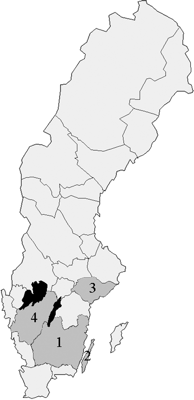
Map of the areas where moose were examined: 1, Småland; 2, Öland; 3, Sörmland; 4,Västergötland.
Seroreactivity to Anaplasma sp. in roe deers
Antibodies against A. phagocytophilum were detected in 91/107 (85%) of the roe deer samples when a cutoff titer of 1:128 was used.
PCR
No rickettsial DNA was detected by PCR of the gltA gene of SFG rickettsiae species of the 113 analyzed tissue samples from the spleen of moose and sheep. All positive and negative controls showed expected outcomes.
Statistical analyses
The CI for each animal species, area, and the total are presented in Tables 1 and 2. No significant statistical difference in seroprevalence between species could be demonstrated.
Discussion
The survey showed that between 17% and 36.9% (CI 19.4[]–27.3) of domestic animals (cats, dogs, and horses) and wild cervids (moose and roe deer) in Sweden had antibodies against Rickettsia spp. tested with R. helvetica as antigen, in a titer at or above 1:64. Even though there is a serological cross-reactivity between the different species, R. helvetica is a likely infectious agent and cause for the reactivity in mammal samples in the present study, because R. helvetica is the only SFG rickettsia generally detected in ticks in Sweden. The corresponding figure for prevalence of antibodies against A. phagocytophilum in the surveyed roe deer samples was 85%. Previous screeening of ticks with PCR from corresponding localities has shown that R. helvetica and A. phagocytophilum occur between 0–15% and 1.5–17.3%, respectively, of the ticks (Severinsson et al. 2010, Wallménius et al. 2012).
The prevalence found of seroreactivity to R. helvetica is higher than previously found in serosurveys of humans in Sweden (2.6–9.7%) (Elfving et al. 2008, Lindblom et al. 2013). One reason for this may be that these animals are more frequently exposed to tick vectors than humans are.
The lowest seroprevalence was detected amongst dogs (17%), which perhaps is influenced by the fact that they represent a group not exposed to ticks to the same extent (Melo et al. 2011) or they have been treated with tick repellents by their owners. However, surveys of dogs in Germany have shown that 78–93.9% of the dogs had antibodies to SFG rickettsiae, with a seroprevalence rate of 66.0% for R. helvetica corresponding to the prevalence rates and distribution of Rickettsia-carrying tick species (Wächter et al. 2015a). Our results, revealing a seroprevalence of 17%, correspond from that perspective with a Rickettsia-infected tick prevalence of 0–15%.
A similar level of seroprevalence (22%) was found in the cats in our study. For comparison, in sero-epidemiological studies of cats and dogs in Japan (Tabuchi et al. 2007) and Australia (Izzard et al. 2010), 0.9% and 59% of the cats and 1.7% and 57% of the dogs were positive to SFG rickettsiae when screened at a 1:20 and 1:50 dilution, respectively, using IFA. Cats has been shown to be seroreactive also to other SFG rickettsiae (R. massiliae, R. conorii, R. felis) and may have a role in the transmission cycle of these microorganisms (Segura et al. 2014).
In our survey, horses showed a higher average seroprevalence (36.5%) to R. helvetica than the other domestic animals. Similar studies in Brazil have shown a serological reactivity between 0–13% up to 38.5% in horses in different areas, in titers equal to or above 1:64 (Tamekuni et al. 2010, Toledo et al. 2011) and 7% in Spain (Lourdes et al. 2014). Horses may be frequently exposed to I. ricinus ticks during the summer period, but we had no data concerning seropositivity in relation to age or previous exposure to tick bites. However, five of 63 horses had titers ≥256, which is indicative of recent or current infection.
The above findings for domestic animals can be compared to the seroprevalence of moose ranging between 13.8% and 40.0% (mean 24%) in the different localities (Table 2). The seroprevalence was not statistically different among the four areas. Because moose and deer often are infested with high numbers of ticks, they may be an important natural host of tick-borne pathogens. In a study from Japan, DNA from R. helvetica was found in the peripheral blood of sika deer in 7.8% of the examined animals, indicating that this species may represent a potential reservoir host of R. helvetica in Japan (Inokuma et al. 2008). In a similar study of 237 roe deer in Denmark, all were PCR negative for R. helvetica (Skarphedinsson et al. 2005), a finding similar to the DNA results in this study on spleen from moose and sheep. The latter samples were also included in another study (Malmsten et al., 2014), in which a total 263 samples were analyzed by PCR showing an overall prevalence of A. phagocytohilum DNA of 26%. Inhibition was not considered as a likely problem or explanation of the results. Because seropositive moose were DNA-negative in the present study, the seroreactivitity probably represents past exposure. The chances of finding a wild animal with an active rickettsial infection is always occasional, and serology therefore represents a better surveillance tool than PCR (Skarphedinsson et al. 2005, Soares et al. 2015).
The samples from roe deer in this study were collected during 1990–1993. In the 20 years that have passed, tick numbers and distribution in Sweden reportedly have increased (Jaenson et al. 2012) and the seroprevalence would therefore be expected to be higher in a current material. There might also be a bias for the Rickettsia prevalence found in roe deer sera as a consequence of the long storage time.
Amongst the roe deer, 85% (91/107) of the serum samples were positive to A. phagocytophilum, which is in the same range as previously reported from Denmark, where 96.6% of 237 examined roe deer were positive. This is also similar to findings in Norway (96%) and Slovenia (94%) and indicates that roe deer are commonly exposed to and infected with A. phagocytophilum (Stuen et al. 2002, Petrovic et al. 2002, Skarphedinsson et al. 2005). On the basis of PCR findings, they are also thought to be competent reservoirs of A. phagocytophilum (Hulinska et al. 2004, Stuen et al. 2013).
The present findings demonstrate that animal species analyzed were exposed to and probably infected with R. helvetica. For serologic identification of the rickettsial species, multiple IFA titers against different rickettsial species antigen are required (Wächter et al. 2015b). Usually homologous antibody titers are higher than heterologous titers and more specific against the infecting rickettsia. We did not use this opportunity because we did not have access to a larger number of rickettsial strains. Because we only had one serum sample for each animal and could not investigate seroconversion, we examined the sera in cutoff titers used in other studies that clearly demonstrate that the animals were exposed to the infection. All positive samples were then titrated to their final titer.
To obtain a more reliable and better understanding of the role of mammals as a reservoir or natural host for Rickettsia spp., it would be necessary to isolate the bacteria from the blood of suspected reservoir animals to clarify their reservoir potential. The role of domestic mammals in the ecology of rickettsial species is of interest because of their close connection to humans and because they may cause the spread of the disease. Data on the presence and modes of maintenance and transmission of R. helvetica and other rickettsial species in a vertebrate reservoir are needed to advance our knowledge in this area.
Footnotes
Acknowledgments
The study was financially supported by grants from the Center of Clinical Research Dalarna (project no. 9486) and Olle Engqvist Byggmästare Stiftelse (11877). We thank the National Veterinary Institute, Uppsala, and the Swedish University of Agricultural Sciences Uppsala, for help providing the serum and tissue samples analyzed in this study.
Author Disclosure Statement
No competing financial interests exist.