
Abstract
To expand the documentation of rickettsioses in Indonesia, we conducted an ectoparasite and small mammal investigation involving four major islands: Java, Sumatra, Sulawesi, and Kalimantan. Coastal and highland regions on each island surveyed were chosen to represent different ecologies in Indonesia. Indication of the presence of Rickettsia spp. was evident in all areas sampled. Typhus group rickettsiae-specific antibodies had significantly higher prevalence among small mammals captured in Java compared to the other islands surveyed (78% in coastal and 50% in highland regions) and the prevalence of spotted fever group rickettsiae-specific antibodies was significantly higher in Kalimantan than the other islands investigated. Hosts and vectors were restricted by Rickettsia spp. but not by coastal or highland regions. Our findings expand the range in which rickettsial pathogens have been documented within the Indonesian archipelago and point to a significant risk to human health.
Introduction
R
Evidence of rickettsioses in the Indonesian archipelago has been reported since the early 1900s. Endemic scrub typhus, murine typhus, and spotted fever group rickettsiae (SFGR) have been documented from the western Indonesian islands (Sumatra and Java) to the eastern Indonesian islands (Gag and Papua) (Richards et al. 1997, Ibrahim et al. 1999, Richards et al. 2002, Richards et al. 2003, Jiang et al. 2006, Barbara et al. 2010). Unfortunately, very little has been reported about their public health impact. The prevalence of murine typhus, scrub typhus, and SFGR diseases in febrile subjects in northeastern Papua were 5%, 3%, and 1%, respectively (Punjabi et al. 2012). In central Java, the prevalence of murine typhus in febrile subjects was 7%, while evidence of other rickettsioses was not found (Gasem et al. 2009). A wide range of atypical symptoms and the lack of reliable diagnostic assays likely lead to the underreporting of documented infections. Here, we studied the risk of rickettsioses on four major islands and compared the prevalence of rickettsioses between different environments on each island. Rodents were trapped in coastal and highland regions. Serum, organs, and ectoparasites were collected from the rodents to determine the prevalence of rickettsioses within hosts and vectors of Rickettsia spp. Our findings reveal a high prevalence in all areas investigated and imply a risk to human health in these regions.
Materials and Methods
Study sites and sample collection
A detailed description of study sites, sample collection, and the diversity of small mammals captured has been described previously (Barbara et al. 2010). The survey was conducted between April 2007 and May 2008 on four islands in Indonesia with coastal and highland regions chosen for each island. The coastal regions consisted of fishing villages, while the highland regions contained farming villages surrounded by forest or conservation areas. The coastal and highland study sites were Loji (8 meters above sea level [asl]) and Cihamerang (910 meters asl) villages in West Java; Air Manis (10 meters asl) and Koto Rantang (890 meters asl) in West Sumatra; Malalayang (35 meters asl) and Kinilow (730 meters asl) in North Sulawesi; and Manggar Baru (7 meters asl) and Bukit Bangkirai (110 meters asl) in East Kalimantan (Fig. 1). Sherman and tomahawk traps were used to capture animals in peridomestic and field areas. A total of 507 small mammals representing 13 different species were captured. Sera and ectoparasites were collected from all animals. In addition, organs (spleen and kidney) were obtained from rats (Rattus sp.), house shrews (Suncus murinus), mice (Mus musculus), and Maxomys sp. A detailed description of study sites, sample collection, and the diversity of small mammals captured has been described previously (Barbara et al. 2010). The study protocols (05AUC07 and 08AUC04) were approved by the United States Naval Medical Research Unit No. 2 Institutional Animal Care and Use Committee in compliance with all applicable federal regulations governing the protection of animal in research.
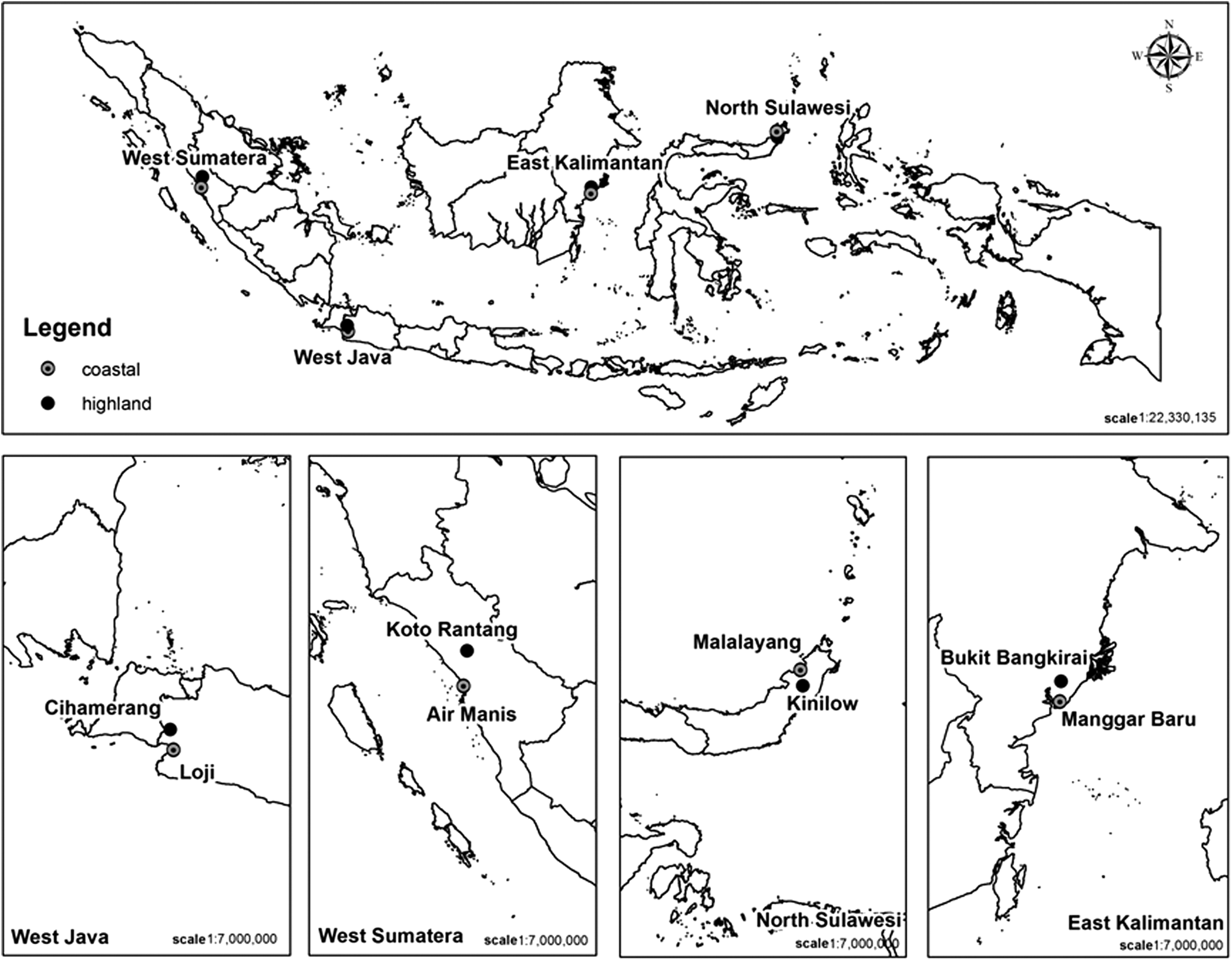
Map of Indonesia with field site locations.
Serology and PCR
Rat and mouse sera were tested by ELISA for scrub typhus group orientiae (STGO), typhus group rickettsiae (TGR), and SFGR group-specific antibodies using O. tsutsugamushi, R. typhi, and R. rickettsii ELISA antigen preparations, respectively, following published procedures (Richards et al. 1997). The net optical density (OD) value for each sample or control was determined by subtracting the OD obtained with phosphate-buffered saline from the OD obtained using the rickettsia antigen. The cutoff was determined by calculating the mean net OD value plus three standard deviations of the negative controls. These values were consistently less than 0.200. However, we used a screening cutoff net OD value of 0.5 (1:100 dilution). Those samples that were positive by screening were further tested by serial four-fold dilutions (1:100 to 1:6400). The sample was considered positive if the sum net OD value of the four-fold dilutions was greater than 1.0 net absorbance. The titer was subsequently determined to be the inverse of the highest dilution that gave a net absorbance of 0.2 or greater.
Nucleic acid was prepared from organs and ectoparasites using the QIAmp DNA extraction kit (Qiagen, Hilden, Germany) following manufacturer's instructions. Five different quantitative real-time PCR assays were performed on each sample to detect Rickettsia genus-specific 17-kDa antigen gene, tick-borne spotted fever group rickettsia-, R. felis-, and R. typhi-specific fragments of ompB and O. tsutsugamushi following procedures described earlier (Barbara et al. 2010, Jiang et al. 2004).
Statistical analysis
Statistical analysis was performed using STATA software (STATA Corp., College Station, TX). Kolmogorov-Smirnov test was applied to compare the prevalence of STGO, TGR, or SFGR-specific antibodies by areas.
Results
Rodents
ELISA results of sera from 357 rodents are shown in Table 1. The prevalence of antibodies reactive against STGO, TGR, and SFGR ELISA antigens ranged from 0% to 25%, 0% to 78%, and 9% to 73%, respectively. TGR infections were significantly more prevalent in Java than on other islands, and SFGR infections were significantly more prevalent on Kalimantan than other islands (p < 0.01). In addition, SFGR-specific antibody prevalence was significantly higher than the prevalence of STGO- and TGR-specific antibodies in Kalimantan (p < 0.01). There were no significant differences in rickettsial antibody prevalence between coastal and highland regions on Java, Sumatra, or Sulawesi, while on Kalimantan the prevalence of antibodies to these pathogen groups were higher in the coastal region (p < 0.01).
SFGR, spotted fever group rickettsiae; STGO, scrub typhus group orientiae; TGR, typhus group rickettsiae.
Rattus tanezumi (Asian house rat) was the most common rodent collected in all areas investigated except in coastal Sulawesi, where both R. tanezumi and Rattus norvegicus (brown rat) were found in similar numbers, and in coastal Kalimantan, where R. norvegicus was the predominant species. ELISA results show that R. tanezumi and R. norvegicus were likely hosts of TGR, whereas STGO- and SFGR-specific antibodies were present in a more diverse range of hosts. STGO-specific antibody prevalence was higher in field rodents and TGR prevalence was higher in peridomestic rodents in each region, but the differences were not statistically significant. The gender of the rodents also was not significantly different in relation to antibody prevalence. Mice were only found trapped on the island of Sulawesi (n = 4) and none had positive antibody responses to rickettsiae. Sera from other species of small mammals captured were not tested by ELISA due to the lack of appropriate secondary antibodies.
The spleen and kidneys from 156 rodents and shrews were chosen randomly for PCR testing based on species, place and time collected. Rattus tanezumi from Java (n = 3) and Sumatra (n = 1) had evidence of R. typhi infection. Maxomys sp. (n = 2) from Sulawesi, Rattus exulans (n = 1) and R. whiteheadi (n = 1) from Kalimantan had evidence of O. tsutsugamushi infection. Of all PCR-positive rodents, only R. tanezumi from Java had antibodies against R. typhi antigen.
Ectoparasites
A total of 2,712 ectoparasites were collected including three genera of fleas [Xenopsylla cheopis (n = 204), Nosopsyllus (n = 3), and Monopsyllus (n = 2)]; two genera of lice [Hoplopleura (n = 257) and Polyplax (n = 285)]; six genera of mites (Leptotrombidium (n = 476), Demodex (n = 1), Echinolaelaps (n = 58), Haemogamassus (n = 5), Laelaps (n = 1231), and Ornithonyssus (n = 105)]; and six genera of ticks [Rhipicephalus (Boophilus) (n = 4), Rhipicephalus (not Boophilus) (n = 18), Amblyomma (n = 14), Haemaphysalis (n = 31), Ixodes (n = 17), and Dermacentor (n = 1)]. Ectoparasites were pooled according to host species, ectoparasite species, place collected, and time collected and tested for the presence of Rickettsia genus-specific 17-kDa antigen gene, tick-borne spotted fever group rickettsia-, R. felis-, and R. typhi-specific fragments of ompB. PCR results from the fleas collected indicated the presence of R. felis and R. typhi and have already been reported (Barbara et al. 2010). The presence of R. typhi DNA was also found in mites (Laelaps) and lice (Poliplax). R. felis DNA was found in the most diverse range of arthropods including mites (Laelaps), lice (Poliplax, Haemogamassus), and ticks (Haemaphysalis, Ixodes). Only ticks were positive for SFGR (Haemaphysalis, Ixodes, and Rhipicephalus (not Boophilus)) and only the Leptorombidium mites tested positive for STGO. In examining the host on which the PCR-positive ectoparasites were found, it is interesting to note that ectoparasites found on shrews were positive for R. typhi, R. felis and SFGR. In addition, mites found on squirrels (Sundasciurus lowii) were positive for O. tsutsugamushi. Table 2 provides the details for the PCR-positive ectoparasites.
R. felis: Rickettsia felis quantitative real-time PCR positive (qPCR+); R. typhi: R. typhi qPCR +; O. tsu: Orientia tsutsugamushi qPCR +; SFG: SFG-specific qPCR +.
Discussion
Serological and molecular results from this study suggested a considerable risk for rickettsial infection in humans throughout Indonesia, since the potential hosts and vectors were found in all areas investigated. There was no difference in rickettsia-specific antibody prevalence between coastal and highland regions on each island, suggesting that ecological and anthropological variations did not affect the distribution of rickettsial pathogens. However, differences were found between the islands. A significantly higher prevalence of TGR-specific antibodies was found in rodents trapped on Java compared with the other islands, while a significantly higher prevalence of SFGR-specific antibodies was found in rodents trapped on Kalimantan. These results suggest that the risk of human infection with these pathogens may differ by island. There was no difference in rickettsial-specific antibody prevalence between peridomestic and field rodents from each region. Our data indicated that rodents were the most important hosts for Rickettsia spp. and that humans might not have any important influence in maintaining the rickettsial diseases in the region.
Spotted fever group rickettsiae were the most prevalent cause of rickettsial infections, as evidenced by the presence of rickettsial group-specific antibodies by ELISA, and we found ectoparasites infected by R. felis (a SFGR) in most of the regions investigated. However, we cannot say which spotted fever group rickettsia (e.g., R. felis or one or more of the tick-borne SFGR) infection(s) resulted in the SFGR-specific antibodies we detected by ELISA as antibodies against SFGR species cross-react in serological assays. As SFGR infections were the most prevalent across the archipelago, further studies are necessary to reveal the importance of non–R. felis SFGR species. In addition, there has been no report of R. felis as a pathogen for human disease although it has been circulating in Java since at least 1994 (Jiang et al. 2006). Its importance for public health may have been masked due to similarity of symptoms with other febrile illnesses and the lack of species-specific diagnostic assays. Moreover, our serological and molecular assays could not distinguish the presence of R. felis–like organisms recently reported as prevalent in Asia, the Americas, and Africa (Odhiambo et al. 2014). The original report that described the presence of R. felis in Indonesia was from two X. cheopis flea pools from Malang, Indonesia and were confirmed by sequencing to be R. felis and not R. felis–like organisms (Odhiambo et al. 2014). Of all the PCR-positive rodents in this study, only one was also serologically positive; as antibodies take time to develop after infection and persist long after the pathogen is no longer present, this is not unexpected.
Rattus tanezumi, R. norvegicus and S. murinus were potential hosts of TGR, while STGO and SFGR had a wider range of hosts. Although R. typhi and R. felis are known as flea-borne rickettsiae, our results show they may also be found in other arthropods such as lice and mites. Xenopsylla cheopis, Poliplax, and Laelaps were the most common fleas, lice, and mites infected with these agents and are therefore potentially the most important vectors of R. typhi and R. felis in Indonesia. Our results indicate that ticks could also be potential vectors of R. felis in Indonesia and this finding has also been reported in other countries (Parola 2011). Chiggers and ticks were restricted vectors of STGO and SFGR, respectively. To our knowledge, we report for the first time the potential vectors/hosts of Rickettsia spp. across Indonesia.
Our findings suggest that the risk for rickettsial infections in Indonesia is high and indicate the existence of various hosts and vectors across the country. Therefore, health care providers should consider rickettsioses as an important component of infectious diseases, particularly in heavily rodent- and shrew-infested areas. Moreover, regular surveillance and use of reliable diagnostic assays to determine rickettsioses in humans are urgently needed.
Footnotes
Acknowledgments
We would like to thank the staff of livestock services and district offices for their support in the field. We also would like to thank Achmad Saim for his supervision in ectoparasite identification.
This work was supported by the Global Emerging Infections Surveillance and Response System, a division of the United States Department of Defense Armed Forces Health Surveillance Center. The views expressed in this article are those of the authors and do not necessarily reflect the official policy or position of the Department of Defense, Department of the Navy, or the U.S. government. Some of the authors are employees of the U.S. government or military service members (SW, MW, IW, UA, CAS, KAB, ALR, and PJB). This work was prepared as part of their official duties. Title 17 U.S. Code section 105 provides that “copyright protection under this title is not available for any work of the United States Government” Title 17 U.S. Code section 101 defines a U.S. government work as a work prepared by a military service member or employee of the U.S. government as part of that person's official duties.
Author Disclosure Statement
No competing financial interests exist.