




Abstract
Toxoplasma gondii is an important intracellular parasite, which can infect endothermic vertebrate animals, including minks (Neovison vison). However, information on T. gondii infection in minks in China is limited. Therefore, we investigated the seroprevalence and risk factors of T. gondii infection in minks in northeastern and eastern China. A total of 1499 mink blood samples were randomly collected from eight cities between March 2014 and January 2015 in northeastern and eastern China, and antibodies to T. gondii were examined using the modified agglutination test. Overall, the seroprevalence of T. gondii infection was 8.14% in the examined minks. The T. gondii seroprevalence was different among cities (ranging from 1.85% in Changchun to 15.75% in Dalian), genders (4.31% in male and 6.22% in female), seasons (spring: 11.64%; summer: 7.34%; autumn: 7.37%; and winter: 7.32%), and ages (young: 5.79%; subadult: 5.03%; and adult: 11.08%). Region and age were considered as risk factors for T. gondii infection. These results provided baseline data for the prevention and control of T. gondii infection in minks in China.
Introduction
T
As an important economic animal, mink (Neovison vison) is one of the three pillars in international fur market, which can provide better marten. Owing to its abundant economic benefit, an increasing number of minks have been raised in China, and these domesticated minks also provided meat for people. The heart of mink can be made “mink heart pill” which have good curative effect for rheumatic heart disease and congestive heart failure, and mink liver can be used for the treatment of night blindness (Pu 2002). Thus, epidemiological investigations of T. gondii infection in minks are necessary and significant because mink is closely associated with humans.
Although several investigations about the seroprevalence of T. gondii infection in minks in other countries were reported (Henriksen et al. 1994, Smith and Frenkel 1995, Smielewska-Łoś and Turniak 2004, Sepulveda et al. 2011, Ahlers et al. 2015), data on the prevalence of T. gondii infection in mink in China are limited. Thus, the present investigation was conducted to determine the seroprevalence of T. gondii infection in minks in northeastern and eastern China and to assess associated risk factors.
Materials and Methods
Ethics statement
This study was approved before its commencement by the Ethics Committee of the Lanzhou Veterinary Research Institute, Chinese Academy of Agricultural Sciences. The blood samples were collected by veterinarians without causing any additional discomfort to the animals.
The study sites
This investigation was carried out in eight cities of four provinces in northeastern and eastern China (Fig. 1). Mudanjiang city locates in Heilongjiang province (121°11′–135°05′E, 43°26′–53°33′N). Changchun and Jilin cities belong to Jilin province (121°38′–131°17′E, 40°52′–46°18′N). Dalian city locates in Liaoning province (118°53′–125°46′E, 38°43′–43°36′N). Yantai, Weihai, Qingdao, and Rizhao cities belong to Shandong province (34°22′–38°23′N, 114°19′–122°43′E) (Fig. 1).
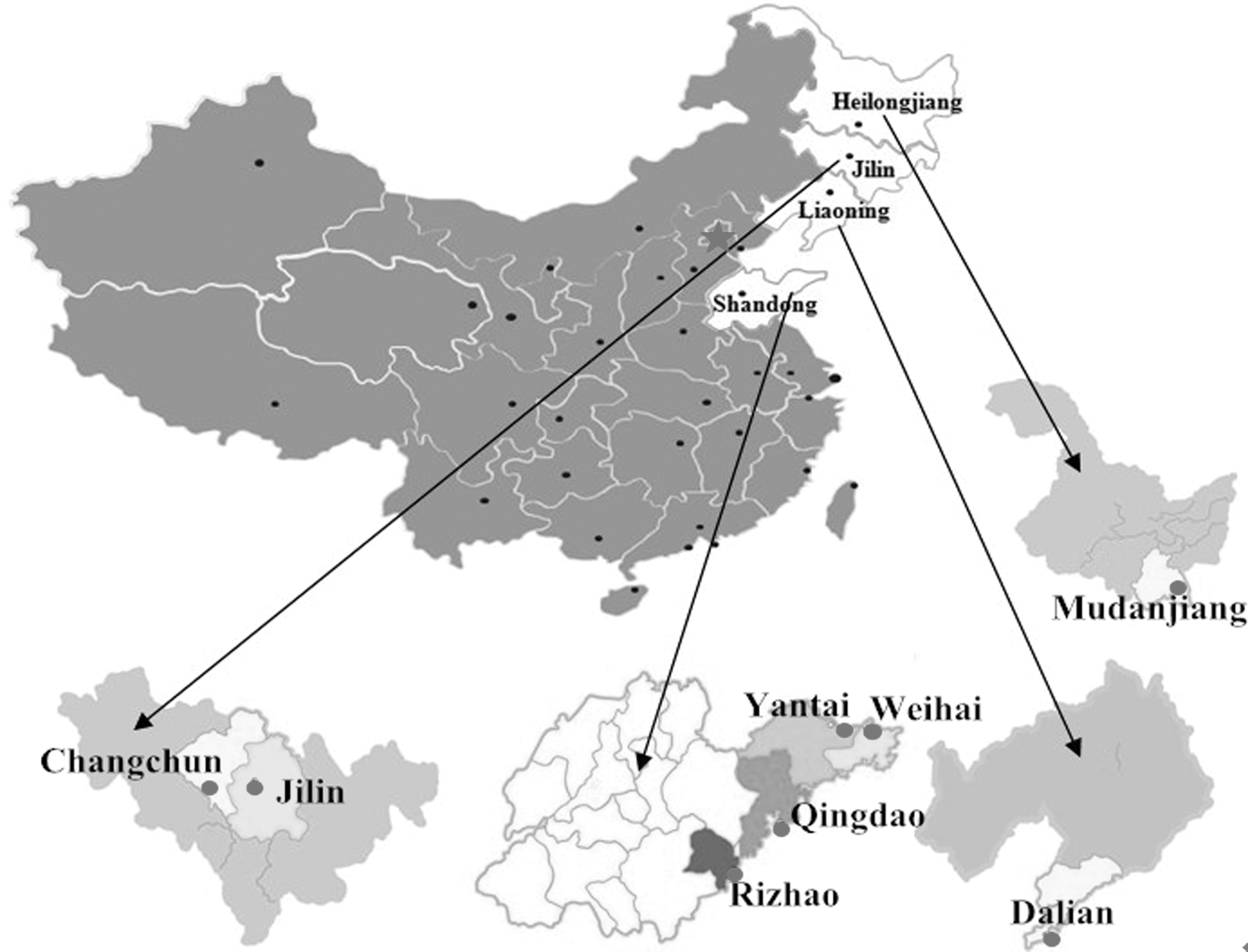
A map of China showing the geographical regions in four provinces where farmed minks were sampled.
Serum samples
A total of 1499 mink blood samples were collected randomly from 16 mink herds between March 2014 and January 2015 from eight cities in the four studied provinces (Table 1). Data of the animals (geographic origin, gender, age, and diet) were acquired from owners. Mink serum was acquired by centrifugation at 2000 g for 10 min and stored at −20°C until further detection.
CI, confidence intervals; OR, odds ratios with 95% CI.
Serological examination
Antibodies to T. gondii were assayed by the modified agglutination test (MAT). The Toxoplasma MAT antigen was provided by the Laboratoire de Parasitologie-Mycologie, Centre National de Référence de la Toxoplasmose, Centre de Ressources Biologiques Toxoplasma, Hôpital Maison Blanche, Reims Cédex, France. The pig serum with the titers greater than 1:200 was used as positive control. The procedures of antibody detection were carried out as described previously (Qin et al. 2014, Lou et al. 2015). In brief, each mink serum was diluted from 1:25 to 1:800, using twofold dilutions in U-bottom 96-well microtiter plates. The antigen was added to each well. Then, the sealed microtiter plates were incubated for 12 h at 37°C. The assay was considered positive when formation of antigen-antibody agglutination layer occurred at dilutions of 1:25 or higher. The positive, negative, and blank controls were included in each test. The results were considered reliable when the positive control titers were 1:200 or greater. Sera with dubious results were re-tested.
Statistical analyses
Exploratory analysis was implemented to reveal the potential relationship and risk factors of T. gondii infection in minks of different regions, genders, ages, and seasons by chi-square analysis. p-Value (p<0.05) was considered as statistically significant. Odds-ratios (OR) with 95% confidence intervals (CI) based on likelihood ratio test were analyzed in the present study. All statistical analyses were performed by SAS (Statistical Analysis System, Version 9.1).
Results and Discussion
A total of 122 (8.14%, 95% CI 6.76–9.52) serum samples from 1499 minks were positive for T. gondii antibody by MAT at a 1:25 cut-off, with antibody titers of 1:25 in 85, 1:50 in 22, 1:100 in 6, 1:200 in 4, 1:400 in 3, and 1:800 in two samples, respectively. The T. gondii seroprevalence in minks was different among cities (ranging from 1.85% in Changchun to 15.75% in Dalian), genders (4.31% in male and 6.22% in female), seasons (spring: 11.64%; summer: 7.34%; autumn: 7.37%; and winter: 7.32%) and ages (young: 5.79%; subadult: 5.03%; and adult: 11.08%) (Table 1).
The overall T. gondii seroprevalence in minks in the present study was 8.14%, which was lower than that in other studies, such as the 13.9% in minks in Poland by latex agglutination test (LAT) (Smielewska-Łoś and Turniak 2004), 70% in minks in Chile by LAT (Sepulveda et al. 2011), 77% in minks in a freshwater ecosystem in United States by MAT (Ahlers et al. 2015), and 66% in minks in Missouri and Kansas in United States by the Sabin–Feldman dye test (Smith and Frenkel 1995), but higher than the 3% in minks in Denmark by LAT (Henriksen et al. 1994). The difference may be due to differences in living environment, geographic region, climatic conditions, the density of cats in farms, and the diet of minks (Cosendey-KezenLeite et al. 2014, Mahmoudvand et al. 2015). Moreover, different sensitivity and specificity of the different detection methods were also considered as an important reason (Dawoud et al. 2009, Hamidinejat et al. 2014, Gu et al. 2015).
No significant effects of gender and season on T. gondii seroprevalence were found in the present study (p > 0.05), although slightly higher seroprevalence was present in spring (Table 1). However, geographical location is revealed as an important risk factor for T. gondii infection in minks (p < 0.01). Among these eight cities, minks in Dalian (15.75%) were nearly ten times (OR = 9.91, 95% CI = 3.35 – 29.32) at risk of acquiring T. gondii infection compared to Changchun (1.85%). Compared to minks in Changchun, minks in Mudanjiang (15.08%) were more than nine times at risk of infecting T. gondii. These results indicated that minks in Changchun had lower opportunity to acquire T. gondii infection. This difference is probably due to food the minks ate. Minks in examined farms in Changchun were fed with dry full ration, whereas minks in Dalian were fed with fresh fish and by-products of pigs, cattle, and goats, which may contain T. gondii tissue cysts (Liu et al. 2012, Xu et al. 2014).
Furthermore, the T. gondii seroprevalences in minks of different ages are different, ranging from 5.03% (95% CI 3.03–7.04) in subadult to 11.08% (95% CI 8.81–13.36) in adult (Table 1). Adult minks (OR = 2.03, 95% CI = 1.20 – 3.44) had more than two times risk of acquiring T. gondii infection than young minks, which had similar seroprevalence with subadult minks. Adult minks have more chance to acquire T. gondii infection by ingesting raw meat containing tissue cysts, or by consuming water and food contaminated with T. gondii oocysts, presenting the highest seroprevalence, which was coincidental with some previous studies (Ranucci et al. 2013, Jiang et al. 2014).
Minks can be infected by T. gondii through a variety of routes; drinking water contaminated by T. gondii oocysts and ingesting raw or undercooked meat containing tissue cysts are possibly the major ways of infection. The significance of these routes of infection in the transmission of T. gondii to minks needs to be explored in further studies to improve the data of risk factors of T. gondii infection in farmed minks.
Conclusion
The present survey revealed an 8.14% seroprevalence of T. gondii infection in minks in northeastern and eastern China. Region and age were considered the risk factors for mink infection with T. gondii. These data indicated that mink is a susceptible animal of T. gondii, and humans may get infected by ingesting raw or undercooked mink meat containing T. gondii tissue cysts. The present results provided baseline data for the prevention and control of T. gondii infection in minks in China.
Footnotes
Acknowledgments
Project support was provided by the Quality Inspection Special Public Welfare Industry Research (grant no. 201410061), the Science Fund for Creative Research Groups of Gansu Province (grant no. 1210RJIA006), and the Agricultural Science and Technology Innovation Program (ASTIP) (grant no. CAAS-ASTIP-2014-LVRI-03). Laboratoire de Parasitologie-Mycologie, Centre National de Référence de la Toxoplasmose, Centre de Ressources Biologiques Toxoplasma, Hôpital Maison Blanche, Reims Cédex, France, is thanked for providing the Toxoplasma MAT antigen.
Author Disclosure Statement
No competing financial interests exist.