
Abstract
Introduction:
Rift Valley fever (RVF) is a vector-borne zoonotic disease caused by phlebovirus in the family Bunyaviridae. In Kenya, major outbreaks occurred in 1997–1998 and 2006–2007 leading to human deaths, huge economic losses because of livestock morbidity, mortality, and restrictions on livestock trade.
Aim:
This study was conducted to determine RVF seroprevalence in cattle, sheep, and goats during an interepidemic period in Garissa County in Kenya.
Methods:
In July 2013, we performed a cross-sectional survey and sampled 370 ruminants from eight RVF-prone areas of Garissa County. Rift Valley fever virus (RVFV) antibodies were detected using a multispecies competitive enzyme-linked immunosorbent assay. Mixed effect logistic regression models were used to determine the association between RVF seropositivity and species, sex, age, and location of the animals.
Results:
A total of 271 goats, 87 sheep, and 12 cattle were sampled and the overall immunoglobulin G seroprevalence was 27.6% (95% CI [23–32.1]). Sheep, cattle, and goats had seroprevalences of 32.2% (95% CI [20.6–31]), 33.3% (95% CI [6.7–60]), and 25.8% (95% CI [22.4–42]), respectively. Seropositivity in males was 31.8% (95% CI [22.2–31.8]), whereas that of females was 27% (95% CI [18.1–45.6]).
Conclusions:
The high seroprevalence suggests RVFV circulation in domestic ruminants in Garissa and may be indicative of a subclinal infection. These findings provide evidence of RVF disease status that will assist decision-makers to flag areas of high risk of RVF outbreaks and prioritize the implementation of timely and cost-effective vaccination programs.
Introduction
R
The Rift Valley fever virus (RVFV) is a Phlebovirus belonging to the family Bunyaviridae. The occurrence of RVF epidemics has been associated with the El Niño/Southern Oscillation phenomenon that leads to heavy and persistent rainfall, causing flooding and hatching of infected eggs of Aedes mosquitoes that transmit the virus to livestock (Anyamba et al. 2010).
The 1997–1998 RVF outbreaks in Kenya caused 170 human deaths in Garissa, which was the epicenter of the outbreak (Woods et al. 2002). A total of 478 human cases were also reported in North Eastern Kenya and southern Somalia (CDC 1998). During the 2006–2007 epidemic period, more than 30,000 livestock and 700 human cases and 158 human deaths were reported in Kenya (WHO 2007, Munyua et al. 2010, Nguku et al. 2010). These estimates were mainly from reports and studies conducted in Garissa and may have underestimated the burden of RVF at the national level.
Although most RVF serological surveys in Eastern Africa focus on epidemics (Woods et al. 2002, WHO 2007, LaBeaud et al. 2008, Nguku et al. 2010), limited research has been done to determine RVFV transmission patterns during the interepidemic periods (King et al. 2010, Gray et al. 2015, Muiruri et al. 2015). The objective of this study was to determine levels of RVFV exposure in goats, sheep, and cattle in the Garissa County, Kenya, during an interepidemic period.
Methods
Study area
The study was performed in Garissa County a semiarid zone in Northeastern part of Kenya, bordering Somalia to the east and Tana River to the west, located between latitude 0° 58′ N and 1° 30′ S and longitudes 38° 34′ E and 41° 05′ E (Fig. 1). It covers ∼33,620 km2, with a population of 623,060 persons and 1.5 million livestock. It has low altitude ranging from 70 to 400 meters above sea level suitable for flooding during heavy rainfalls.
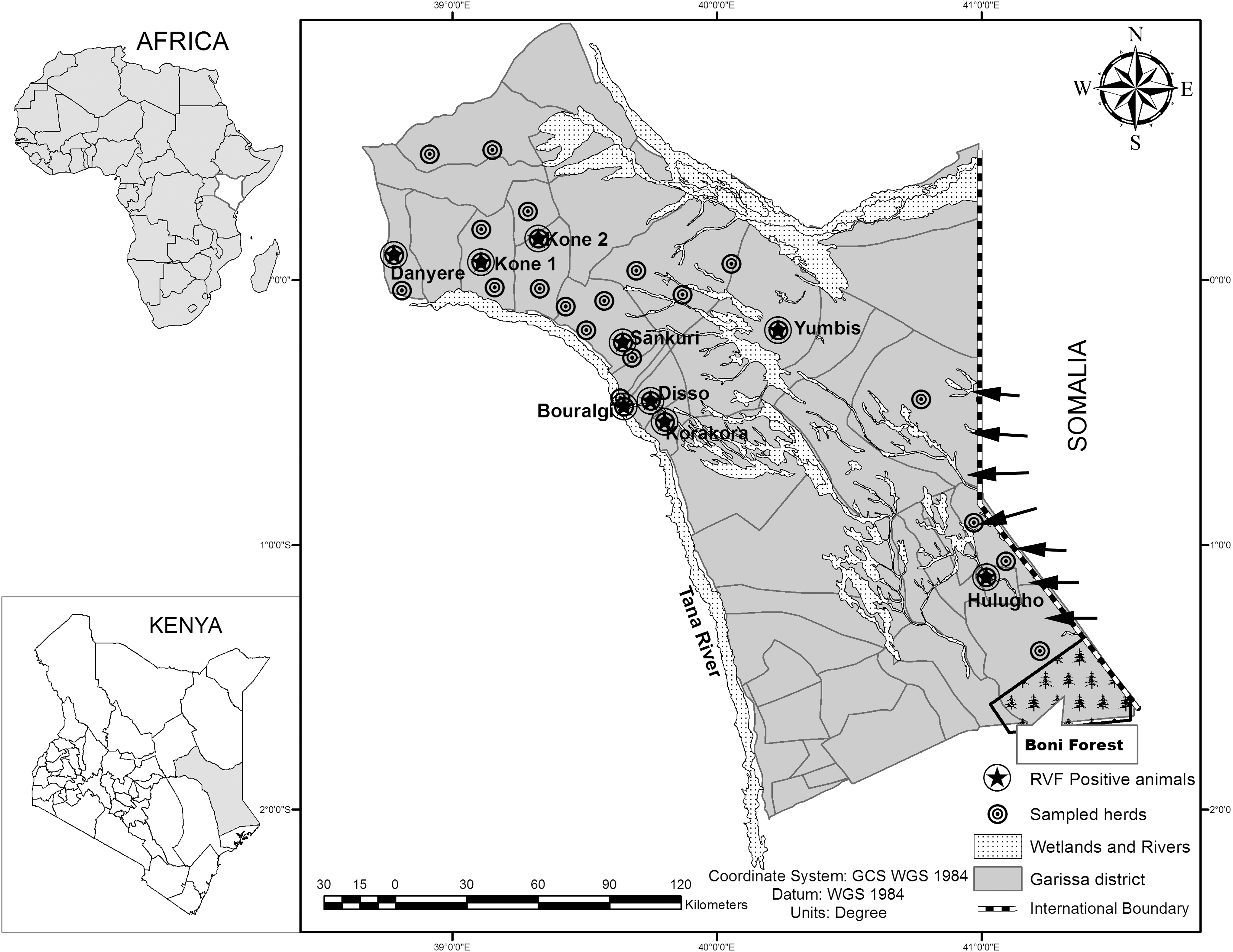
Geographical location of study sites in Kenya indicating all sampled herds (bull pointed) and the spatial distribution of RVF seropositive animals (starred) in Garissa, livestock trekking routes (black arrows). RVF, Rift Valley fever.
Study design and sample size determination
A cross-sectional study design was used in Garissa County, where the sample size was determined based on methods for estimating a proportion of diseased animals that have a defined precision in the entire animal population (Dohoo 2009). With a priori RVFV seroprevalence (P) of 50%, maximum allowable error of 10% (P = 50 ± 10), and confidence level of 95%, the required sample size was estimated to be 384 animals. This was increased to 400 to improve the precision of the prevalence estimates.
Selection of the sites and sampling of animals
In July 2013, eight divisions in the County that were affected by the 2006–2007 RVF outbreaks were purposively selected for the survey. These were Danyere, Kone, Sankuri, Korakora, Bouralgi, Disso, Yumbis, and Hulugho divisions. A total of 300 villages were sampled by using a probability proportionate to size approach, where 30 villages were finally selected based on their mosquito breeding sites suitability and presence of artificial animal watering points and high livestock populations. To avoid reporting anti-RVFV antibodies false positives, the exclusion criterion was based on animals that have been recently vaccinated against RVF. We relied on guidance by the local veterinarians and animal technicians who assisted in identification of animal herds that had not been vaccinated against RVF for inclusion and random sampling. Using a global positioning system, receiver coordinates of all sampled herds in villages and distribution of water bodies were recorded for traceability. Five milliliters of whole blood was collected from all the animals by jugular venipuncture into plain vacutainer tubes, this was transported to the Garissa laboratory and kept at room temperature for 20 min. Centrifugation was done for 10 min at 2419.2 g. Sera were recovered as aliquots into cryovials and stored at −80°C until testing at KEMRI laboratories
Serological testing
We investigated the presence of RVFV antibodies by using a commercial indirect competitive enzyme-linked immunosorbent assay (cELISA, ID Screen®; IDVet Innovative Diagnostics, Grabels, France). In brief, 50 μL of prediluted serum samples and controls (positive and negative) was added into the wells of the coated plate and 50 μL of RVF sample dilution buffer was added to each well containing the controls or samples. Each plate was then covered with an adhesive cover plate and incubated for 1 h at 37°C. Three washes were performed with wash solution before 100 μL of conjugate was added and incubated for 30 min at 21°C. The three wash steps were repeated and 100 μL of substrate was added to each well and incubated for 15 min at 21°C, then 100 μL of stop solution was added. The results were read within 30 min after stopping the reaction at 450 nm on a microplate reader (ELX800-USA; Biokit). Validation of the test was done when the mean value of the negative control optical density (ODNC) >0.7 and that of the positive control (ODPC) was <30% of the ODNC (ODPC/ODNC <0.3). Seropositivity was detectable as suspect or negative (S/N%) value of ≤40% (Ellis et al. 2014; See Supplementary Data; Supplementary Data available online at
Statistical analysis
Univariable logistic regression analysis was used to determine the association between the putative risk factors and the serostatus for RVF for each animal. We estimated the effects of the exposure variables on the distribution of the outcome variable while controlling for other variables (covariates). Variables for which an association (p ≤ 0.2) was detected were included as predictors in a generalized mixed effect multivariable logistic regression model. The factors meeting this criterion were species, age, sex, and location, and formed the maximum saturated model. By using a backward stepwise elimination process, we assessed the factors and their interactions and retained them in the model only when p ≤ 0.05. The final fitted individual models were evaluated by including location (sampling site) as a random effect to adjust for possible clustering of RVF seropositivity within herds. Intracluster correlation coefficient (ICC; ρ, rho) describing the degree of similarity among seropositive animals in each location was estimated. Adjusted odds ratios (ORs) for seropositivity were estimated using a “lme4” package in R. All analyses were performed using R statistical software version 3.1.3 (R Development CoreTeam 2015).
Ethical considerations
Ethical approval for this study protocol was obtained from the institutional review board of the Faculty of Veterinary Medicine at University of Nairobi (Protocol approval number UON/PHPT/VEP/2012/003). The animal restraint and sampling were designed to be less invasive to animals and appropriate personal protective equipment was used to minimize injury and infection to animal technicians and veterinary surgeons who conducted the blood sampling according to the World Organization for Animal Health (OIE) guidelines for use of animals in research and education (OIE 2014).
Results
Cross-sectional survey
A total of 415 animals were sampled, 45 samples were discarded because of leakages and compromise in quality during transportation. Seropositivity for 370 (89.2%) of the samples was analyzed as stratified by location, species, and age as presented in Table 1.
Seroprevalence by location, sex, and species
The overall RVFV antibody seroprevalence for 370 animals was 27.6% (95% CI 23–32.1). The seropositivity for cattle, sheep, and goats was 33.3% (4/12), 32.2% (28/87), and 25.8% (70/271), respectively. We recorded a unique local maximum seroprevalence of 76.9% (10/13) in goats from Hulugho in the southern part and 45.5% (5/11) in sheep from Bouralgi in the west-central part (Table 2 and Fig. 1). The variance between locations from the logistic regression model was 0.09 for 370 animals and we estimated the ICC ρ of 0.02 within locations.
CI, confidence interval; SP, seroprevalence.
The overall seroprevalence for all male species was 31.8% (95% CI 18.1–45.6) and for all female species was 27% (95% CI 22.2–31.8). Female goats had seroprevalence of 26.3% (95% CI 20.8–31.9) whereas male goats had seroprevalence of 21.4% (95% CI 6.2–36.6). Female and male sheep had a seroprevalence of 30.1 (19.6–40.7) and 42.6 (16.9–68.8) respectively. Goats >1-year-old had seroprevalence of 31.9% (95% CI 25.7–38.2) whereas sheep >1-year-old had seroprevalence of 35.5 (95% CI 24.8–46.3). Sheep ≤1-year-old had prevalence of 9.1% (95% CI −7.9 to 26.1) compared with goats 1.8% (95% CI −1.7 to 5.3) as indicated in Table 3.
Host factors influence on seropositivity
A mixed effect logistic regression model in which location was a random effect was used to investigate RVFV antibody prevalence association with host factors (age and sex). Owing to lack of accurate animal demographics and to control for confounding, age was categorized as ≤1 year and >1 year. Seropositivity increased with advanced age, animals >1-year-old had an 18-fold likelihood to be seropositive than animals ≤1-year-old, OR 18.24 (95% CI 5.26–116.4), p = 0.0001 (Table 4).
Reference level.
Significance level.
OR, odds ratio; RVF, Rift Valley fever.
Discussion
Here we report the presence of RVFV IgG antibodies in domestic ruminants from Garissa County in Kenya, 6 years after the 2006–2007 outbreak. Although the overall seroprevalence of RVF in sheep, cattle, and goats in this study area was 27.6%, earlier studies in the same and neighboring areas reported high seropositivity of about 70% in small ruminants in Ijara in 2010, this may be attributed to differences in time of sampling (Lichoti et al. 2014).
Garissa has suitable ecological conditions forming a suitable niche for mosquito survival and hence virus persistence in the environment (Woods et al. 2002); it has forested areas such as Boni, with a rich hydrological network of Tana river and wetlands or dambos, less pervious soil types, and vast flat areas that form floodplains during prolonged rainy seasons, this provides conducive mosquito breeding grounds and a large ruminant population provides hosts for potential virus amplification (Hightower et al. 2012).The spatial distribution of seropositivity was spread across the study sites with observed aggregation near wetlands, indicating spatial dependency in northwestern, central, and southern parts of Garissa (Fig. 1). Longitudinal studies in Garissa indicated circulation of RVFV that was likely to be related to livestock migration to forested areas (Lichoti et al. 2014, Owange et al. 2014, Arum et al. 2015).
High RVF seroprevalence has been demonstrated in humans living in Garissa and the neighboring Lamu County, and our findings confirm the high prevalence of RVFV in livestock, which may serve as surrogate indicators for disease in humans (Gray et al. 2015, Muiruri et al. 2015). Garissa is the main route of livestock trade into Kenya from Somalia (Fig. 1); the trekking of live animals may be responsible for the spillage of infection to Somalia and reintroduction from Somalia, thus creating an RVF endemic foci, this has been observed in Sahrawi territories and the Comoros Islands, where RVF outbreaks have been associated with transboundary livestock trade (Di Nardo et al. 2014, Roger et al. 2014)
The role of sex and age in RVF seropositivity has been examined. In Madagascar, IgG antibodies were significantly higher in males than in females in both cattle and small ruminants, but increased with the age of the animals (p < 0.0001) (Jeanmaire et al. 2011). However, in Garissa we found no significant difference in seroprevalence according to sex for all animal species (p = 0.68), despite the overall prevalence in male animals being 42.6% (CI 16.9–68.8) compared with that in females being 30.1% (CI 19.6–40.7).
The main limitations to this study were logistic challenges that led to our inability to sample more animals; these findings may have limited generalizability, especially for cattle seropositivity because of smaller sample sizes. We can, therefore, only draw inferences on RVFV seropositivity for a small population in a limited geographic scale and the estimated ICC values should be interpreted with caution. IgM ELISA and PCR were not conducted; this might have provided information on acute RVFV infections, whereas plaque reduction neutralization test was not performed to rule out false positives and cross immunoreactivity with other arboviruses.
Conclusion
The high RVFV seroprevalence in ruminants in Garissa is indicative of viral circulation despite no occurrence of outbreaks or lack of observable clinical signs of RVF, this may be indicative of a subclinal RVF infection. More surveillance studies are required during the interepidemic period for early RVF outbreak detection and for enhancement of cost-effective-targeted vaccinations in high-risk areas of Kenya.
Footnotes
Acknowledgments
We are indebted to the Kenya Directorate of Veterinary Services for permission to undertake this study and its entire staff in Garissa County and to Drs. Jackson Kinyua, Rashid Mohammed, and Stephen Gathogo for logistical support during field sampling. We appreciate Dr. Penina Munyua of CDC-Kenya for critical revision of this article.
Author Disclosure Statement
No competing financial interests exist.
References
Supplementary Material
Please find the following supplemental material available below.
For Open Access articles published under a Creative Commons License, all supplemental material carries the same license as the article it is associated with.
For non-Open Access articles published, all supplemental material carries a non-exclusive license, and permission requests for re-use of supplemental material or any part of supplemental material shall be sent directly to the copyright owner as specified in the copyright notice associated with the article.