
Abstract
Toxoplasma gondii is a protozoan parasite with both public health and veterinary importance. The aims of our study were to estimate the T. gondii seroprevalence in sheep in Latvia and to evaluate potential risk factors for seropositivity. The blood samples investigated were collected from 1039 sheep from 84 farms between January 2012 and April 2013. The sera were tested for immunoglobulin G antibodies against T. gondii using an in-house enzyme-linked immunosorbent assay. Antibodies against T. gondii were detected in 179 (17.2%) of the sheep, and at least one seropositive sheep was found on 37 (44.0%) of the farms. The seroprevalence was significantly higher in adult (over 1 year old) sheep than in juveniles—the adult sheep had 1.8 times higher odds to test seropositive than did the juveniles. The seroprevalence was significantly higher in Latvian Dark Headed sheep than in crossbreed and German Merino sheep. Sheep from large herds (with more than 100 sheep), as well as those from farms with cattle, had higher odds to test seropositive, whereas the presence of poultry on the farm appeared as a protecting factor. The results show that sheep in Latvia are commonly exposed to T. gondii.
Introduction
T
Sheep (Ovis aries) are economically important domestic animals in many countries, and they are often raised for human consumption and wool production. Sheep farming is gaining popularity in Latvia; there are 83,655 sheep on 4425 farms, and most (79.0%) of the farms have 20 or fewer sheep (
Sheep are commonly infected with T. gondii, and the meat of infected sheep is considered an important T. gondii infection source for humans (Dubey 2009). The known T. gondii infection routes for sheep include ingesting sporulated oocysts from contaminated feed or pastures and transplacental infection (Dubey 2009, Innes et al. 2009). The risk factors associated with ovine T. gondii infections include the age of the sheep and the management of the farm; for example, the presence of cats on the farm can increase the risk of infection (Skjerve et al. 1998, Stimbirys et al. 2007, Vesco et al. 2007, Cavalcante et al. 2008, Cenci-Goga et al. 2013). Clinical signs of ovine toxoplasmosis include fever and respiratory distress, as well as early embryonic death and resorption, mummification, stillbirth, neonatal death, and weak lambs (Dubey 2009).
The data on T. gondii infections in sheep from the Baltic States are limited, and locally relevant risk factors are largely unknown. In Lithuania, the seroprevalence was 42.1% (n = 354) (Stimbirys et al. 2007). Two available estimates from Latvia were both obtained from limited sample size; 5.6% by the complement fixation test (n = 107) and 45.0% by the latex agglutination test (n = 20) (Eglīte and Keidāns 2000). The aims of our study were to estimate T. gondii seroprevalence and evaluate potential risk factors for T. gondii infections in sheep in Latvia.
Materials and Methods
Collection of samples and background information
A total of 1039 ovine blood samples were collected from 84 farms (1–246 samples from each farm) between January 2012 and April 2013. All of the farms reportedly fed the animals outdoors on a pasture from April to December.
The samples were collected during the State program for Surveillance of Animal Infection diseases (
To evaluate potential risk factors associated with T. gondii infections, information regarding the age, sex, and breed of each sheep, herd size, presence of cattle or poultry on the farm, and the geographical location of the farm were collected from the Agricultural Data Center database (
Enzyme-linked immunosorbent assay
The sera were analyzed with a modified indirect in-house enzyme-linked immunosorbent assay (ELISA) for the presence of anti-T. gondii antibodies. We used a protein extract of T. gondii RH strain tachyzoites (European Union Reference Laboratory for Parasites, Rome, Italy) as the antigen. Total protein lysates were obtained by incubating purified T. gondii tachyzoites in RIPA buffer (150 mM NaCl, 1% NP-40, 0.5% sodium deoxycholate, 0.1% SDS, 50 mM Tris–HCl pH 8.0) for 20 min on ice, followed by centrifugation at 13,000 g to remove insoluble material.
Flat bottomed microtiter plates were coated with the antigen (10 μg/mL in 0.05 M carbonate buffered saline, pH 9.6) and incubated overnight at 4°C. Wells were blocked with solution of 0.5% bovine serum albumin (BSA, US Biological, Salem, Massachusetts) in 0.01 M phosphate-buffered saline (PBS, Sigma-Aldrich Chemie GmbH, Taufkirchen, Germany) for one hour at 37°C and then washed thrice with 0.05% polyoxyethylenesorbitan monooleate (Tween-20, US Biological, Salem) in PBS. Serial dilutions of sera (1/10; 1/50; 1/100) and of conjugate (1/10,000; 1/20,000) were tested, and the dilutions 1/10 and 1/10,000, respectively, were selected for the study.
Samples were analyzed in duplicate. The sera were diluted 1/10 in a solution of 1% BSA in PBS and then incubated for 30 min at 37°C. Wells were washed thrice with 0.05% PBS/Tween-20. Anti-sheep IgG peroxidase (Sigma-Aldrich Chemie GmbH, Taufkirchen, Germany) was diluted 1/10,000 in a solution of 1% BSA in PBS and incubated for one hour at 37°C. Wells were washed thrice with 0.05% PBS/Tween-20, and the color reaction was developed with 3, 3′, 5, 5′ tetramethylbenzidine (US Biological, Salem) peroxidase substrate for 15 min at 37°C. Reaction was stopped with 1 N HCl, and optical density (OD) was measured at 450 nm using an ELISA reader (Labsystems Multiskan MS, Vantaa, Finland).
The positive and negative controls were pool of five positive ovine sera and pool of five negative ovine sera, as defined using a commercial indirect ELISA (ID Screen® Toxoplasmosis Indirect, ID VET, Montpellier, France). The mean OD of the positive control and of the negative control was determined for 20 replicates of the controls on the same plate. Then, the controls were included on each plate, and a corrected OD value was calculated for each sample as described by Lind et al. (1997): corrected OD = (OD of the sample—OD of the negative control) × (mean OD of the positive control/OD of the positive control) + mean OD of the negative control. The cutoff for seropositivity was the mean OD of the negative control + thrice the standard deviation.
Repeatability of the in-house ELISA was evaluated by calculating the coefficient of variation (CV) for 48 repetitions of positive control and 48 different positive samples. The CV was expressed as a percentage.
Moreover, a subset of the samples (n = 44) were analyzed using a commercial indirect ELISA (ID Screen Toxoplasmosis Indirect, ID VET, Montpellier, France), according to the manufacturer's instructions. The samples were tested in dilution 1:40, and sera with S/P/% ≥50% were considered positive. To evaluate the agreement among the two tests, interrater agreement (kappa) was calculated and kappa values (k) were considered as follows: poor agreement (k < 0.20); fair agreement (k = 0.21–0.40); moderate agreement (k = 0.41–0.60); good agreement (k = 0.61–0.80); or very good agreement (k = 0.81–1.00).
Statistical analyses
The sample size available for the study was evaluated to be sufficient for an estimate of animal-level seroprevalence. With expected seroprevalence of 10–45% and ignoring clustering, the sample size needed for an estimate of seroprevalence was calculated to be 139–381 using open source software OpenEpi v.2.3.1 (Dean et al. 2015). Clustering was taken into account by not settling for the minimum sufficient sample size.
Age was used as a continuous variable and as divided into two age groups, juvenile (up to 1 year) and adult (older than 1 year). The herd size was divided into two groups, up to 100 sheep and more than 100 sheep. The breeds and geographical regions (Kurzeme, Zemgale, Vidzeme, and Latgale) were used as dummy variables.
The confidence intervals (CIs) of the prevalence estimates and the two-tailed p values of the two-by-two table comparisons were calculated using the Mid-P Exact in open source software OpenEpi v.2.3.1 (Dean et al. 2015). p < 0.05 was considered statistically significant. Multivariable logistic regression analyses with Stata 13.1 (StataCorp, College Station) were used to evaluate the combined effects of the variables. The multivariable models were built using forward selection and backward elimination.
Results
Evaluation of the in-house ELISA
The CV for positive controls was 9.5%, and the CV for positive samples was 10.2%. The interrater agreement between the in-house ELISA and the commercial ELISA was good; the kappa value was 0.78.
Seroprevalence
The overall T. gondii seroprevalence was 17.2% (95% CI: 15.0–19.6). On 44.0% (95% CI: 33.7–54.8) of the farms, there was at least one seropositive animal. Farms with at least one seropositive animal were found in different parts of the country (Fig. 1).
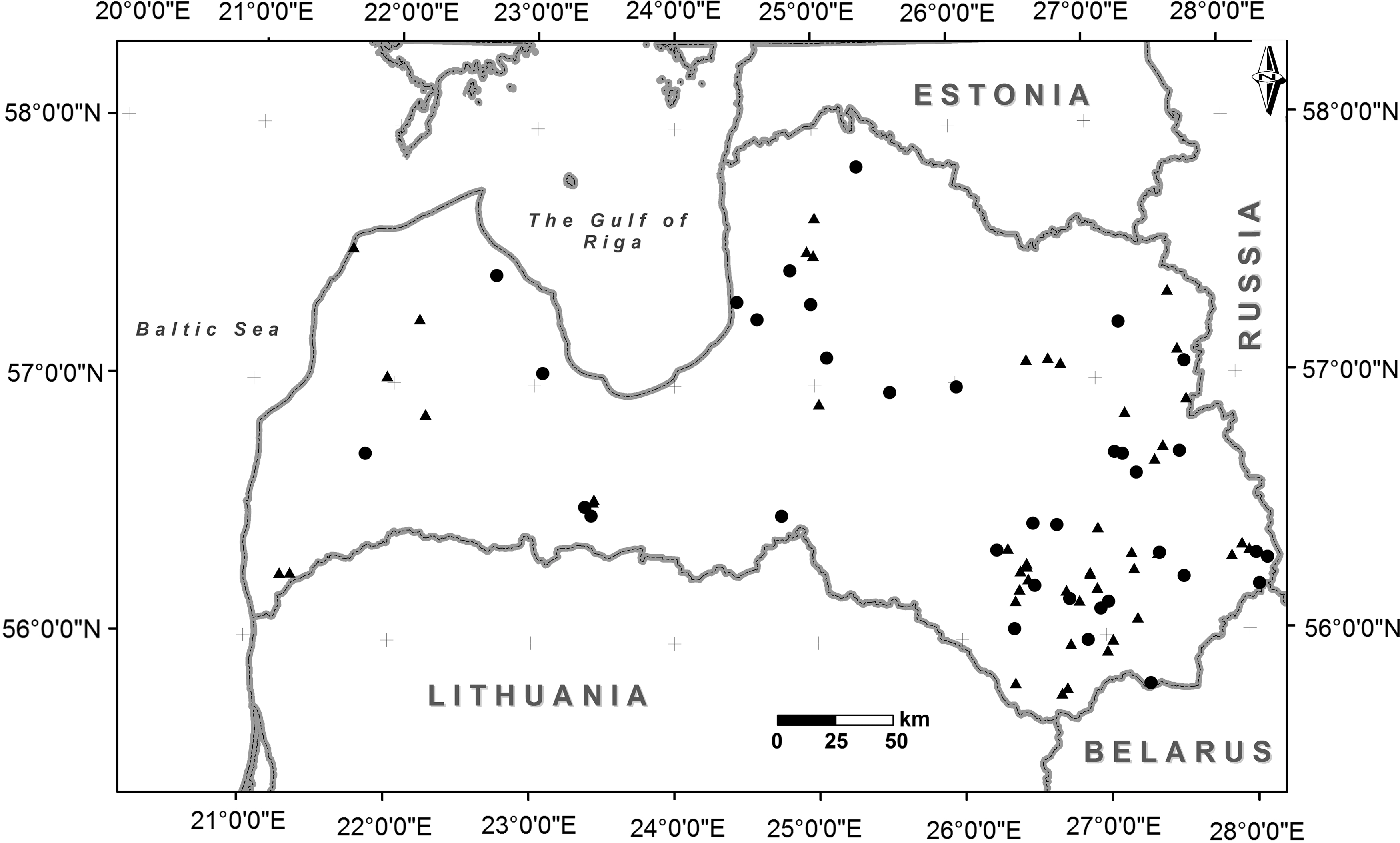
Map of Latvia showing the location of the sheep farms included in the study. From each farm, samples from 1 to 246 sheep were tested for the presence of antibodies against Toxoplasma gondii. Circles indicate farms where at least one T. gondii seropositive sheep was detected. Triangles indicate farms where no T. gondii seropositive sheep were detected.
Risk factors
The age of the sheep ranged from 0.1 to 14.3 years. The mean age was 3.4 years and median was 2.9 years. The youngest seropositive lamb was 4 months old, and its mother was seronegative. There was a significant difference (p < 0.05) between the seroprevalence in juvenile and adult sheep (Table 1). In the univariable analysis, adult sheep had 1.9 (95% CI: 1.1–3.1) times higher odds of testing seropositive than the juvenile sheep. When the age of the sheep was used as a continuous variable, a simple univariable model suggested that the odds of testing seropositive increased by 9.2% (95% CI: 2.4–16.5) for each year of age—the pattern observed was however fluctuating (Fig. 2).
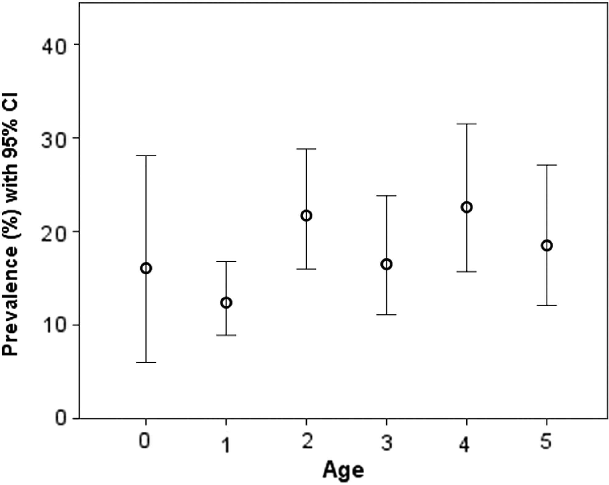
T. gondii seroprevalence in sheep in Latvia, by age in years.
Significant difference between the seroprevalences of the two groups.
Significant difference between the seroprevalence in Latvian Dark Headed sheep and Crossbreed sheep and Latvian Dark Headed sheep and German Merino sheep.
The seroprevalence did not differ by sex, but differences between the breeds were observed (Table 1).
The seroprevalence was significantly higher (p < 0.001) in sheep from herds with more than 100 animals than in sheep from smaller herds (Table 1). Sheep from farms that also housed cattle had significantly higher (p < 0.05) seroprevalence than those from farms without cattle, whereas sheep from farms that also housed poultry had significantly lower (p < 0.001) seroprevalence than those from farms without poultry. There were no significant differences in the seroprevalence between the regions.
Two multivariable logistic regression models were built (Table 2), and the results of these models were in agreement with the results of univariable analyses. Adult age was a significant risk factor (odds ratio 1.8), while being of crossbreed or of German Merino breed were protective factors compared with the Latvian Dark Headed sheep. Belonging to a larger herd, as well as living on a farm that also housed cattle, was a risk factor (odds ratios 1.8 and 1.6, respectively), whereas living on a farm with poultry appeared as a protective factor (odds ratio 0.4).
The breeds were included as dummy variables; Latvian Dark Headed sheep was used as the reference breed.
Discussion
The serological results illustrate that T. gondii infections are widespread in sheep in Latvia. The strengths of our study include the sufficient sample size for estimating the animal-level seroprevalence and the collection of samples from different parts of the country, which increases the generalizability of the results. The observed proportion of farms with at least one positive animal should be interpreted cautiously, as the sample was not optimal for a farm-level estimate. Although the sample was a convenience sample, no major sampling bias was anticipated, and the sample can be considered to represent the local sheep population well. Agreement between the in-house method we used and the commercial ELISA was good. It should nevertheless be emphasized that the serum concentration we used can be regarded as high, which may lead to nonspecific reactions. Serum dilution 1/10 is generally not an advisable dilution for ELISA, despite that it appeared to be better than 1/50 or 1/100 for this in-house method. Moreover, BSA as a serum protein could cause nonspecific reactions.
The overall T. gondii seroprevalence (17.2%) in Latvian sheep was significantly lower than that reported in Finland (24.6%, p < 0.001), Lithuania (42.1%, p < 0.001), and the Czech Republic (59.0%, p < 0.001) (Stimbirys et al. 2007, Bártová et al. 2009, Jokelainen et al. 2010). The seroprevalence was similar to that detected in Norway (16.2%) (Skjerve et al. 1998). The results of our study differed significantly (p < 0.001 and p < 0.01) from the two previously reported seroprevalence estimates from Latvia (Eglīte and Keidāns 2000). However, the results are not directly comparable because different study designs and methods were used.
Adult age was a significant risk factor for seropositivity. This suggests that the infections were typically acquired over time whereby older animals have had a longer time for parasite exposure. This is in accordance with other observations and their interpretation (Tenter et al. 2000, Dumétre et al. 2006, Stimbirys et al. 2007, Dubey 2009).
In Latvia, sheep are kept on a pasture most of the year; as a result, they are at risk of T. gondii infection if the environment is contaminated with sporulated oocysts. Natural water sources may also be available for the animals. Sporulated oocysts of T. gondii are highly resistant to environmental conditions (Dubey 2010). The oocysts remain infectious in moist soil or sand for up to 18 months (Frenkel et al. 1975). Latvia is located between the 55° and 58° parallels north, and the climate is suitable for long survival of oocysts. The annual average temperature is +5.9°C, average precipitation is 667 mm, and the average relative humidity is 81%. During the coldest winter months (January, February), the average minimum temperature is −7.7°C. Recently, an increase of precipitation has been observed (Latvian Environment, Geology and Meteorology Centre,
The seroprevalence was highest in Latvian Dark Headed sheep (Table 1). In Latvia, these sheep are widely raised for meat, wool, and fur (
Sheep from farms that also housed cattle had higher odds of testing seropositive, whereas the presence of poultry on the farm appeared as a protecting factor in the multivariable model. However, because the number of farms was limited in the sample, these results should be interpreted cautiously. The epidemiology of the parasite on farms raising several host species has received little research attention, while the presence of cats on the farms is a risk factor (Skjerve et al. 1998, Vesco et al. 2007, Cavalcante et al. 2008, Cenci-Goga et al. 2013). Unfortunately, information on the presence of cats on the farms and whether it was associated with raising other animals on the same farms was not available for the present study.
Sheep from larger herds had higher odds of testing seropositive, which suggests that management of larger farms, in particular, fails to stop and may help the life cycle of T. gondii. For example, feeding concentrate has been associated with the infection (Tzanidakis et al. 2012).
T. gondii was recently ranked fourth among zoonotic parasites evaluated for their global relevance as foodborne pathogens (FAO/WHO 2014). Seropositive sheep can be assumed to harbor T. gondii cysts in their tissues (Dubey and Jones 2008), which is why mutton should be only consumed after it has been thoroughly cooked.
Conclusions
Our study demonstrates that sheep are commonly exposed to T. gondii in Latvia. Higher seroprevalence was observed in older animals, indicating that the infections were acquired and implying that the agricultural environment is contaminated with oocysts. The results also indicate that, if consumed undercooked, the meat of Latvian sheep can be a potential source of T. gondii for other hosts, including humans.
Footnotes
Acknowledgments
The authors thank Furio Spano and Simona Cherchi for their help in developing the ELISA and for providing the antigen. The authors also thank the staff of the Institute of Food Safety, Animal Health and Environment “BIOR” for their cooperation and positive attitude toward the study and Maris Nitcis from the Institute of Life Sciences and Technology, Daugavpils University, for creating the map.
Author Disclosure Statement
No competing financial interests exist.