
Abstract
Alkhumra hemorrhagic fever virus (AHFV) is an emerging novel flavivirus that was discovered in Saudi Arabia in 1995. The virus has since caused several outbreaks in the country that resulted in case fatality rates ranging from 1% to 25%. Meager information has been published on the ultrastructural features of the virus on cells under in vitro or in vivo conditions. The present electron microscopic study examined and compared the intracellular growth of the AHFV on the LLC-MK2 cells and brain cells of new born Wistar rats, inoculated intracerebrally. The cytopathological changes in both cell systems were noted, and localization of the virus particles in different cellular components was observed. Both apoptotic and lytic cell interactions were seen in the electron micrographs of both the LLC-MK2 and the rat brain cells. The results were discussed in relation to similar situations reported for other virus members of the genus Flavivirus.
Introduction
A
AHFV causes acute febrile illness with hepatitis, hemorrhagic manifestations, and encephalitis. A case fatality rate as low as 1% was reported from Najran and as high as 25% was reported from Makkah (Madani 2005, Madani et al. 2011). Phylogenetical analysis of AHFV showed close relationship with the Kyasanur Forest disease virus (KFDV) (Mehla et al. 2009). As the KFDV is transmitted by ticks (Bhat and Goverdhan 1973), AHFV was classified as a member of the tick-borne encephalitis (TBE) group of viruses of the genus Flavivirus (King et al. 2012).
The purpose of this study was to describe the ultrastructural features and the intracellular growth of AHFV on cells under in vivo and in vitro conditions, as visualized under the electron microscope (EM), and to note the fate of the infected cells after infection.
Materials and Methods
Alkhumra hemorrhagic fever virus
AHFV (AHFV/97/Nj/09/SA) used in this study for inoculation of the newborn Wistar rats and LLC-MK2 cells was originally isolated from a patient's blood during an outbreak of the disease in Najran, Southern Saudi Arabia (Madani et al. 2012). A titer of 108.2 TCID 50/mL was used to inoculate the newborn rats and the LLC-MK2 cells.
The cell culture
In this study, the LLC-MK2 cell line, which is extracted from the Rhesus monkey kidney epithelium cells, was used to grow the AHFV. The choice of this cell line was based on a previous study, demonstrating its superiority over other cell lines for growth of AHFV (Madani et al. 2016).
Preparation of the AHFV from the LLC-MK2 cell culture for EM
The methods used in this study for preparation of the LLC-MK2 cell monolayers, their inoculation with the AHFV, their harvest, fixation for EM, and examination under the EM were the same as previously described (Madani et al. 2017). In brief, the LLC-MK2 monolayers were grown in three 75 cm2 Corning tissue cell culture flasks at 37°C in Eagle's minimum essential medium (EMEM) supplemented with 10% fetal calf serum (FCS; Sigma, St. Louis, MO). When the monolayers were 70% confluent, each flask was inoculated with 1 mL of the prototype AHFV in the form of undiluted newborn rat brain suspension, passage 1, and then incubated at 37°C for 1 h to adsorb. This was followed by the addition of 15 mL of EMEM containing 2% FCS and incubation at 37°C. The monolayers were observed daily under an inverted microscope for the presence of discernible cytopathic effect (CPE). When the CPE affected 30% or greater of the monolayer, the three flasks were removed from the incubator and the media were collected and pooled. The cell sheet from the surface of each flask was gently scraped by a cell scraper (Falcon, New Jersey). Fifteen microliters of the pooled media were added to the scraped cells in each flask. The flasks were then shaken gently, and the contents of the three flasks were pooled. This pool was spun in a cooled bench centrifuge (Eppendorf, 5702, UK) at 141.98 g (1000 rpm) for 10 min. The pellet was resuspended in 5 mL phosphate buffer saline (PBS) pH 7.4 and fixed in 2.5% glutaraldehyde in PBS pH 7.4 for 2 h at room temperature. This was followed by three successive rinsings in distilled water, fixation in PBS-1% osmium tetroxide for 20 min, prestaining en bloc with 2% uranyl acetate for 10 min, and rinsing with distilled water. This was followed by dehydration in an ethanol series as follows: 50% ethanol for 3 min, 70% ethanol for 3 min, 90% ethanol for 3 min, and two changes of 100% ethanol for 3 min each. The dehydration process was followed by two changes of propylene oxide for 5 min each. This was followed by infiltration with a mixture of propylene oxide and epoxy (PELCO® Eponate 12™ Kit; Ted Pella, Inc., Redding, CA) with a final infiltration in pure resin and final embedding in fresh epoxy resin. Thin sections (70–90 nm thick) were cut from the block, stained in 4% uranyl acetate and Reynold's lead citrate, and examined by using a transmission electron microscope (TEM; Philips CM-100). Sections of noninoculated LLC-MK2 cells were prepared and treated as described earlier and examined under an EM.
AHFV inoculation into newborn Wistar rats
The methods used in this study for inoculation of the newborn Wistar rats were the same as previously described (Madani et al. 2017). In brief, 11 in-house bred, 2-day-old Wistar rats were used in the experiments. All the inoculations and keeping of the inoculated rats were performed in a biosafety level-3 facility. Seven rats were inoculated intracerebrally with the AHFV, as described by Madani et al. (2014). The remaining four rats were injected intracerebrally with EMEM and kept as controls. All the rats were observed daily for any clinical manifestations or death. The brains of dead or moribund rats were harvested, placed in 2.5% Glutaraldehyde, and processed for EM as hereunder described.
Preparation of the virus from the newborn rat brains for EM
The methods used in this study for preparation of the brains from Wistar rats for electron microscopic examination were the same as previously described (Madani et al. 2017). Briefly, the brains from dead, moribund, and the control baby rats were removed and fixed in 2.5% glutaraldehyde in PBS pH 7.4 for 2 h at room temperature. Further fixation, embedding in fresh epoxy resin, thin sectioning (70–90 nm thick), staining in 4% uranyl acetate and Reynold's lead citrate, and examination under a TEM (Philips CM-100) were as previously described (Madani et al. 2017).
Results
The control rats remained healthy until the end of the experiment. All the inoculated rats succumbed to infection initially, with mild neurologic signs starting on day 3 postinoculation. By days 5–7, the disease signs progressed to irritability, tremors, convulsions, spastic paresis, and opisthotonus. By day 7 postinoculation, 71% of the inoculated rats died and the remaining moribund rats were euthanized.
The EM micrographs of the inoculated LLC-MK2 and baby rat brain cells showed both lytic and apoptotic interactions. Figure 1 shows micrographs of inoculated LLC-MK2 and rat brain cells that underwent lysis and apoptosis. In the cells that showed lytic interaction, there was loss of cell membrane integrity, disorganization of the cellular contents, cytoplasmic clumping, and mitochondrial swelling (Fig. 1). The nuclei in these cells showed swelling with karyolysis. In both types of cells that underwent apoptosis, the nucleus was dense and condensed with a preserved outline. The cytoplasm showed fragmentation with a conserved cell outline.
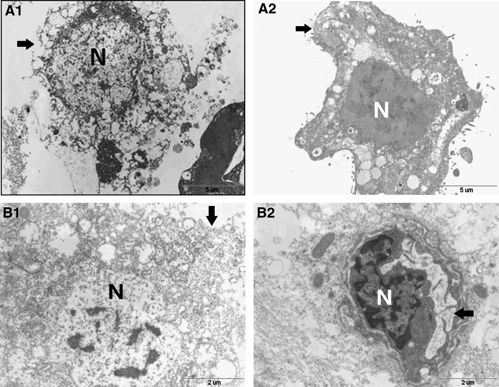
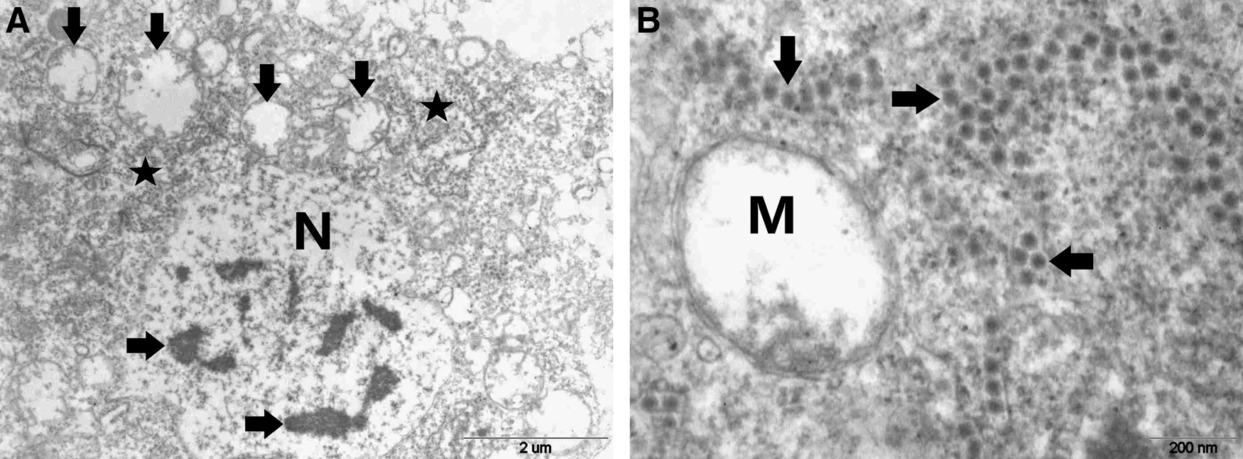
Figure 2 shows a magnified image of a rat brain cell that underwent lysis. There is an ill-defined nuclear membrane and clumped chromatin (Fig. 2A). There are clumps of virus particles in the cytoplasm, whereas the nucleus and mitochondria (Fig. 2B) are devoid of virus particles. Figure 3 shows ultrastructural changes seen on the rough endoplasmic reticulum (RER) on an AHFV-infected rat brain cell. The RER was hypertrophic and contained virus particles within its dilated cisternae. EM micrographs from sections of both AHFV-inoculated rat brain and LLC-MK2 cells were studded with numerous cytoplasmic vesicles containing various quantities of spherical virus particles (Fig. 4). No virus particles were seen in the nucleus or the mitochondria (Fig. 4). Figure 5 shows AHFV-infected rat neuron containing numerous virus-laden RER vesicles with intact and ruptured membranes. Figure 6A shows electron micrograph of part of an infected rat brain cell with virus particles scattered within the RER. Some are arranged as virus arrays; a magnification of such arrays is shown in Figure 6B.
Shows part of an AHFV-infected LLC-MK2 cell that underwent lysis,
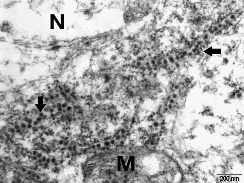
Electron micrograph of part of a rat brain cell section showing virus particles inside dilated cisternae of the RER (arrows) between a mitochondrion (M) and the nucleus (N). RER, rough endoplasmic reticulum.
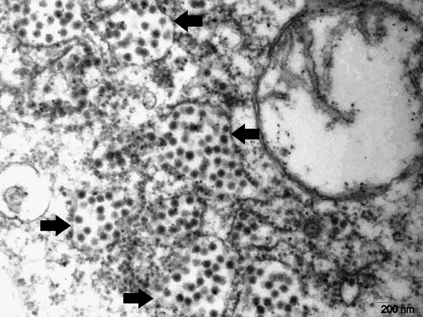
Virus particles inside cytoplasmic vesicles of LLC-MK2 cells (black arrows) and mitochondria devoid of virus particles (white arrows).
Electron micrograph of part of an AHFV-infected rat brain cell showing many virus-laden vesicles with intact and ruptured membranes (arrows).

Electron micrograph of an AHFV-infected rat brain cell showing
Discussion
This study provides insight on the infectivity of AHFV in cells under in vivo (newborn rats) and in vitro conditions (LLC-MK2 cell line), as visualized under the EM. Observations were made on different cellular organelles of the infected cells. The types of CPE brought about by the virus infection and localization of the virus particles in both types of cells were noted.
The noninoculated cells in both cell systems did not show any cytopathological changes. The results indicated that CPE was seen in both cell systems. Apoptosis and cell lysis were observed in both in vitro and in vivo cells. The ultrastructures seen in apoptosis or cell lysis, in either cell system, did not vary. However, a thorough examination of cell sections from both in vivo and in vitro systems indicated dominance of the number of the apoptotic cells over the lytic cells in the in vitro system, as compared with that in the in vivo system, likely due to the possibility that the microglia macrophages of the in vivo system (Neumann et al. 2009) could have cleared most of the apoptotic cells (Roy et al. 2014). Such a situation is lacking in the LLC-MK2 in vitro cell system that are nonphagocytic (Barrias et al. 2010). As a result, more apoptotic cells were seen in the in vitro system as compared with the in vivo system. This was also reported by Hirano et al. (2014), who studied infectivity of the West Nile virus (WNV), Japanese encephalitis virus (JEV), and the TBE virus in mice brain cells and found that the apoptotic cells were rarely seen in their examined mouse brain electron micrographs.
The electron micrographs of both AHFV-infected rat brain and LLC-MK2 cells showed dilatation of the cisternae of the RER that were studded with large quantities of virus particles. This observation indicates that the RER is the site of multiplication of AHFV. This is consistent with what has been reported for other members of the genus Flavivirus such as the Dengue virus (DV) (Fernandez-Garcia et al. 2009), the WNV (Samuel 2002), the Yellow Fever Virus (Ishak et al. 1988), and the JEV (Hase et al. 1990), where the RER acts as the crucial cell organelle that supports their multiplication (Pastorino et al. 2010, Inoue and Tsai 2013).
In this study, the electron micrographs showed that the RER, in both in vivo and in vitro cells, were few in numbers as compared with the noninoculated controls. This was probably due to their obliteration and degeneration by the AHFV during the process of its multiplication. This was also observed for other viruses (Hase et al. 1990), where it was observed that the RER, at the late stages of infection, was completely obliterated, leaving the rarified cytoplasm devoid of it.
In this study, the nucleus was devoid of virus particles. This was expected because AHFV, as a member of the genus Flavivirus, which has a single-stranded positive-sense RNA (ss+sense RNA), does not replicate in the nucleus (Pastorino et al. 2010). An interesting feature that was seen predominantly in micrographs from sections of the infected baby rat brain cells was the presence of precisely arranged arrays of virus particles that were spread over the RER. The locations of these arrays could have been multiplication sites. Such features were also seen for other flaviviruses, for example, DV, WNV, JEV (Uchil and Satchidanandam 2003), and Zika virus (Lednicky et al. 2016).
In this study, the electron micrographs showed the presence of numerous swollen mitochondria, in both the in vivo and in vitro cell systems, which were found in close proximity to the sites where plenty of virus particles were collecting in the cytoplasm (multiplication sites). This observation was also reported for other flaviviruses where the authors suggested that the virus had been utilizing the energy in these mitochondria for its multiplication requirements (EL-Bacha & Da Poian, 2013).
From this study, it could be concluded that AHFV produced severe CPE in both the rat brain cells and the LLC-MK2 cell line, as visualized by the EM. The virus multiplication was cytoplasmic, involving the RER, which had undergone hypertrophy and dilatation of its cisternae. The nucleus and mitochondria were devoid of virus particles; however, micrographs of the mitochondria showed swelling in their lumen, indicating activity toward supplying the virus with its requirements. Cell lysis and apoptosis were seen in both the infected LLC-MK2 and rat brain cells. Precisely arranged virus particle arrays were seen, predominantly, in the infected rat brain cells. Features of the intracellular growth of the AHFV in the cells under study were highly comparable with those reported for other members of the genus Flavivirus.
Footnotes
Acknowledgments
The authors thank Sheikh Mohammed Hussein Al-Amoudi for funding this research and the Scientific Chair for Viral Hemorrhagic Fever at King Abdulaziz University, Jeddah, Saudi Arabia. They also thank Mr. Helmy Abdullah, Mr. Abdullah Al-Salawaty, and Mrs. Ola Alahmadi for their excellent technical assistance, and Mr. Saad Alhusaiki for his assistance in preparing the images. This study is one of the research products of the Scientific Chair of Mohammad Hussein Alamoudi for Viral Hemorrhagic Fevers, King Abdulaziz University, Jeddah, Saudi Arabia. The sponsor, Sheikh Mohammad Hussein Alamoudi, had no involvement in the study design, in the collection, analysis, and interpretation of data, in the writing of the article, or in the decision to submit the article for publication.
Authors' Contributions
T.A.M. and E.M.E.A. conceived and designed the study and wrote the article; E.M.E.A. performed isolation and titration of Alkhumra hemorrhagic fever virus (AHFV), designed and performed the procedure for preparing the virus for electron microscopy (EM), and examined the virus under the EM; S.M.J. facilitated the EM examination, and processed the fixed AHFV samples for examination under the EM; A.M.H. assisted in the preparation of LLC-MK2 cell culture and in the preparation, cultivation, and titration of the virus; H.A. assisted in the inoculation of the rats with the AHFV and harvest of their brains and in the arrangement, setting up, performing the EM examination, and interpretation of micrographic images; S.S.A. assisted in the interpretation of micrographic images and discussion; H.M.S.A. and E.I.A. critically revised the article; and all authors read and approved the final article.
Ethical Approval
King Abdulaziz University's policy on the care and use of laboratory animals was followed. Ethical approval was obtained from the Research Ethics Committee at the Faculty of Medicine, King Abdulaziz University, Jeddah, Saudi Arabia.
Author Disclosure Statement
No competing financial interests exist.