
Abstract
Background:
Chickens are considered potential reservoirs for human extraintestinal infections with pathogenic Escherichia coli. However, information about genetic relatedness between E. coli from healthy chickens and human patients is still limited.
Methods and Results:
In this study, clinical samples from patients with extraintestinal infections and healthy broiler chickens were collected from geographically related locations in Egypt during the 2nd half of 2015. The recovered isolates were tested for susceptibility against β-lactam antimicrobials and screened for the presence of extended-spectrum β-lactamases (ESBLs) and virulence genes; clonal and phylotypes were also determined. Forty-eight percent (48/100) and 31.3% (50/160) of human and chicken samples were positive for E. coli, respectively. Although only 4% (2/50) of the chicken isolates were resistant to the tested β-lactams, over 58% of human E. coli isolates (28/48) exhibited resistance to cefotaxime. For β-lactamases, 52.1%, 33.3%, 20.8%, and 6.25% of human E. coli were positive for bla CTX-M, bla TEM, bla OXA, and bla CMY, while bla TEM, bla OXA, and bla CMY were found in 32%, 4%, and 34% of chicken isolates, respectively. Low frequencies of virulence genes within human and chicken E. coli isolates were detected by PCR. The majority of E. coli isolates harboring β-lactam resistance genes from human and chicken sources belonged to phylogroup C and B1, respectively. Using pulsed-field gel electrophoresis (PFGE), some E. coli grouped based upon source; however, most clusters contained isolates from both humans and chickens.
Conclusions:
The above findings suggest that although no single clone appeared to be circulating among E. coli isolates from human and chicken, some shared characteristics exist among isolates from both sources. Increased study will aid to track the dissemination of β-lactam-resistant E. coli from healthy chickens to humans for implementation of effective intervention strategies.
Introduction
E
Extraintestinal disease-causing lineages in humans are thought to have originated primarily from the intestinal tract of the host (Manges and Johnson 2015); however, genetic similarity between human ExPEC and APEC strains suggests the significant role of chickens as potential reservoirs for human extraintestinal infections (Mora et al. 2009, Johnson et al. 2012, Manges and Johnson 2012). Generally, chicken-derived E. coli strains are transmitted directly to humans through contaminated meat and/or indirectly through the contaminated environment (Hammerum and Heuer 2009, Jakobsen et al. 2010).
Antimicrobial resistance is one of the global threats that affects human and animal health particularly in developing countries (Adenipekun et al. 2016). The improper and overprescription of antimicrobials in conjunction with the lack of adequate regulations in Egypt have led to the exacerbation of the antimicrobial resistance problem (Dooling et al. 2014, Dahshan et al. 2015). Egypt is listed among the countries with high prevalence of antimicrobial resistance, especially extended-spectrum β-lactamases (ESBLs) (Bouchillon et al. 2004). ESBL producers secrete enzymes, β-lactamases, which confer resistance to penicillins, cephalosporins, and monobactams, but are still inactivated by β-lactamase inhibitors (Paterson and Bonomo 2005). β-lactam resistance is often mediated by transmissible plasmids with a possibility of distribution within bacterial strains and/or among different genera (Pfeifer et al. 2010).
The continuous administration of antimicrobials for prophylactic measures and growth promotion in chicken farms at subtherapeutic doses produces selective pressure against both pathogenic bacteria and gut microbiota (Frye and Jackson 2013). The commensal E. coli in the chicken gut develop resistance either due to chromosomal mutation or the acquisition of resistance traits from mobile genetic elements (e.g., plasmids, transposons, and integrons) (von Wintersdorff et al. 2016). As food animals acquire the resistance determinants, this may explain the implication that commensal E. coli have as potential reservoirs that transmit resistance genes to humans through food sources (Smet et al. 2010, Schjørring and Krogfelt 2011).
Because resources are limited for a national surveillance program to monitor development of antimicrobial resistance in developing countries, local studies are needed that focus attention on the present state of resistance in specific bacteria for potential use in mitigation strategies (Ayukekbong et al. 2017).
To date, there is no study in Egypt that has investigated genetic relatedness between E. coli isolates from healthy chickens and extraintestinal disease-causing lineages in humans. This study aimed to determine the coexistence of antimicrobial resistance, virulence-associated genes, and phylogenetic groups in E. coli isolates from patients with extraintestinal infections and healthy chickens. The clonality of these isolates was also determined using pulsed-field gel electrophoresis (PFGE).
Materials and Methods
Sample collection
Ninety-eight nonrepeated E. coli isolates from human patients (n = 48) and healthy chickens (n = 50) were included in this study. Mid-stream urine samples were collected aseptically from patients with UTIs admitted to Suez Canal University Hospitals (SCUH), Ismailia, Egypt. Sputum and pus samples were taken in sterile containers from patients who showed symptoms of extraintestinal infections (pulmonary and wound infections). The clinical samples from patients (n = 100) of both sexes and ages ranging from 15 to 70 years old were included in this study. Clinical assessment of the patient's condition was done by medical history and physical examination. An informed consent was taken from each patient to participate in this study. The board of SCUH approved collection of clinical samples from patients before sampling.
Broiler farms at Dakahliya (n = 6) and Sharkia (n = 4) Governorates, Egypt, were included in this study. Approximately, 20% of poultry production in Egypt is localized in Sharkia and Dakahlia Governorates according to the official reports from the Egyptian Ministry of Agriculture and Central Agency for Public Mobilization and Statistics (CAPMAS). Data from each farm, including age and size of the flock, hygienic measures, and administered antibiotics, were recorded. Forty healthy chickens (four chickens per farm), showing no evidence of illness, were collected randomly from the enrolled farms. A total of 160 samples encompassing four (liver, spleen, intestine, and cloacal swab) samples from each chicken were taken. Each sample was packaged separately in a sterile polyethylene bag, and then transported immediately to the laboratory of Hygiene and Zoonoses Department, Faculty of Veterinary Medicine, Mansoura University, for the conventional isolation of E. coli.
Human and chicken samples from geographically related locations, with distances ranging from 60 to 120 Km, were collected over a 6-month period from June to December, 2015. The process of conventional isolation and identification of E. coli from human and chicken clinical samples was done according to standard bacteriological procedures (Collee et al. 1996). All E. coli isolates were confirmed with the VITEK®2 System using the VITEK 2GN cards (bioMérieux, Durham, NC) according to the manufacturer's directions.
Antimicrobial susceptibility testing
Minimum inhibitory concentration (MIC, μg/mL) for E. coli was determined by broth microdilution using the Sensititre™ semiautomated susceptibility system (TREK Diagnostic Systems, Inc., Westlake, OH) and the Sensititre ESBL panel CMV2DW according to the manufacturer's guidelines. Interpretation of results was done according to Clinical and Laboratory Standards Institute (CLSI) (CLSI 2016). Antimicrobials and their breakpoints were as follows: aztreonam (≥16 μg/mL), ceftazidime (>16 μg/mL), cefotaxime (≥4 μg/mL), cefepime (≥16 μg/mL), cefquinome (≥16 μg/mL), imipenem (≥4 μg/mL), and piperacillin/tazobactam (≥128/4 μg/mL). Phenotypic confirmation of ESBL production was determined using cefotaxime/clavulanic acid and ceftazidime/clavulanic acid. The reference strains, E. coli ATCC 25922, Pseudomonas aeruginosa ATCC 27853, Enterococcus faecalis ATCC 29212, and Staphylococcus aureus ATCC 29213 were used as quality controls for MIC.
DNA template preparation
Briefly, 1 mL of the enriched bacterial culture was centrifuged at 8000 g for 2 min. The pellet was resuspended in 1 mL nuclease-free water, homogenized, and boiled for 15 min. The resulting supernatant of the boiled lysates after centrifugation was used as DNA template (Ramadan et al. 2016).
Detection of β-lactam resistance and virulence genes
E. coli isolates from patients and healthy chickens were screened for the presence of β-lactam resistance genes: bla CTX-M, bla TEM, bla SHV, bla OXA, and bla CMY and virulence-associated genes: ompT (outer membrane protein), iss (serum resistance), papC (P fimbriae), kpsMII (group 2 capsular polysaccharide), and sfaA (S fimbriae). The primer sequences and amplified products are summarized in Table 1. Each uniplex PCR reaction in a final volume of 15 μL consisted of 2 μL DNA template, 7.5 μL of 2 × PCR Master Mix, 1.5 μL of forward primer (10 μM), and 1.5 μL of reverse primer (10 μM). PCR cyclic conditions were as follows: 95°C for 5 min, 30 cycles of 95°C for 30 s, annealing at specific temperature (Table 1) of each primer for 30 s, and extension at 72°C for 45 s. Amplified products were electrophoresed, stained, and visualized under ultraviolet light.
Phylogenetic typing of E. coli
The revised Clermont E. coli phylotyping method was performed to assign human and chicken E. coli isolates into one of the eight phylogroups A, B1, B2, D, C, E, F, and Clade I (Clermont et al. 2013).
Pulsed-field gel electrophoresis
Clonal relatedness between E. coli isolates from humans and chickens was determined using PFGE according to the procedures described previously (Ribot et al. 2006). DNA plugs were exposed to enzymatic digestion with 10 U of XbaI (Roche Molecular Biochemicals, Indianapolis, IN) and then fragments separated using the CHEF-DRII PFGE system (Bio-Rad, Hercules, CA) for 19 h at 6 V with a ramped pulsed time of 2.16–54.17 s at 14°C. Cluster analysis was determined using BioNumerics software and Dice coefficient, and the unweighted pair group method (UPGMA). Optimization settings for dendrograms were 2% with a position tolerance of 1%. Clusters were identified as having ≥55% similarity.
Statistical analysis
The variation in frequencies of β-lactam resistance genes, virulence-associated genes, and phylotypes among E. coli isolates from human patients and healthy chicken was assessed using Chi-square (χ2 ) test at a probability value p < 0.05.
Results
The overall occurrence of recovered E. coli from the clinical samples of human patients and healthy chickens was 48% (48/100) and 31.3% (50/160), respectively. Of the fifty E. coli isolates from chicken samples (one isolate per each sample), 19 isolates were from intestine, 13 from liver, 11 from spleen, and 7 from cloacae. Of the seven antimicrobials tested, 29 human E. coli isolates exhibited resistance to at least one of the examined antimicrobials with the highest frequencies against cefotaxime (58.3%; 28/48). Lower resistance (4.2%; 2/48) and (12.5%; 6/48) was detected with imipenem and piperacillin/tazobactam, respectively (Table 2). Converse to the human E. coli isolates, 96% (48/50) of the chicken isolates did not exhibit any resistance to the antimicrobials tested in this study. One chicken isolate showed resistance to aztreonam, cefotaxime, and cefquinome, while the other was resistant to cefotaxime only.
Phenotypic confirmation of ESBL in the isolates was performed using cefotaxime and ceftazidime, each individually combined with a β-lactamase inhibitor, clavulanic acid. Of the cefotaxime-resistant E. coli from humans, approximately 86% (24/28) were ESBL positive and only one E. coli from chickens (Supplementary Table S1; Supplementary Data are available online at
Human and chicken E. coli were also investigated for the presence of β-lactam resistance genes using PCR. Approximately, 45% and 55% of non-ESBL-producing isolates from humans (11/24) and chickens (27/49) carried at least one β-lactamase-encoding gene, respectively (Supplementary Table S1). The overall prevalence of bla TEM, bla OXA, and bla CMY in chicken isolates was 32%, 4%, and 34%, respectively. The respective prevalence of bla CTX-M, bla TEM, bla OXA, and bla CMY from human isolates was 52.1%, 33.3%, 20.8%, and 6.25%. No amplicons for bla SHV were identified from both chicken and human isolates. Furthermore, no chicken isolates possessed bla CTX-M. The distribution of bla CTX-M and bla OXA (χ 2 = 6.458, df = 1, p < 0.05) was significantly higher among human than chicken isolates. However, bla CMY was detected at higher frequencies (χ 2 = 11.61, df = 1, p < 0.05) in chicken isolates compared to human isolates. Screening for virulence-associated genes in both chicken and human isolates showed that 18%, 12%, and 2% of the chicken isolates harbored ompT, iss, and papC, respectively. No amplified products for kpsMII and sfaA were detected among chicken isolates. The only virulence-associated gene identified in human isolates was papC with an overall percentage of 18.8%. For phylotyping, the higher distribution of phylotypes (B1, A, and C) with statistically significant association (χ 2 = 25.4841, df = 1, p < 0.05) was determined in chicken (47/50, 94%) compared to human (23/48, 47.9%) isolates (Table 3).
The association of β-lactamase-encoding genes and phylogroups of E. coli isolates from both sources is presented in Table 4. Phylogroup C was identified in 36%, 50%, and 100% of human E. coli isolates harboring bla CTX-M, bla OXA, and bla CMY, respectively. For chicken isolates, higher percentages of isolates carrying at least one β-lactam resistance gene belonged to phylotype B1.
From the PFGE analysis of the human and chicken isolates (Fig. 1), 14 clusters (A-N) of different PFGE patterns with at least 55% similarity were obtained. The majority of clusters contained E. coli isolates from both human and chicken sources; however, five clusters included isolates from either chickens (F and I) or humans (K, L, and N). Only three clusters (G, J, and M) were composed of >10 isolates per cluster. Chicken and human E. coli isolates were approximately represented in clusters G and M equally, whereas cluster J was mainly dominated by chicken isolates (93.3%). In clusters D, F, and J, the respective chicken E. coli isolates (no. 78 and 80), (91, 92, and 93), and (53 and 76) had identical PFGE patterns. Meanwhile, only one clonal pair of human E. coli isolates (no. 13 and 15) had identical PFGE patterns in cluster E.
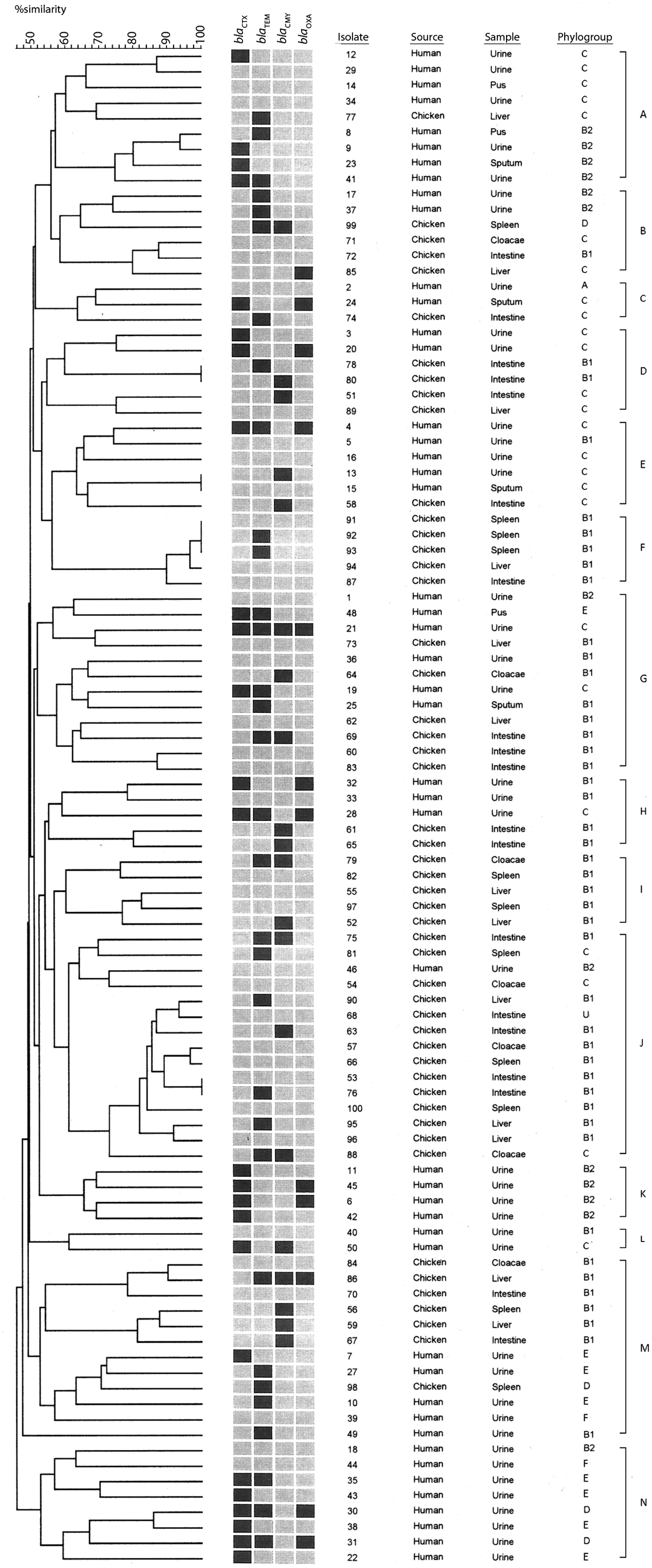
PFGE patterns, β-lactam resistance genes, isolate source, and phylotypes of Escherichia coli isolates from human patients and healthy chickens. Analysis of PFGE was performed using Dice coefficient and the UPGMA. Fourteen clusters (A–N) of different PFGE patterns were determined with a similarity not less than 55%. Clonal isolates 100% identical were identified in four clusters: D (isolate no. 78 and 80), E (isolate no. 13 and 15), F (isolate no. 91, 92, and 93), and J (isolate no. 53 and 76). Black and gray squares represent positive and negative PCR results for β-lactam resistance genes. PFGE, pulsed-field gel electrophoresis; UPGMA, unweighted pair group method. Figure 1 can be viewed in great detail online at
Discussion
The high level of ESBLs among E. coli causing extraintestinal infection, particularly UTIs, poses a great challenge to public health. Horizontal gene transfer of β-lactam resistance genes, disseminated by mobile genetic elements, has led to the broadening of resistance to include other antimicrobial classes, leaving few therapeutic choices (Frye and Jackson 2013). Although studies have shown that chicken-derived E. coli contributed to UTIs in humans with a probability of resistance genes disseminating through the food chain (Manges and Johnson 2012, Berg et al. 2017), information about antimicrobial resistance of E. coli from healthy chicken in Egypt is still limited. In this study, the antimicrobial resistance profiles, phylogenetic and clonal diversity, between E. coli from patients with extraintestinal infections and healthy chicken were determined.
High frequencies of resistance among human E. coli isolates against cefotaxime, aztreonam, and cefquinome in this study were not surprising. β-lactams, especially third-generation cephalosporins, are considered the major prescribed antimicrobials in Egyptian hospitals (Talaat et al. 2014). Resistance to imipenem, one of last resort therapy for treating ESBL-producing Enterobacteriaceae, was also detected, but in low frequency. Our results also showed low prevalence of ESBLs among chicken E. coli. Variable prevalence of ESBLs among healthy chicken E. coli isolates from different studies could be related to the variation of farming system, regime of drug administration, and isolation method of bacteria (Jakobsen et al. 2010, Reich et al. 2013, Koga et al. 2015).
In this study, high occurrence of bla CTX-M among E. coli isolates from human patients was similar to that previously reported in Egypt (Abdallah et al. 2015, Elsherif et al. 2016). Predominance of bla CTX-M is commonly associated with multidrug resistance (MDR) due to the propensity of CTX-producing E. coli to exhibit resistance to different classes of antimicrobials other than β-lactams (Pitout and Laupland 2008, Ruppé et al. 2009, Adwan et al. 2014, Seyedjavadi et al. 2016).
The bla CMY gene, which mediates resistance to cephamycins, oxyimino-cephalosporins, as well as β-lactamase inhibitors, was detected in approximately one-third of the examined chicken isolates. The existence of this gene in chicken isolates that displayed no resistance to the examined β-lactams could be explained by inability of genes to express their function due to mutations in the gene sequence and/or a defective gene (Jackson et al. 2013). Gene expression analysis provides a significant clue to identify the interrelationship between resistance genes and phenotypic profiles (Suzuki et al. 2014, Hidano et al. 2015).
Knowing the phylogenetic characterization of E. coli and their precursors could possibly identify the contributing role of different strains to diseases (Clermont et al. 2013). E. coli strains have been differentiated into commensal (A and B1) and pathogenic (B2 and D) phylogroups (Clermont et al. 2000). With the improvement of genomic data for E. coli, new phylogroups C, E, F, and Clade I were assigned; phylogroup C is closely related to other commensal phylogroups (A and B1), while E and F phylogroups are considered pathogenic, and clade I belongs to Escherichia cryptic Clade I (Clermont et al. 2013). The dominance of phylotype B1 (74%) among chicken E. coli isolates in this study with the absence of B2 agreed with that previously reported by Ghodousi et al. (2015) and Rasmussen et al. (2015). With extraintestinal infections in humans, the majority of E. coli is assigned to phylotype B2 and less commonly to phylotype D (Johnson et al. 2003, Ewers et al. 2007, Pitout 2012). Significantly, the designation of 47.9% of human E. coli isolates into phylogroups B1, A, and C in this study suggested the possible association of commensal E. coli with extraintestinal infections after the acquisition of virulence traits (Moreno et al. 2006a, Ewers et al. 2007).
Our findings displayed high frequencies of genes encoding antimicrobial resistance among the commensal phylotypes, B1 in chicken isolates and C in human isolates. Many previous studies have reported that β-lactam resistance genes were frequently associated with E. coli belonging to commensal phylogroups (Mnif et al. 2012, Huber et al. 2013, Chakraborty et al. 2015), while others determined that the majority of β-lactam-resistant E. coli belonged to phylogroup B2 and D (Mosquito et al. 2015). Although there are controversial views about the existence of antimicrobial resistances in different phylogroups that require further investigation, it is plausible to assume that continuous and prolonged exposure of commensal E. coli, designated to phylogroups A, B1, and C, to antibiotics during the treatment of pathogenic bacteria, led to the resurgence of antimicrobial resistances among these commensals (Moreno et al. 2006b).
In addition to the phylogenetic diversity among the E. coli from humans and chickens, the two groups of isolates were also genetically diverse using PFGE. Heterogeneous populations of E. coli from both humans and food animals have been observed previously (Bergeron et al. 2012, Melo et al. 2015). This was seen in the 14 different clusters in the dendrogram and the number of distinguishable PFGE patterns. On the other hand, very few isolates were characterized as identical as three groups of E. coli from chickens had similar PFGE patterns, which may indicate they were clonal. Overall, PFGE results suggested that no single clone was circulating between the human and poultry population or even within the same populations in this study, as would be expected in a foodborne outbreak.
Conclusions
The above findings indicated that healthy chicken-derived E. coli shared some of the antimicrobial-resistant determinants, β-lactamases genes, with the E. coli isolates from patients with extraintestinal infections. Although PFGE results revealed that both isolate sets were not clonal, an undefined relationship existed among these isolates as some of the clusters were composed of both human and chicken E. coli. This could be due to close physical contact between the two groups, resulting in exchange of E. coli populations. The adoption of strict hygienic measures in poultries and implementation of antimicrobial resistance surveillance that includes healthy chicken are needed.
Footnotes
Acknowledgment
Science & Technology Development Fund, Egypt (STDF) has supported Dr. Ramadan's travel to the U.S.
Authors' Contributions
HHR conceived, designed the experiments, performed molecular analysis of E. coli, analyzed data, and wrote the manuscript draft. CRJ conducted the data analysis of PFGE and contributed to the writing of manuscript draft. SAT participated in human samples collection and conventional isolation of E. coli. AAM performed isolation of E. coli from chicken samples. JBB and TAW performed the technical part of antimicrobial susceptibility testing, and PFGE. All authors approved the final version of the manuscript for publication.
Disclosure Statement
No competing financial interests exist.
The use of commercial products in this article does not constitute recommendation by the United States Department of Agriculture (USDA).
References
Supplementary Material
Please find the following supplemental material available below.
For Open Access articles published under a Creative Commons License, all supplemental material carries the same license as the article it is associated with.
For non-Open Access articles published, all supplemental material carries a non-exclusive license, and permission requests for re-use of supplemental material or any part of supplemental material shall be sent directly to the copyright owner as specified in the copyright notice associated with the article.