
Abstract
Borrelia burgdorferi as a causative agent of Lyme disease is transmitted by Ixodes spp. ticks to humans and animals. Sheep is considered a natural reservoir for B. burgdorferi and plays a pivotal role in disease transmission and the expansion of natural foci. An epidemiological investigation of B. burgdorferi in sheep is essential for prevention and control of Lyme disease. In this study, we developed a recombinant outer surface protein C (OspC)-based ELISA for serological study of B. burgdorferi in sheep with a specificity and sensitivity of 84.4% and 86.2%, respectively. A total of 972 collected serum samples from the Northeast China regions in 2015 and 2016 were determined with positive rates of 5.8% and 12.2%, respectively. Thus, specific pathogen-free sheep were infected with B. burgdorferi SZ strain to study on the secretion of specificity antibody against OspC. It revealed that specific antibody was detected on day 5 postinoculation and sustained in a high level for ∼28 days, the peak occurred at ∼13 days. Taken together, the result indicated that the established ELISA is capable for clinical diagnosis and epidemiological study on B. burgdorferi in sheep at the early stage of infection and detecting the specific antibody during the secretion period.
Introduction
L
There was report that B. burgdorferi sensu lato and vertebrate hosts have a specific association (Humair and Gern 2000). Besides humans, Lyme illness also affects animals, including dogs, cattle, birds, and sheep (Zhioua et al. 1994, Ogden et al. 1997, Gaumond et al. 2006), which are main hosts of B. burgdorferi. Serological investigations have confirmed that positive antibodies to B. burgdorferi in sheep are present in Italy, France, and China (Ciceroni et al. 1996, Cabannes et al. 1997, Li et al. 1997). It was reported that no obvious clinical symptoms have been found on B.burgdorferi infected sheep. However, the animals play a pivotal role in the maintenance of Lyme borreliosis cycles and transfer pathogens among cofeeding ticks as a vehicle (Ogden et al. 1997). Sheep is likely to be a threat to farm owners and associated workers. Therefore, it is worth developing a rapid and inexpensive assay for early detection and protect high-risk individuals from the disease.
Several methods are available for detection of B. burgdorferi, including in vitro cell culture, conventional polymerase chain reaction (PCR), real-time PCR, and loop-mediated isothermal amplification (LAMP). It takes longer times and lower sensitivity via in vitro examination. Molecular biological techniques such as PCR, LAMP, and real-time PCR are more efficient and sensitive than isolation of pathogens. LAMP and real-time PCR assay are available to provide high sensitivity and specificity with simultaneous detection during the test; however, LAMP has intrinsic disadvantages, including higher target selectivity and cross-contamination. Target-specific probes designed in TaqMan probe-based real-time PCR decrease the possibility of contamination during the amplification process, however, the cost of high-precise instruments is not affordable in developed countries and limits their practical use.
Outer surface protein C (OspC) of B. burgdorferi is expressed when B. burgdorferi enters a warm-blooded animal (Grimm et al. 2004, Pal et al. 2004, Ramamoorthi et al. 2005). It involves the early immune response during Lyme borreliosis and thus considered a valuable candidate during clinical diagnosis and vaccine study (Fingerle et al. 1998, Liang et al. 2004). Therefore, we established an OspC-based ELISA method for detection of B. burgdorferi and for epidemiological investigation of the pathogen among sheep.
Materials and Methods
Bacterial strain culture and isolation
The SZ strain (B. garinii) of B. burgdorferi was isolated from a specimen of the tick Dermacentor sp. collected in Maoershan town, Heilongjiang Province, China, by our laboratory. The sample was stored at −80°C with glycerin (50%) until use. After thawing on ice, the strain was cultured in BSK-H medium at 33°C. When its growth reached log phase, it was harvested by centrifugation at 12,000 × g for 25 min. The precipitate was washed three times with PBS by centrifugation at 5000 × g for 25 min. Finally, the collected precipitate was used for immunization of experimental sheep.
Sheep and sera
Three 8- to 9-month-old Chinese sheep (Nos. 14, 15, and 16) were purchased from Hebei Province, China. PCR and isolation were used to determine that these sheep were not infected with B. burgdorferi and thus available to be used in the B. burgdorferi infection experiment.
The precipitate prepared was suspended in 12 mL of PBS and then mixed with 12 mL of Freund's complete adjuvant. Each animal was infected subcutaneously with 8 mL of the liquid mixture. Serum samples from these three sheep were collected before immunization. After immunization, serum samples were collected at 4-day intervals in the first month, 6-day intervals in the second month, once a week in the third month, twice a month in the subsequent fourth and fifth months, and once in the sixth month. These samples were stored at −20°C until used in the study of changes that occurred in antibodies against OspC in the sheep.
Serum samples collected from 180 of 8–9-month-old sheep were purchased from B. burgdorferi-free farms in Hebei province in 2013 and were used to evaluate the specificity of the ELISA. The mixture of these sera was used as serum-negative control.
Two hundred ten samples from our laboratory that were known to be positive for B. burgdorferi were used to evaluate the sensitivity of the ELISA. The mixture of these sera was used as the serum-positive candidates.
A total of 972 serum samples from field-grazing sheep in Northeast China regions (Heilongjiang, Jilin, and Liaoning provinces) were randomly collected from May to July 2015 (498 specimens) and in July 2016 (474 specimens), respectively.
ELISA procedure
The expression, purification, and reactogenicity detection of the recombinant protein OspC were studied previously (Wilske et al. 1993). Based on this protein, we developed a new ELISA. The optimum concentration of recombinant antigen OspC, serum, and conjugate was determined using different concentrations of each reagent (antigen 1.5 μg/mL, serum 1:200, and conjugate 1:8000). The procedure was as follows: Microplates (Nunc, Denmark) were coated with 1.5 μg/mL of recombinant antigen OspC in 0.1 M carbonate buffer (100 μL/well), pH 9.6, at 37°C for 1 h and then at 4°C overnight. After washing three times with PBST (PBS containing 0.1% Tween 20), the plates were blocked with 2% gelatin in carbonate buffer (150 μL/well) at 37°C for 30 min. After drying the plates, the samples, positive and negative serum controls, and blank (PBST) were added in duplicate (100 μL/ well) to the plates and then kept at 37°C for 1 h. After washing three times with PBST, a horseradish peroxidase conjugate of monoclonal anti-goat/sheep IgG (A-9452; Sigma) dilution was added to each well (100 μL), and then, the plates were kept at 37°C for 1 h. After washing two times with PBST and two times with PBS, 50 μL of TMB (T0440, purchased from Sigma Company) was added to each well, and plates then were kept at room temperature for 12 min. Fifty microliters of 0.1 M H2SO4 was added to each well, and the plates then were read at 450 nm using an ELISA automat (Model 680; Bio-RAD).
Determination of threshold
The 210 positive serum samples and the 180 negative serum samples from sheep were tested using the developed ELISA. Results were expressed as percentage of the specific antibody mean rate (AbR%) using the following formula: AbR% = (sample mean optical density [OD] − negative control OD)/(positive control mean OD − negative control OD) × 100%. The specificity and sensitivity of the ELISA were calculated using the following formulas: Sp (specificity) = number of negative serum/the total number of negative serum × 100%; Se (sensitivity) = number of positive serum/the total number of positive serum × 100%. The positive threshold was determined according to the value of the specificity and sensitivity of the ELISA.
Cross-reaction experiment
To test the cross-reaction of the ELISA for B. burgdorferi with other pathogens that infected sheep, we used sheep serum samples that were positive for Babesia (collected from Heilongjiang, Jilin, and Liaoning), rotavirus, Mannheimia haemolytica, Bibersteinia trehalosi, Anaplasma, and Toxoplasma. All samples mentioned above were collected and stored in our laboratory and tested with the recombinant protein OspC to determine the specificity of the developed ELISA method.
Serological investigation
A total of 972 (2015 and 2016) sheep serum samples were measured using the developed ELISA to investigate the infection rate and to study the epidemiology of B. burgdorferi among sheep in the Northeast China region. The sera collected over a span of 6 months from sheep that were experimentally infected with B. burgdorferi SZ strain were used to study the changes that occurred in antibodies (IgG) against OspC.
Results
Threshold determination for the ELISA method
The positive threshold, specificity, and sensitivity of the ELISA were determined using 210 pathogen-positive serum samples from our laboratory and 180 negative serum samples collected in Hebei province. The OD value of each sample was calculated as AbR% and then divided into 13 classes (e.g., class 1: <–10%; class 2: from −10% to −5%; class 3: from −5% to 0, until class 13: >45%). The positive threshold values of 10%, 15%, 20%, 25%, and 30% were calculated using the above formulas, and the corresponding specificities and sensitivities were 73.3% and 92.4%, 79.4% and 90.5%, 84.4% and 86.2%, 87.2% and 81.9%, and 91.1% and 77.1%, respectively (Fig. 1, Supplementary Table S1; Supplementary Data are available online at

Determination of positive threshold value of OspC-based ELISA. The OD value of 210 pathogen-positive serum samples from our laboratory and 180 negative serum samples collected from Borrelia burgdorferi-free farms in Hebei province was calculated as AbR% and then divided into 13 classes with each 5% in a range from below −10% to upper 45%. AbR%, antibody mean rate; OD, optical density; OspC, outer surface protein C.
Specificity test of the developed ELISA
No cross

Specificity test of OspC-based ELISA. No cross-reaction detected using the developed ELISA to Babesia (collected from Heilongjiang, Jilin, and Liaoning provinces), rotavirus, Mannheimia haemolytica, Bibersteinia trehalosi, Anaplasma, and Toxoplasma. The result was presented by anti-OspC-specific antibody mean rates (AbR%).
Anti-OspC antibody secretion survey in sheep
Serum samples from the three experimentally inoculated sheep were collected within 6 months after infection, and the AbR value was measured by the established ELISA method (Fig. 3). The seroconversion of the experimentally infected livestock occurred at day 5 postinoculation, and the highest level of antibodies was attained after ∼13 days. High secretion level sustained for ∼28 days and a specific antibody can be detected until 61 days of inoculation.
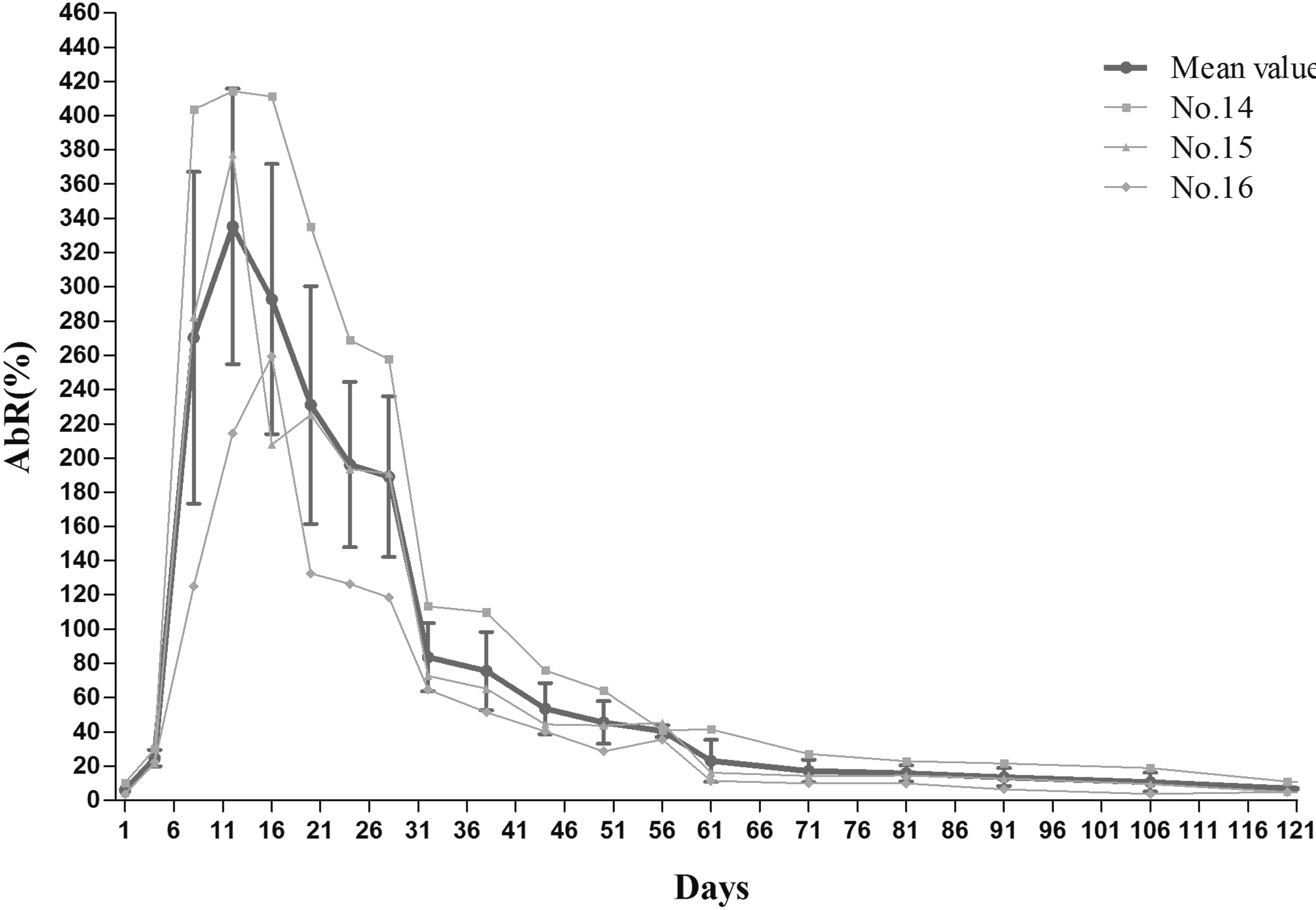
Investigation of anti-OspC antibody secretion by the developed ELISA. The gray curves presented three experimentally infected sheep antibody secretion levels, and the black curve revealed average values with standard deviations of the antibody secretion level during the survey period via AbR values. Antibody was detected by day 5 postinoculation, the peak occurred at ∼13 days and continued to secrete in a high AbR level for ∼28 days. After 61 days of inoculation, the antibody AbR value was below positive threshold determined by the ELISA.
Serological investigation of field serum samples in Northeast China regions
The results of the 498 (collected in 2015) and 474 (acquired in 2016) sheep serum samples showed that the total infection rate was 5.8% and 12.2%, respectively. The positive rate for the 122 samples from Jilin province in 2015 was 5.7% and it was 6.3% and 5.4% from the 208 samples in Heilongjiang province and 168 specimens in Liaoning province, respectively. In contrast, 14.2%, 13.5%, and 8.6% positive rates were identified from 189 samples in Heilongjiang province, 133 samples in Jilin province, and 152 samples in Liaoning province during 2016, separately (Table 1).
The three provinces, Heilongjiang, Jilin, and Liaoning, are regarded as Northeast China regions.
Discussion
In this study, we developed an ELISA to detect B. burgdorferi in sheep. It is rapid, simple and cost-effective method to avoid of complicated amplification process during the B.burgdorferi diagnosis. The developed ELISA method was available to apply for multiple samples simultaneously during the epidemiological study. The acquired data provided useful information on clinical diagnosis. Easy standardization and simple operation of the assay enabled its wide use in field and laboratory pathogen detection, especially in developing regions. Our results showed that the established ELISA had a specificity and sensitivity of 84.4% and 86.2% at a 20% positive threshold.
OspC was chosen as the antigen to develop the ELISA for the detection of B. burgdorferi in sheep. This is partly because the protein plays a vital role in B. burgdorferi survival in mammalian hosts. It has also been shown that OspC is required at the early stage of mammalian infection (Liang et al. 2002, Grimm et al. 2004, Stewart et al. 2006, Tilly et al. 2006). The protein is expressed when ticks begin feeding or immediately after needle inoculation and is not detectable when B. burgdorferi is found in a host (Schwan et al. 1995, Montgomery et al. 1996, Schwan and Piesman 2000, Liang et al. 2004). Therefore, antibodies against OspC have the capacity to diagnose B. burgdorferi in the early stages of infection.
The investigation on experimentally infected sheep via established ELISA showed that the specific anti-OspC antibody was produced by day 5 postinoculation, the peak occurred at ∼13 days, and continued to secrete in a high AbR level for about 28 days. The antibody can be detected until 61 days of inoculation. These results confirmed that OspC is expressed after needle inoculation and present at the first few weeks after infection. Based on anti-OspC antibody secretion dynamics, epidemiological surveillance appropriates for the serum sample collection after 1 month of tick bites on sheep. A small animal model on B. burgdorferi infection was well studied, however, the phenomenon of OspC expression during an ∼1-month period in the study and antibody secretion level in natural infection is not fully understood. Therefore, a further analysis of ruminants during spirochete growing by tick bites is worthy of investigation.
Although B. burgdorferi was isolated in China more than 20 years ago, epidemiological study of B. burgdorferi infection on sheep is still unknown. In this study, our goal is to develop a rapid and reliable method for the detection of B. burgdorferi among sheep in an inexpensive manner for clinical use in Lyme disease control and pathogenic investigation in China and other neighboring developing countries reported with the same pathogen infection cases. The results from 498 (collected in 2015) and 474 (collected in 2016) sheep serum samples collected from the northeast region revealed a total positive rate of 5.8% (29/498) and 9.7% (48/474), respectively. The infection rates of samples from Heilongjiang, Jilin, and Liaoning regions were 6.3%, 5.7%, and 5.4% in 2015 and 14.3%, 13.5%, and 8.6% in 2016, separately (Table 1). The increased positive rate might be due to temperature change and cultivation in rural areas recently. Thus, B. burgdorferi was prevalent in sheep in the region, which indicated that sheep are considered a host for B. burgdorferi transmission and infection and the Northeast China region is likely to be natural epidemic foci of B. burgdorferi. The field sample examination thus showed a temporal increase of B. burgdorferi infection in sheep during 2016 in comparison with the previous year. Furthermore, sheep feeding is popular in the Northeast China regions due to large areas of grassland and forest, in which also is the natural habitat of wild animals. Potential contact between wild species and livestock induces the exposure of tick bite and the associated tick-borne diseases. However, shepherd, farm and forest workers in this region are rarely cautious about tick prevention measures. A family-oriented farming system with poor organization, lack of knowledge, and a low-efficient clinical diagnostic method leads to the annual report of B. burgdorferi infection in China.
Conclusion
The ELISA method that we developed is suitable for clinical diagnosis and epidemiological study on field serum samples with high specificity and sensitivity. It is available to identify B. burgdorferi (SZ strain) at the early stage of infection among sheep herds and examine specific antibody during a certain period of secretion phase. The results of this study provide useful information on the epidemiology of B. burgdorferi in sheep. The Northeast China region is considered the natural epidemic foci of B. burgdorferi. Individuals especially forest workers and shepherds in the areas should notice disease prevention measures to protect themselves from severe illness caused by the infection from tick bites. Animal transport from this region must be under quarantine regulation to prevent expansion of the foci of B. burgdorferi.
Footnotes
Acknowledgment
This work is supported by the National Natural Science Foundation of China (31372438, 31201911).
Author Disclosure Statement
No conflicting financial interests exist.
References
Supplementary Material
Please find the following supplemental material available below.
For Open Access articles published under a Creative Commons License, all supplemental material carries the same license as the article it is associated with.
For non-Open Access articles published, all supplemental material carries a non-exclusive license, and permission requests for re-use of supplemental material or any part of supplemental material shall be sent directly to the copyright owner as specified in the copyright notice associated with the article.