
Abstract
There is no information on rickettsial diseases in domestic animals in Bhutan. This study provides preliminary serological data on exposure of domestic animals to Rickettsia, Orientia, and Coxiella. Animal sera were collected opportunistically from Bhutan and tested in the Australian Rickettsial Reference Laboratory for IgG antibodies against spotted fever group (SFG) and typhus group (TG) Rickettsia, scrub typhus group (STG), and Q fever (QF). Of the 294 animals tested, 136 (46%) showed serological evidence of past exposure to one or more rickettsiae: 106 (36%), 62 (21%), 45 (15%), and 11 (4%) being positive against SFG Rickettsia, Orientia, TG Rickettsia, and Coxiella, respectively. Dogs appeared to exhibit the highest seropositivity against SFG (55%) and TG Rickettsia (45%), horses against STG (91%), while goats were mostly positive for Coxiella (9%). Dogs also appeared to have high risk of being exposed to SFG Rickettsia (odd ratios [OR] 5.71, 95% confidence interval [CI] 3.02–10.80, p < 0.001), TG Rickettsia (OR 48.74, 95% CI 11.29–210.32, p < 0.001), and STG (OR 6.80, 95% CI 3.32–13.95, p < 0.001), but not against QF (OR 1.95, 95% CI 0.42–8.95, p = 0.390). Differences in seropositivity rates between animal species may have been significant for SFG, TG, and STG, but not for QF. The differences in the seropositivity rates of the four infections between districts appeared to be significant for TG and STG, but not for SFG and QF. The seropositivity rates of domestic animals to the four rickettsial infections were consistent with similar studies on the human population in the same areas and appear to demonstrate a high prevalence of exposure to rickettsiae in Bhutan. These preliminary findings constitute baseline data for Bhutan. The findings of this study call for an increased human-livestock sector collaboration in rickettsial diseases research aimed at developing diagnostic and therapeutic guidelines and formulating preventive and control measures through a One Health approach.
Introduction
R
Bhutan is a small Himalayan kingdom situated between India in the south and China in the north. It has an estimated human population of 770,000 in 2016 (National Statistics Bureau 2016) living in 20 districts. In a serosurvey among 864 healthy individuals, sampled equally from eight selected districts in Bhutan, an overall seroprevalence of 49% against rickettsioses was detected, represented by 22.6%, 15.7%, 3.5%, and 6.9% against STG, SFG, TG, and QF, respectively (Tshokey et al. 2017). As of 2016, Bhutan's livestock population largely comprised cattle (303,374) followed by yaks (49,617), goats (39,513), cats (33,866), pet dogs (28,630), horses (18,890), pigs (15,324), and sheep (11,277) (Livestock Statistics 2016). There are few buffaloes (532) and the poultry population (1,038,553) has increased recently due to the mushrooming of poultry farms all over the country. There are currently no data on rickettsioses in domestic animals in Bhutan. This preliminary study was undertaken to determine the serological evidence of rickettsial infections in domestic animals in Bhutan.
Materials and Methods
Study design, location, and animal sampling
This study was an opportunistic sampling of animals and not representative of the whole country or all animal species. It was conducted in the areas where a human seroprevalence study (Tshokey et al. 2017) was carried out as part of the same project between January and April 2015. For the human study, eight districts from Bhutan's 20 districts were selected through a probability proportionate to size method. From each district, a rural and an urban area were selected by the same method, resulting in 16 sampling sites in total (8 urban and 8 rural). Blood samples were collected from domestic animals in those same areas by trained livestock staff. Serum was stored at −70°C, until shipment to the Australian Rickettsial Reference Laboratory (ARRL) for serological testing. For this animal study, a sample size was not predetermined since it was a preliminary study designed only to demonstrate evidence of exposure of domestic animals to rickettsial pathogens in the areas where a high human rickettsial seroprevalence had been detected.
Serological testing
Serum samples were shipped at room temperature to the ARRL, a nationally accredited diagnostic laboratory, for rickettsial serological testing. Testing for IgG antibodies was carried out by indirect microimmunofluorescence assay (IFA) (Graves et al. 1991) using in-house prepared rickettsial antigens; SFG Rickettsia consisted of Rickettsia australis, Rickettsia honei, Rickettsia conorii, Rickettsia africae, Rickettsia rickettsii, and Rickettsia felis antigens; TG Rickettsia consisted of R. prowazekii and R. typhi antigens; STG consisted of O. tsutsugamushi (Gilliam, Karp, and Kato strains) and O. chuto antigens, and QF using C. burnetii phase I and phase II antigens of the Nine Mile strain. Known positive and negative control animal sera for the specific animal species being tested were included on each slide during testing. For some animal species where no definite positive and negative controls were available (such as yaks), certain assumptions were made by using known positive and negative control human sera to ensure the quality of the antigens, conjugates, and the test procedure, even though a nonhuman serum was being titrated.
IFA slides were coated with antigens from SFG and TG Rickettsia, Orientia, and Coxiella (phase I and II). Fluorescein-labeled IgG antibodies against the animal species tested (anti-bovine, anti-dog, anti-goat, anti-horse, anti-cat) from KPL were used to detect the IgG antibody-antigen complexes following established protocols in the ARRL and a previous related study (Muleme et al. 2016). Samples were initially screened at low dilutions (1:32 for Rickettsia and Orientia and 1:80 for Coxiella) and titrated to end-point titer when positive. Serum samples were pipetted onto the fixed antigens on the slide wells in duplicates and incubated for 40 min at 37°C. Unbound antibodies were removed by washing in 10% phosphate-buffered saline (PBS). Following this, dried slides were treated with secondary conjugated antibodies and incubated under similar conditions. Unbound conjugated antibodies were washed off with 10% PBS and dried slides were observed under an ultraviolet light microscopy with FITC filters at 400 × magnification. Positive samples were identified by the presence of bright green fluorescence, while negative samples lacked any fluorescence. Based on similar past studies, antibody titers of ≥1:64 (Nanayakkara et al. 2013, Cunha et al. 2014) against any of the SFG, TG, and STG antigens were considered positive for the rickettsial group agents, and antibody titers of ≥1:160 (Muleme et al. 2016) against C. burnetii phase I or II or both were considered positive for QF.
Statistical analysis
Data were entered into an Excel spreadsheet and analyzed with STATA software version 14 (StataCorp). Descriptive statistics were carried out to calculate the frequencies and percentages of each of the variables such as animal species sampled from each district and seropositivity rates. Chi-squared or Fischer's exact test was used to explore the association between seropositivity and different study variables considering p values of <0.05 statistically significant. Univariate logistic regression analysis was attempted with seropositivity status of each rickettsial disease (seropositive vs. seronegative) as an outcome and the animal species and study sites as an explanatory variable. The odds ratios (OR) and the corresponding 95% confidence intervals (CIs) and p values were calculated. Variables with a p value of <0.05 were considered to be significantly associated with the seropositivity of the rickettsial disease.
Ethics approval
This study was approved by the Council for RNR Research of Bhutan (CoRRB), Ministry of Agriculture and Forests, Royal Government of Bhutan, through approval no. CORRB/TCO/D-2/732.
Results
A total of 294 domestic animals from 7 species were opportunistically sampled from an urban and rural study site of the 8 districts (Table 1). More than half, 164 (56%) of the animals were sampled from rural areas and the remainder from urban areas.
Of the 294 sampled animals, 136 (46%) showed serological evidence of past exposure to one or more rickettsial agents. Individually, 106 (36%), 62 (21%), 45 (15%), and 11 (4%) were positive for IgG antibodies against SFG Rickettsia, Orientia, TG Rickettsia, and Coxiella, respectively. Among those seropositive against more than one agent, the commonest was dual seropositivity against SFG and TG (15%), followed by SFG and STG (13%). The highest antibody titers observed against antigen groups for SFG, TG, and STG was 1:512 and for Coxiella was 1:320 (Table 2). The distribution of the 136 seropositive cases within the approximate location of the sampling sites is shown in Fig. 1. These seropositivity rates in domestic animals when compared to seropositivity in humans (Tshokey et al. 2017) from the same study sites (Table 3) showed that rural Samtse and rural Chukha districts in the south-western Bhutan had higher SFG prevalence and rural Trongsa and rural Zhemgang in central Bhutan had higher STG exposure in both human and animal populations. In addition, although generally of low prevalence in both human and animal populations, QF seemed to be more prevalent in rural Mongar, rural Punakha, and rural Chukha districts (Table 3).
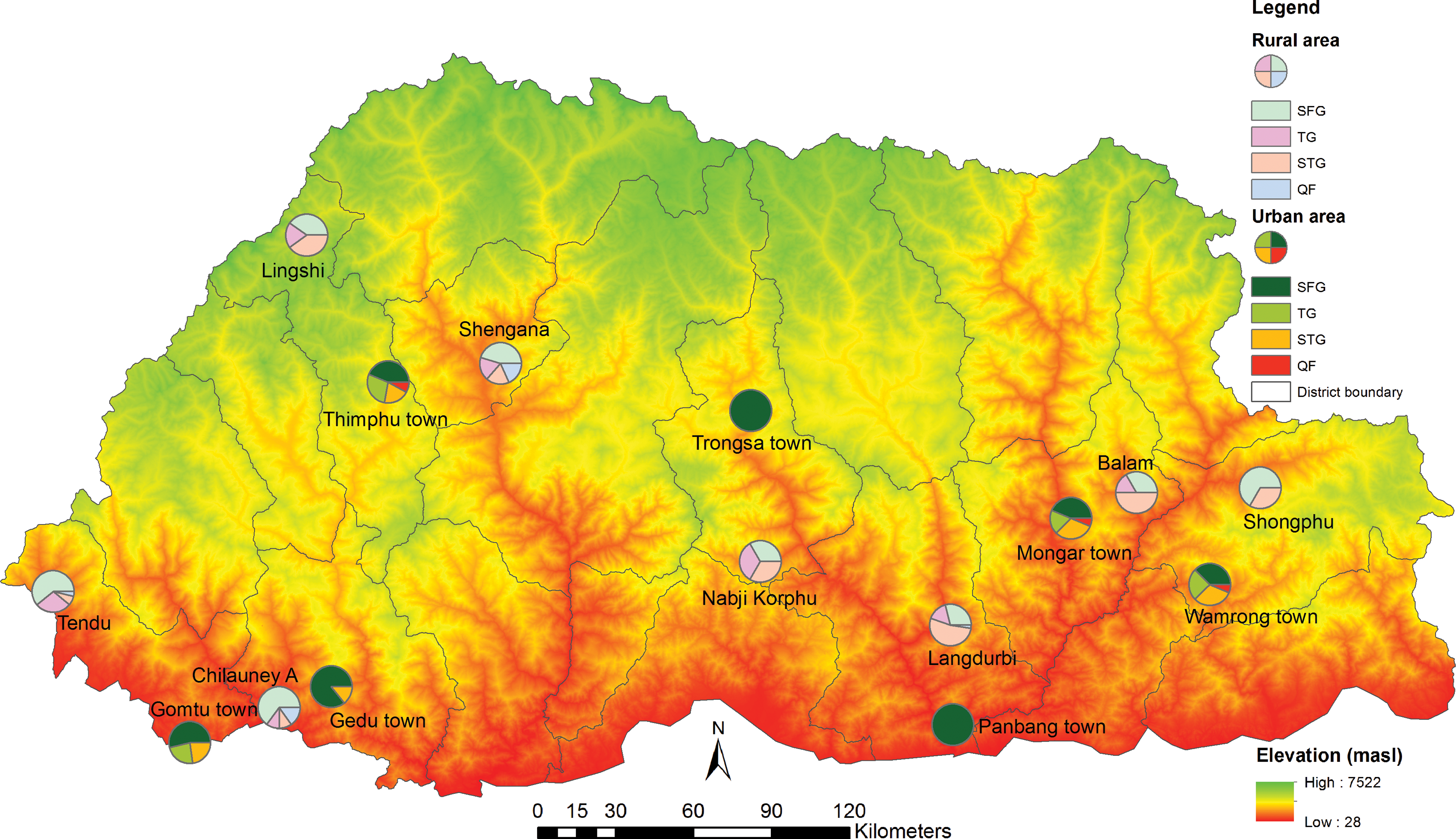
Map of Bhutan showing pie charts demonstrating proportion of animals seropositive against SFG, STG, TG, and QF in each urban and rural sampling area. Seronegative animals are not shown on the map. Pie chart location provides an approximate location of sampling sites. QF, Q fever; SFG, spotted fever group; STG, scrub typhus group; TG, typhus group. Color images available online
Sera were screened at a dilution of 1:32 (SFG, TG, and STG) and 1:80 (C. burnetii) and titrated to end-titer if positive. A titer of ≥1:64 (for SFG, TG, and STG) and ≥1:160 (for C. burnetii) was taken as positive.
SFG, spotted fever group; STG, scrub typhus group; TG, typhus group.
Human data from Tshokey et al. (2017).
QF, Q fever.
Seropositivity rates against the four different rickettsial infections among the sampled animal population of different species are presented in Table 4. Only positive sera are presented in this table. Dogs exhibited the highest seropositivity against SFG Rickettsia (55%) and TG Rickettsia (45%); horses against STG (91%), and goats against QF (9%). Differences in seropositivity rates between animal species may have been significant for SFG, TG, and STG, but not for QF. The differences in the seropositivity rates of the four infections between districts may have been significant for TG and STG, but not for SFG and QF (Table 4). These comparative seropositivity data between different animal species and different study sites should be interpreted with caution due to the overall small sample size and variable number of each animal species sampled from each study sites.
p value <0.05 indicates significant differences in seropositivity rates between different animal species or the eight study sites (districts).
Given the unequal number of animal species sampled from each study site, logistic regression analysis would have been questionable and thus data are not presented in detail. However, for a closer comparison, a few comparisons with high odds ratios were probably significant and worth mentioning. Compared to other animals, dogs appeared to have a high risk of being exposed to SFG (OR 5.71, 95% CI 3.02–10.80, p < 0.001), TG (OR 48.74, 95% CI 11.29–210.32, p < 0.001), and STG (OR 6.80, 95% CI 3.32–13.95, p < 0.001), but not against QF (OR 1.95, 95% CI 0.42–8.95, p = 0.390). In addition, the differences in seropositivity against STG between animals from urban and rural areas appeared to be significant with rural animals at double the risk of being exposed compared with urban animals (OR 2.07, 95% CI 1.14–3.78, p = 0.017). This difference was not seen for SFG, TG, or QF.
Discussions
This is the first serological study of rickettsial diseases and QF in domestic animals in Bhutan. A high seropositivity of domestic animals against rickettsial infections especially SFG (36%) and STG (21%) was observed. The findings of this study were consistent with the findings of a seroprevalence study of the same infections in the human population (Tshokey et al. 2017) from the same study sites. While the seropositivity of SFG was higher in animals, STG was higher in the human population. This could be due to differences in vectors and their exposure risks; tick infestation of domestic animals being high in Bhutan favoring tick-borne SFG in animals, while human exposure to rodents (host for mites) would possibly explain human mite-borne STG. This difference also possibly suggests that the extent to which human disease occurs may not always be proportionate to exposure in animals although animals and their arthropod vectors act as the reservoirs for these zoonoses. The observed dual seropositivity against SFG and TG of 15% was likely due to cross-reacting antibodies between the two rather than true exposure to both groups. However, dual seropositivity of about 13% against SFG and STG reveals the true proportion of exposure to both these agents in domestic animals since SFG and STG antigens are distinct and serological cross-reactions are unlikely. The key message from this and the related human study is that STG and SFG are widely prevalent in both the human and animal population in Bhutan, with some likely hot-spots. Findings on TG Rickettsia and QF were less convincing in both the human and animal populations due to low seropositivity rates.
There is overall limited data on animal seroprevalence to Rickettsia and Orientia, especially in Asia, compared to Coxiella which have been studied widely. Nevertheless, studies on Coxiella have concentrated on goats, sheep, cattle, and horses but no other animal species. The seropositivity rate of SFG (36%) among domestic animals in this study was higher than that reported in Mongolia (≈20%). In the Mongolian study, seroprevalence rate was 30%, 13%, 21%, 35%, and 2% for cattle, goats, sheep, horses, and camels, respectively (von Fricken et al. 2018). Although data would be specific to the study sites rather than nationally representative, the prevalence of SFG in Bhutanese dogs (55%) was similar to Sri Lankan dogs (≈42%), whereas seropositivity against STG appeared to be higher in Bhutanese dogs (45%) than Sri Lankan dogs (24%) in the areas studied (Nanayakkara et al. 2013). These findings suggest that dogs may act as an important reservoir of rickettsial infections in Bhutan.
In the case of Coxiella, seropositivity rate was low in the sampled domestic animals in this study (4%) as well as in the human population (7%) in the same areas (Tshokey et al. 2017). The seropositivity of QF (≈4%) in this study appeared lower than that reported in studies from India at 14% in animals although these had reproductive disorders (Vaidya et al. 2010); at 14% in Tibetan sheep in China (Yin et al. 2015b) and 14% in free-range yaks in China (Yin et al. 2015a). Unlike sheep and yaks in China, none of the few sheep and yaks in the current study showed evidence of exposure to Coxiella. A meta-analysis on C. burnetii in horses world-wide reported a seroprevalence of 16%, although none of the 122 cases of equine abortion, stillbirth, or neonatal foal death was positive for Coxiella DNA (Marenzoni et al. 2013). The few Bhutanese horses in this study did not have evidence of exposure to Coxiella. With this apparently low prevalence of QF, for Bhutan, a developing country with numerous other priorities, investing in QF vaccines for domestic animals and veterinarians may not merit immediate consideration. This preliminary data, however, need to be confirmed with larger nationally representative studies. Nevertheless, other activities aimed at adequate case identification and management are likely to be important with clear strategies for prevention and control through close human-animal sector collaboration.
Although some apparently significant findings have been seen in this first study in animals, percentage seropositivity results and their significance should be interpreted with caution due to wide variations in the number of animals sampled from each area and within each species. For instance, yaks being found only in the highlands, the 10 yaks tested were from one specific area in Thimphu district only and 10 of the 11 horses tested were from the one sampling area in Zhemgang district. However, these preliminary findings are the baseline data for future research into the epidemiology of rickettsial diseases in Bhutanese domestic animals.
In Bhutan, although some human-livestock sector collaborations were initiated recently, there are no ongoing collaborative activities specific to research and management of rickettsioses between the two sectors. Currently, diagnostics is limited to the use of rapid immunochromatographic test kits for scrub typhus alone in the human sector with none available in the livestock sector for rickettsial diseases. Clinical management, prevention, and control guidelines are nonexistent in both sectors. This study has brought together key personnel from both the sectors and the data generated should prompt taking these typical “One Health” collaborative activities further.
This study has significant limitations, the main one being the opportunistic sampling strategy. Assumptions were made during testing due to unavailability of known positive and negative controls for some animal species (e.g., yaks). The number of animals sampled from each district or sampling areas and within each animal species was not uniform between sampling areas (no fixed sample size), thereby making comparisons contentious. For some animal species, there were only a few animals sampled and consequently few or no seropositive cases obtained.
Conclusion
In conclusion, this study presents preliminary data on rickettsial diseases in Bhutanese domestic animals and these results constitute baseline data for Bhutan. A high proportion of different animal species were seropositive against SFG and STG, but with fewer animals seropositive against TG and QF. These findings were consistent with the seroprevalence of the same infections in human population in the same areas of Bhutan. Findings from this study and related studies in the human population appear to demonstrate high prevalence of exposure to rickettsiae in both human and animal populations in Bhutan. They may be causally related but further studies would be needed to confirm it. These results call for increased collaboration between the human and animal sectors in rickettsial diseases research aimed at developing diagnostic and treatment guidelines and formulating prevention and control measures through a One Health approach.
Footnotes
Acknowledgments
In Bhutan, we thank the district veterinary officers and livestock technical staff of the eight study districts for their assistance during sample collection. We also acknowledge the kind cooperation of the owners of the animals sampled for the study. In the ARRL, we thank Dr. Mythili Tadepalli and Ms. Chelsea Nguyen for laboratory assistance. We also thank the following pathologists from New South Wales Health Pathology (Australia) who provided financial support toward the research: Drs. M. Formby, A. Cotty, B. Young, T. de Malmanche, H. Tran, B. Bhagwandeen, and the late Dr. B. Murugasu. The University of Newcastle (Australia) has contributed to this work by providing a PhD scholarship to the senior author.
Author Disclosure Statement
No conflicting financial interests exist.