




Abstract
Hepatitis E virus (HEV) has been isolated from humans and several animals' species. During the last years, the knowledge of HEV infection dramatically changed and enriched. The aim of this study was to estimate the seroprevalence of HEV in industrial pigs in different districts of Southern Bulgaria. Three hundred sixty swine serum samples were tested for anti-HEV IgG antibodies. The samples were collected from four industrial farms from three districts of Southern Bulgaria. HEV-specific antibodies in porcine serum were detected by enzyme-linked immunosorbent assay (PrioCHECK HEV Ab porcine). The overall HEV seroprevalence was 60.3%. The seropositivity varied widely depending on age groups and investigated farms. The overall prevalence in weaners was 25%, in fattening pigs 75.8%, and in group of sows was found the highest HEV positivity of 80%. The occurrence of HEV positivity in sows and fattening pigs presented odds ratio (OR) = 17.200 (95% confidence interval [CI]: 8.8–33.7) and OR = 11.342 (95% CI: 6.1–21.0), respectively, compared to weaners. The study indicated that HEV is widespread in industrial farms in Bulgaria and presented high seroprevalence in pigs. The results found that HEV seropositivity showed age dependency. The National Health Authorities should raise awareness of HEV and its zoonotic potential.
Introduction
Hepatitis E virus (HEV) has been isolated from humans and several animals' species. According to the World Health Organization (WHO) database, 20 million human HEV infections are estimated worldwide annually, leading to 3.3 million human cases with clinical presentation, resulting in ∼44,000 deaths in 2015 (WHO 2018). HEV is classified into family Hepeviridae, divided in two genera: Orthohepevirus and Piscihepevirus (ICTV 2017). Orthohepevirus included four species: Orthohepevirus A (Genotypes: HEV-1–HEV-8), Orthohepevirus B (Avian HEV Genotype 2), Orthohepevirus C (Germany Rat HEV, Vietnam Rat HEV and Ferret HEV), and Orthohepevirus D (Germany Bat HEV) (ICTV 2017, Capai et al. 2018, Pepovich et al. 2019). Genus Piscihepevirus has only one species—Piscihepevirus A, and one genotype—Cutthroat Trout HEV (ICTV 2017, Capai et al. 2018). HEV-3, HEV-4, and HEV-7 are zoonotic and infect both humans and animals. HEV-3 has been found in humans, swine, wild boar (ICTV 2017), wild deer (Wild deer HEV Subtype 3b, GenBank accession no. AB189071) (Tei et al. 2003), rabbit (Rabbit HEV Subtype 3ra, GenBank accession no. FJ906895) (Zhao et al. 2009), and mongoose (Mongoose HEV Subtype 3a, GenBank accession no. AB591734) (Nidaira et al. 2012). HEV-4 has been isolated from humans and swine (ICTV 2017). One report demonstrated a human chronic HEV infection caused by HEV-7 (Human HEV 7, GenBank accession no. KT818608), and also HEV stains were isolated from camels and were categorized into genotype 7 (DcHEV 7, GenBank accession no. KJ496143 and KJ496144) (Woo et al. 2014, Lee et al. 2016).
HEV infection in swine is asymptomatic; although histological liver changes were observed by autopsy (Halbur et al. 2001), the infection could be developed in all age groups: weaners, fattening pigs, and sows, and the main mechanism of HEV transmission among pigs is fecal-oral. Bouwknegt et al. (2008) reported that one HEV-infected pig could potentially infected other 8.8 (95% confidence interval [CI]: 4–19) pigs during the acute phase of infection.
In Bulgaria, the first evidence of human HEV infection was reported in 4 out of 53 (7.5%) cases with acute HEV infection in 1995 (Teoharov et al. 1995). Investigations of human HEV infections in Bulgaria have been started since 2014 (Teoharov et al. 2014, Baymakova et al. 2016, Bruni et al. 2018, Cella et al. 2019), whereas first preliminary data for swine HEV infection in Bulgaria were published in 2018 (Pishmisheva et al. 2018). That report presented an overall seroprevalence of anti-HEV antibodies in swine of 40% (34 positive samples out of all 85 sera) in both investigated districts Pazardzhik and Sliven (Pishmisheva et al. 2018). Up to date, no data are available for HEV prevalence among pigs in other areas of the country.
The aim of this study was to estimate the seroprevalence of HEV in industrial pigs in different districts of Southern Bulgaria.
Materials and Methods
Areas and pigs collection
Three hundred sixty pigs (n = 360) were enrolled from four industrial farms (Kalchevo, Yambol, Pishtigovo, and Kostinbrod) in three districts of Southern Bulgaria—Yambol, Pazardzhik, and Sofia (Fig. 1). Animals were divided in three age groups: weaners, fattening pigs, and sows. An equal number of samples (n = 30) were taken from each age group on each farm. Consequently, a total of 90 samples were tested from each farm. Pigs showed no clinical signs at sampling time point; unfortunately, sex had not been recorded. The pigs' selection in farms, respectively, in age groups was done randomly. The investigated farms were located among plains (∼42°48′N and 42°69′N Latitude and 23°32′E and 26°51′E Longitude), and the climate is subtropical/continental (average annual temperature 13–15°C and precipitations about 650 mm/m2).
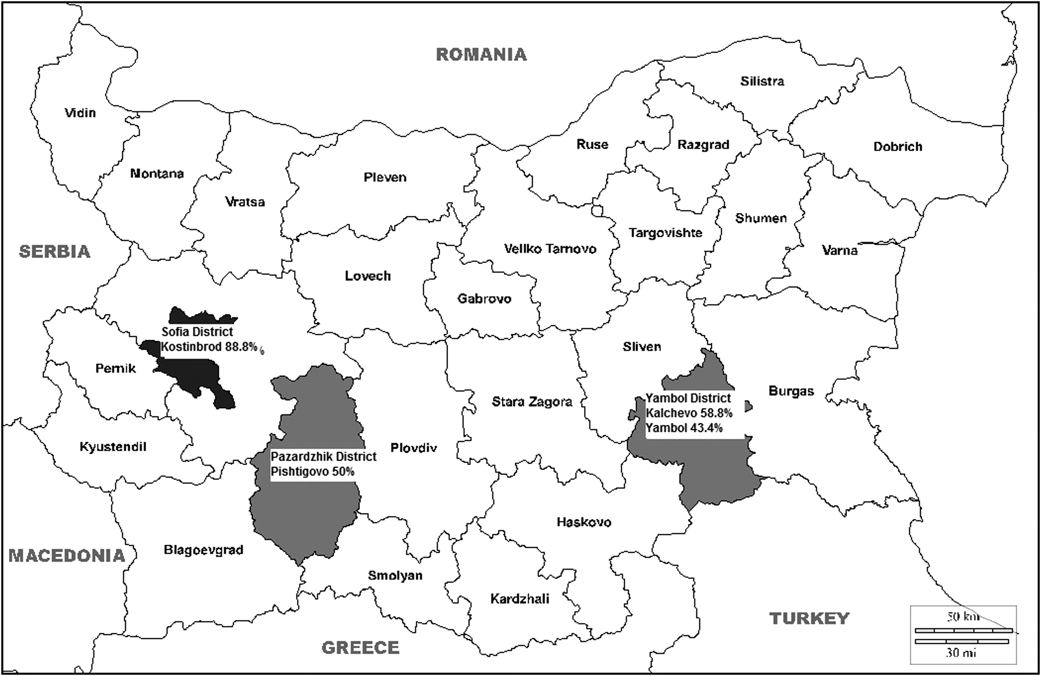
Geographic distribution of HEV infection in pigs from Southern Bulgaria. HEV, hepatitis E virus.
Sampling
Swine blood samples (up to 5 mL per individual) were taken by puncture of the sinus ophthalmicus. Blood collection tubes without anticoagulant were kept at room temperature (20°C) until visible clot retraction, centrifuged at 1500 × g for 10 minutes, and the serum was separated and kept at −20°C until processing.
HEV serological assay
The serum samples were tested for HEV antibodies in Laboratory of Infectious Diseases, Faculty of Veterinary Medicine, Trakia University, Stara Zagora, Bulgaria. A commercial enzyme-linked immunosorbent assay (PrioCHECK HEV Ab porcine; Mikrogen GmbH, Neuried, Germany) was used. The PrioCHECK HEV Ab porcine is a diagnostic test for detection of HEV-specific antibodies in porcine serum and meat juice samples. Microtiter plate is coated with recombinant HEV antigen of the open reading frame 2 (ORF2) and ORF3 of genotypes HEV-1 and HEV-3. The test has 91.0% sensitivity and 94.1% specificity. The cutoff value was calculated according to the manufacturer's instructions. The cutoff is calculated as mean optical density 450 (OD450) of the cutoff control multiplied with 1.2. Results obtained above or equal to the cutoff are considered positive. Results between the OD450 of the cutoff control and the cutoff are doubtful. Results obtained below the cut-off are negative.
Ethics statement
The study was approved by the Ethics Committee in Animal Experimentation and Animal Welfare at Trakia University, Stara Zagora (Bulgaria), and was conducted according to the ethical principles of animal experimentation, adopted by the Bulgarian Ministry of Agriculture, Food and Forestry.
Statistical analysis
HEV-positive results among different swine age groups and farms were compared by the chi-squared test. Binary logistic regression was used to evaluate the risk of positive results according to age group. Statistical analysis has been performed by Excel 2007 (Microsoft, Redmond, WA) and SPSS Statistics 19.0 (IBM Corp., Armonk, NY). A p-value <0.05 was considered statistically significant.
Results
Positive results for anti-HEV IgG were detected in 217 (60.3%) of all 360 tested sera (Table 1). The overall seropositivity in weaners, fattening pigs, and sows was 25% (mean ± SD: 25.0 ± 30.9; 95% CI: 5.2–55.2), 75.8% (mean ± SD: 75.8 ± 36.2; 95% CI: 40.3–111.4), and 80% (mean ± SD: 80.0 ± 17.6; 95% CI: 62.7–97.3), respectively. The highest HEV seropositivity in age group weaners was found in Kostinbrod (21/30, 70%). The lowest HEV seropositivity in age group fattening pigs was estimated in Yambol (7/30, 23.3%). In age group of sows, the highest HEV seropositivity was presented in Kostinbrod (29/30, 96.6%). The overall prevalence of anti-HEV antibodies in each farm was as follows: in Kalchevo—58.8% (53/90), in Yambol—43.3% (39/90), in Pishtigovo—50% (45/90), and in Kostinbrod—88.8% (80/90). Based on age groups and different farms, the chi-squared test showed significant differences in HEV seropositivity between the age groups and farms (Table 1). Five percent of all samples were assessed as doubtful.
Seroprevalence of Hepatitis E Virus Infection by Age Groups in Pigs from Southern Bulgaria
df, degrees of freedom; HEV, hepatitis E virus.
To estimate the risk for HEV seropositivity, the odds ratio (OR) in different age groups was performed by binary logistic regression. The OR of anti-HEV antibody occurrence in fattening pigs and sows was determined by comparing to group weaners (Table 2). We found that the odds of HEV infection was nearly 11 times higher in fattening pigs and 17 times higher in sows than in the group of weaners.
Logistic Regression Showing the Relationship Between Hepatitis E Virus-Positive Pigs and Age Group
CI, confidence interval; NA, not applicable; OR, odds ratio; PE, parameter estimate; SE, standard error.
Discussion
Moderate and high HEV seropositivity among swine were observed in different countries from Southeastern Europe. The estimated anti-HEV positivity in different districts in Romania was 42.7% (65/145) and 49.3% (34/69) (Savuta et al. 2007, 2008), Serbia—34.6% (109/315) (Lupulovic et al. 2010), Greece—80% (76/96) (Siochu et al. 2009), and Croatia—32.9% (469/1424) (Jemersic et al. 2017). In West-European countries of the Mediterranean Basin, the prevalence of swine HEV infection was lower compared to the Balkan Peninsula. In Spain was observed a positivity of 25% (15/60) and 20.4% (233/1141) in different settings and years (Pina et al. 2000, Jimenez de Oya et al. 2011), France—31% (2035/6565) (Rose et al. 2011), and Italy—50.2% (714/1422) (Martinelli et al. 2011). Countries from Western Europe reported relatively high swine HEV seroprevalence. In Switzerland was found 58.1% HEV seropositivity (1161/1999; 95% CI: 55.9–60.2) (Burri et al. 2014), Germany—49.8% and 68.6% (Baechlein et al. 2010, Wacheck et al. 2012), Netherlands—72% in conventional farms (191/265) (Rutjes et al. 2014), Belgium—73% (95% CI: 68.8–77.5) (Thiry et al. 2014), England—85.5% (219/256) and 92.8% (584/629) (Banks et al. 2004, Grierson et al. 2015), and Scotland—61.4% (108/176) (Crossan et al. 2015). Scandinavian countries also had a high prevalence of swine HEV. In Sweden the seropositivity was assessed as 58% (Banks et al. 2004), Denmark—73.2% (Breum et al. 2010), Finland—86.3% (Kantala 2017), and Norway—90% (137/153) (Lange et al. 2017).
In North America, the swine HEV seroprevalence varied widely. In Canada, the reported seropositivity was 18.1% (129/712) in one study and another found 59.4% (594/998) (Meng et al. 1999, Yoo et al. 2001). In USA, the situation was similar and depended on studies—34.5% (29/84) and 68.9% (202/293) (Meng et al. 1997, Withers et al. 2002). The estimated anti-HEV positivity in Mexico was 59.4% and 80% (Cooper et al. 2005, Garcia-Hernandez et al. 2017).
In Asia, the prevalence of HEV antibodies in pigs varied widely. In China, the reported seroprevalence was 78.8% (330/419) and 78.9% (490/621) (Wang et al. 2002, Li et al. 2011). In contrast, Thailand reported 30.7% (23/75) serum samples positive for swine HEV (Meng et al. 1999). Others Asian countries published data in moderate to high range as South Korea—40.7% (57/140) (Meng et al. 1999), Indonesia—72% (71/99) (Wibawa et al. 2004), Taiwan—37.1% (102/275) (Hsieh et al. 1999), and India—42.9% (122/284) (Arankalle et al. 2002).
As shown, the prevalence of HEV antibodies was quite variable between countries, also in different areas in the same countries. This variability could be influenced by study design, diagnostics methods, and tested population. Nevertheless, it seems that pigs could be infected with HEV and be a zoonotic risk for transmission.
The seroprevalence in our study (60.3%) is similar to data found in Switzerland (58.1%) (Burri et al. 2014), Scotland (61.4%) (Crossan et al. 2015), Sweden (58%) (Banks et al. 2004), Canada (59.4%) (Yoo et al. 2001), and Mexico (59.4%) (Garcia-Hernandez et al. 2017). Sows had the highest seroprevalence (80%) in our study. Martinelli et al. (2011) found 70.6% HEV prevalence in sows, also the highest seroprevalence in their study, whereas Danish investigation presented 73.2% positive sows for HEV-IgG (Breum et al. 2010). The results of our study showed an age-dependent seroprevalence (OR = 11.342, p < 0.001; OR = 17.200, p < 0.001). A similar relationship was observed in another report (Breum et al. 2010). Consequently, HEV occurs in all age groups and pigs could be infected in nursery and fattening periods most likely, corresponding to natural HEV infection (Pavio et al. 2010). Most pigs become infected in 6–8 weeks, virus in feces has peak in 12–14 weeks and decline until 20–22 weeks (Pavio et al. 2010). HEV-IgG appears in 8–9 weeks, increase, and approximately all infected pigs in 22–24 weeks could have HEV-IgG (Pavio et al. 2010). Moreover, Bouwknegt et al. (2009) presented evidence of HEV RNA in urine. They found positive sample in urine of a contact-infected pig at 65 days after the beginning of virus fecal shedding (Bouwknegt et al. 2009). The study highlighted the urine as a potential source for HEV infection among pigs and supposed that contact pathway could be better for transmission of HEV than intravenous inoculation (Bouwknegt et al. 2009). Probably the pig farm density, the husbandry conditions, model of pig farming, animal contact with the environment, sewage system, and water facility could be some reasons for high HEV seroprevalence in pigs. Jemersic et al. (2017) found the highest HEV prevalence in regions with the highest density of pigs and wild boars. Garcia-Hernandez et al. associated the high seroprevalence with short production cycle of fattening and high pig population in farms (Garcia-Hernandez et al. 2017). In our study, the included farms were typical industrial farms with high density of animals, shorter farming for fattening, many herds, and close contact among pigs. These factors probably contributed to the high HEV seroprevalence in pigs. Further projects should be planned to evaluate swine HEV seroprevalence in whole country, molecular analysis for sequences, and human HEV seroprevalence in blood donors.
Conclusions
In conclusion, the preliminary results presented in this study showed a high prevalence of HEV antibodies in pigs in Southern Bulgaria. The evidence for HEV infection in pigs highlights the concerns for zoonotic transmission. National Health Organizations should enhance the control in farms and slaughterhouses, obligatorily record human HEV infection, promote risk factors for transmission in risk population groups, and improve the knowledge about infection among physicians, veterinarians, and general practitioners. Further researches could be realized to a thorough understanding of situation of HEV infection in Bulgaria.
Footnotes
Acknowledgment
This study was funded by the Trakia University, 6000 Stara Zagora, Bulgaria (grant no. 01/2017).
Authors' Contributions
I.T. and R.P.—study design, data collection, data interpretation, article preparation, literature search, and funds collection; M.B. and M.C.—study design, statistical analysis, data interpretation, article preparation, and literature search; T.K.—statistical analysis; P.M. and K.K.D.—data collection and funds collection; K.G.—laboratory analysis; and M.P. and L.P.—funds collection. All authors read and approved the final version of the article.
Author Disclosure Statement
No conflicting financial interests exist.