
Abstract
Anaplasma phagocytophilum is the causative agent of a disease known as tick borne fever in sheep, although fever is not always present. Due to inconclusive clinical signs, diagnosis is based on the cytological or molecular detection of the microorganism in blood and/or the determination of antibodies against A. phagocytophilum. The aim of the study was to determine the alterations caused by the presence of antibodies and/or the antigen of A. phagocytophilum in the blood cell count and morphology in sheep. Cytology and indirect immunofluorescence assay were performed for detection of antibodies and the antigen of A. phagocytophilum, respectively. The samples were divided into four groups depending on the result of the antigen and antibody detection. The samples that were only positive for antigen detection had mild anemia, leukopenia (lymphopenia), and thrombocytopenia. The samples that were positive in both assays had anemia, leukopenia (neutropenia and lymphopenia), and thrombocytopenia. Samples that were positive only for antibody detection had mild leukopenia. Morphological findings in infected sheep included band neutrophils, toxic neutrophils, reactive lymphocytes, and activated monocytes. The hematological findings along with cytological and serological tests can contribute to the assessment of the stage of the disease. A combination of leukopenia and thrombocytopenia raises a strong suspicion of the disease. When the microorganism and antibodies are simultaneously present, sheep are more susceptible to secondary complications. The first reported morphological findings and the quantitative hematological alterations are indicative of an inflammatory reaction, antigenic stimulation, and stress.
Introduction
A
In sheep, the disease is known as tick-borne fever since an abrupt high fever and inappetence may be observed in infected sheep. Depression, respiratory signs, and diarrhea as well as abortions have been also associated with the presence of A. phagocytophilum in sheep (Gokce and Woldehiwet 1999b). However, the absence of any clinical sign, including fever, is not uncommon, while susceptibility of infected sheep to secondary infections due to A. phagocytophilum-induced immunosuppression has also been reported (Gokce and Woldehiwet 1999c, Woldehiwet 2008).
Due to this nonspecific clinical picture, diagnosis relies on detection of parasite inclusions within infected leukocytes mainly in the acute phase of the disease as well as molecular testing (Shabana et al. 2018) by detection of the DNA of A. phagocytophilum using specific primers (Drazenovich et al. 2006).
Furthermore, serology has been used in various studies (de la Fuente et al. 2005, Amusategui et al. 2006, Moretta et al. 2019) to detect the prevalence of exposure to A. phagocytophilum in certain geographic areas. The evolution of antibodies against A. phagocytophilum in ruminants for 21 (Stuen and Artursson 2000) up to 480 days (Paxton and Scott 1989) after experimental infection has also been reported.
The main hematological abnormalities that have been reported in either experimental or naturally occurring infection by A. phagocytophilum concern the leukocytes (Ogden et al. 1998, Gokce and Woldehiwet 1999a), more predominantly the polynuclear cells and to a lesser extent the lymphocytes (Batungbacal et al. 1982) and monocytes. The platelet count is also affected (Gokce and Woldehiwet 1999a). The time course of the above hematological abnormalities has been evaluated mainly after experimental infection (Gokce and Woldehiwet 1999a, Stuen et al. 2011). However, in these studies, most data are generated by hematology analyzers and cell morphology evaluation was not reported. Moreover, there is paucity of reports on blood cell morphological alterations such as the presence of band neutrophils, toxic leukocytes, reactive lymphocytes, and monocytes in blood smears.
The aim of the study was to investigate the alterations caused by the presence of antibodies or the antigen of A. phagocytophilum (by serological and/or cytological examination, respectively) in the blood cell count and morphology in sheep.
Materials and Methods
Before the onset of the study, the minimum required total sample size was calculated using the General Linear Multivariate Model with Wilks Likelihood Ratio procedure with the GLIMMPSE software (Kreidler et al. 2013). The type I error rate was set at 0.05, the desired detectable difference in blood cell counts among groups was set at 10%, and the standard deviation was set at 10. The mean scale and variability factors were set at 1 and 2, respectively. The results of the analysis revealed that a minimum sample size of 48 animals (12 per group, power = 0.803) was required.
A total of 138 sheep of Chios breed from 6 Greek dairy farms were included in the study. All included sheep met the following criteria: (1) presence of ticks, (2) absence of other ectoparasites such as fleas and lice, (3) deworming at least 2 months before their selection in the study, and (4) no cytologic or serologic evidence of concurrent tick-borne infections (Borrelia burgdorferi, Babesia sp., and Theileria sp.).
Blood was collected by jugular venipuncture with an 18-G needle into two vacuum tubes (BD, Franklin Lakes, NJ), one without anticoagulant for serum and one containing EDTA for whole blood. The samples were transferred to the Diagnostic Laboratory, School of Veterinary Medicine, Faculty of Health Sciences, University of Thessaly, Greece, placed in a cooler with ice packs, avoiding direct contact with the tubes.
The packed cell volume (PCV) value was determined using the microhematocrit method (Bull et al. 2000). PCV was measured as the height of the red cell column in the tube after centrifugation. The buffy coat was extruded from the centrifuged blood for smear making.
Blood smears were also made from each sample, dried, and stained with Giemsa. The blood smears were used for assessment of leukocyte and platelet counts, as previously described (Katsogiannou et al. 2020). Published reference intervals (Oikonomidis et al. 2018) for the hematological variables of the Chios breed were used for interpretation of data. A 200-cell, manual, leukocyte differential count was also performed, and the blood cell morphology was also evaluated in the same smears. Band neutrophils, toxic neutrophils, reactive lymphocytes, and activated monocytes were identified and scored based on a well-defined protocol for hematologic data reporting (Weiss 1984).
Serum samples were separated from whole blood samples collected in tubes without the anticoagulant by low-speed centrifugation, transferred to plastic vials, and kept frozen at −20°C until tested for IgG antibodies against A. phagocytophilum, using an indirect immunofluorescence antibody test (IFAT). The IFAT was performed using commercially available slides coated with A. phagocytophilum-infected cells (MegaFLUO®; ANAPLASMA ph., Horbranz, Austria) and commercial rabbit fluorescein isothiocyanate-conjugated anti-sheep IgG (Sigma-Aldrich, St. Louis, MO), with a cutoff value of 1/40 (Stuen et al. 2013).
Based on results of the antibody or antigen detection assays, sampled animals were allocated into four groups; (1) group A: sheep with the presence of the A. phagocytophilum antigen, (2) group B: sheep with both antigen and antibody presence, (3) group C: sheep with antibodies against A. phagocytophilum, and (4) group D: sheep found negative in both assays.
Differences in blood cell counts (white blood cells [WBCs], neutrophils, band neutrophils, lymphocytes, monocytes, eosinophils, basophils, and platelets) and PCV values among the four groups were analyzed using the statistical software, IBM SPSS 25. The normality of data was evaluated with the Kolmogorov–Smirnov test and homogeneity of variances with Levene's test. One-way analysis of variance was run to determine the significance of differences among groups for each parameter that was normally distributed. Post hoc comparisons were performed using the Bonferroni test when equal variances were assumed and the Tamhane T2 test when variances were unequal. The data for band neutrophils were analyzed with Kruskal–Wallis and Mann–Whitney U tests due to non-normal distribution. The results are expressed as means
All procedures were performed according to the ethical standards in the Helsinki Declaration of 1975, as revised in 2000, as well as the national law and after receiving approval from our Institutional Animal Use Ethics Committee.
Results
Of the 138 samples tested, 17 were antigen positive and antibody negative (group A), 16 were positive in both assays (group B), and 16 were positive for the presence of antibodies and negative for the presence of the antigen (group C), while 89 were negative in both assays (group D).
As shown in Table 1, the mean values of PCV, platelets (PLT), and WBC were significantly lower (p < 0.05) in group B, compared with all other groups, and in group A than those of groups C and D; no significant difference (p > 0.05) was detected between groups C and D.
Mean ± Standard Error for the Hematocrit (Packed Cell Volume), White Blood Cell Count, and Platelet Count in Peripheral Blood of Sheep from Different Groups and Comparison Between the Groups
Figures with different superscripts are indicative of a statistically significant difference.
Group A: sheep with the presence of the Anaplasma phagocytophilum antigen, group B: sheep with both antigen and antibody presence, group C: sheep with antibodies against A. phagocytophilum, and group D: sheep found negative in both assays.
PCV, packed cell volume; SE, standard error; WBCs, white blood cells; PLTs, platelets; RI, reference interval.
The mean values of each WBC subpopulation in the four groups are presented and significant differences among them are shown in Table 2. A significantly lower mean segmented neutrophil count between group B and each of all the other groups was also observed. On the contrary, a significantly higher band neutrophil count between group B and each of all the other groups was also observed. Finally, the mean lymphocyte and monocyte counts of group A and group B were significantly lower compared with group C and group D.
Mean ± Standard Error for Differential White Blood Cell Counts in Peripheral Blood of Sheep from Different Groups and Comparison Between the Groups
Figures with different superscripts are indicative of a statistically significant difference. Group A: sheep with the presence of the A. phagocytophilum antigen, group B: sheep with both antigen and antibody presence, group C: sheep with antibodies against A. phagocytophilum, and group D: sheep found negative in both assays.
The main findings of the hematological variables of sheep belonging to the group of antigen-positive animals and the group of animals positive in both assays were anemia, thrombocytopenia, and lymphocytic leukopenia. Moreover, in the group of antibody-positive animals and the group of animals positive in both assays, neutropenic leukopenia was more prominent.
The percentage of sheep presenting each of the above findings within each group is presented and significant differences in percentages among groups are demonstrated in Figs. 1 and 2.
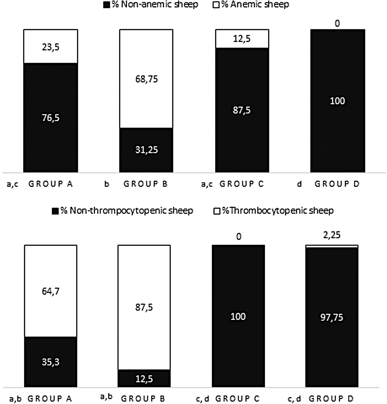
The percentage of sheep presenting anemia and thrombocytopenia within each group and significant differences in percentages among groups.

The percentage of sheep presenting leukopenia, neutropenia, and lymphopenia within each group and significant differences in percentages among groups.
Regarding blood cell morphology, blood smear microscopic evaluation revealed the presence of reactive lymphocytes in seven sheep of group A (41.2%) and activated monocytes in four sheep (23.5%) of the same group. Likewise, reactive lymphocytes in seven sheep (43.75%) and activated monocytes in six sheep (37.5%) were observed in group B. A significantly lower percentage of sheep (12.5%) with only activated monocytes compared with groups A and B was observed in group C. Neither reactive lymphocytes nor monocytes were observed in group D (Fig. 3). Finally, neutrophil toxicity with diffused basophil cytoplasm, scored 1+ (Weiss 1984), was observed in the blood smear of a sheep belonging to group B.

The percentage of sheep that reactive lymphocytes and activated monocytes were detected in their blood smear, within each group and the significant differences in percentages among groups
Discussion
The presence of the A. phagocytophilum antigen without detectable antibodies is indicative of acute infection. Previous studies on the course of antibodies after experimental infection revealed that antibodies can be detected for the first time at day 5 after the infection (Paxton and Scott 1989) in some cases, but mainly at day 7 after the infection, with the highest antibody concentration at about day 14, as has been graphically presented (Stuen and Artursson 2000, Stuen et al. 2009), while the microorganism was detectable from day 3 postinfection (Gokce and Woldehiwet 1999a, Whist et al. 2002). Antibody response has been found to remain at least until 480 days after infection (Paxton and Scott 1989), whereas the microorganism could not be detected in blood smear cytological testing 17 days after the infection (Whist et al. 2002) and in some animals after 175 days (Stuen et al. 1998). The evolution of antibodies after experimental infection and detection of A. phagocytophilum in the blood smear in the course of infection are depicted in Fig. 4. In the same figure, the most probable corresponding position of the infection and/or exposure state of sheep of each group is annotated. Regarding group D, where sheep were found negative in both assays, it can be assumed that either the sheep have never been infected before, excluding an infection in the past with full recovery, or they are in a very early stage of infection.

The period when antibodies against Anaplasma phagocytophilum are detected using IFAT and the period that A. phagocytophilum in blood smear is detected.
Regarding the findings of hematological variables, a lower mean PCV and higher percentage of anemia were observed in sheep found positive in both assays (group B). Anemia was also observed in few animals that were positive either for the antigen or antibody detection. These observations are in accordance with findings of a former study, where the lowest value of the hematocrit was found at day 10 (Gokce and Woldehiwet 1999a), a time point that falls within the period that sheep are expected to be positive for both the antigen and antibodies, as depicted in Fig. 4.
Thrombocytopenia and lower mean PLT count were observed in sheep that were positive for both the antigen and antibodies and, to a lesser extent, in those that were positive only for the antigen. Similarly, the lowest PLT count has been observed between the 2nd and 10th day after experimental infection with A. phagocytophilum (Brun-Hansen et al. 1998, Whist et al. 2002). Among the possible causes of thrombocytopenia, the most possible ones are regarding lymphocyte activation during the acute phase of infection (Brun-Hansen et al. 1998) and the delayed maturation or release of thrombocytes. The latter direct effect of A. phagocytophilum on the bone marrow is strongly supported by the fact that it infects cells of the megakaryocytic lineage (Foster and Cameron 1968, Bakken et al. 1996).
Neutropenia was observed in nearly all sheep found positive in both tests in the present study and in few sheep that tested positive only for antibodies against A. phagocytophilum. The imperceptible neutrophil count alteration in the early stage of infection could be attributed to prolongation of neutrophil half-life that leads to delayed apoptosis due to increased cytokine production (Yoshiie et al. 2000, Scaife et al. 2003). The observed neutropenia at a later stage is, at least partially, the result of cell rupture caused by the parasite. Neutrophil recovery occurs over a period of many weeks with obvious consequences on sheep immune competence and progression of the disease (Larsen et al. 1994).
The mean value of band neutrophils was significantly higher in the group of sheep that were positive for both antigen and antibody detection. In ruminants, the number of mature neutrophils stored in the bone marrow is limited. Thus, in acute inflammatory disease, disorders in the maturation of neutrophils result in the release of band or toxic neutrophils into the blood circulation, as was observed in the present study (Harvey 2012).
The lower eosinophil count in sheep positive for the antigen detection assay and/or antibody has been previously attributed to the rupture of parasitized eosinophils (Whist et al. 2002, Ogden et al. 2003). Moreover, stress and acute infections are often associated with eosinopenia (Jain 1993, Brun-Hansen et al. 1998).
Lymphopenia was observed in sheep positive at least for antigen detection, which is in accordance with previous reports (Gokce and Woldehiwet 1999a, Whist et al. 2002). The significant decrease of lymphocytes mainly concerns circulating B lymphocytes rather than T lymphocytes as was found after experimental infection in sheep using immunoglobulin M and agglutinin for the identification of B and T lymphocytes, respectively (Batungbacal et al. 1982). According to this study, the lymphocyte count decreased from day 7 and returned to normal a week later. These observations were confirmed using monoclonal antibodies, where it was found that A. phagocytophilum causes a decrease in different subpopulations of B (CD4+, CD8+) and T lymphocytes (Woldehiwet 1991). A reduction in lymph, lymphoid tissue of the spleen, and lymph nodes has also been histologically found in animals with anaplasmosis (Hudson 1950). Additionally, an impaired response of lymphocytes of infected sheep to mitogens and inhibition of neutrophil adhesion and phagocytosis have also been reported (Foster and Cameron 1970, Woldehiwet 1987a, 1987b).
Reactive lymphocytes were detected in sheep positive for antigen detection as a result of antigenic stimulation. Although morphological abnormalities have not been previously reported in sheep with anaplasmosis, reactive lymphocytes were found after natural and experimental infection of dogs with Ehrlichia canis and their presence was attributed to antigenic stimulation (Gianopoulos et al. 2016). However, this finding is not specific for Anaplasma since lymphocytes can be stimulated by various antigens and the same holds for activated monocytes (Weiser 2012).
Monocytes seem to be less infected WBCs (Ogden et al. 1998). A lower monocyte count was observed in sheep positive in both assays. Monocyte count reduction has been reported after experimental infection with A. phagocytophilum and it coincided with the induction of fever, while an elevation in monocytes was observed after clinical remission (Tuomi 1967). In inflammatory conditions, an increase in monocyte count is considered to be a result of increased production by the bone marrow to cover the augmented need for phagocytosis (Jain 1993).
Regarding the total WBC count, leukopenia was observed in sheep positive at least in one assay and mainly in sheep positive for antigen detection. The lowest WBC count and the highest percentage of affected animals were reported for sheep positive in both methods. Based on the results, the most prominent leukopenia coincided with the presence of the bacterium in blood. Leukopenia has been previously reported after experimental infection with A. phagocytophilum (Gokce and Woldehiwet 1999a, Whist et al. 2002). Reduction in the WBC count reflects alterations in the number of the different WBC subpopulations, predominantly the neutrophils and lymphocytes.
Concerning the stage of infection, it seems that reduction of blood cells is observed in the acute phase of the disease when sheep are positive in both assays.
Pancytopenia is probably the result of peripheral sequestration, consumption, or destruction of the normal blood elements (Borjesson et al. 2009). In an experimental study in mice, pancytopenia was attributed to the perturbation of normal hematopoiesis and the shift of the bone marrow cell population (Lepidi et al. 2000).
The main limitation of this study was the absence of following up that would give us information on the progression of infection and/or disease. However, this was not possible as it was neither ethical nor acceptable to the farmers to leave the animals untreated.
Conclusions
In this study, quantitative changes in all blood cells, as well as changes in the morphology of WBCs, were found. The results of the complete blood count in combination with cytological and serological tests for detection of the microorganism or antibodies, respectively, can contribute to the assessment of the stage of the disease. From a clinical point of view, the compromised cellular immunity, found during the simultaneous detection of microorganisms and antibodies, shows that during this period, the animals are more susceptible and therefore need to be monitored for early treatment of secondary complications. In addition, the combination of leukopenia and thrombocytopenia is the most important laboratory finding that raises suspicion of the disease.
The quantitative and first reported hematological findings are indicative of an inflammatory reaction, antigenic stimulation, and stress. These findings add to the knowledge of the pathogenetic mechanisms of A. phagocytophilum infection in sheep and highlight the value of the complete blood cell count and evaluation of cell morphology in monitoring disease progression.
Compliance with Ethical Standards
All procedures were performed according to the ethical standards in the Helsinki Declaration of 1975, as revised in 2000, as well as the national law and after receiving approval from our Institutional Animal Use Ethics Committee.
Footnotes
Author Disclosure Statement
The authors declare that they have no conflicts of interest.
Funding Information
No funding was received for this work.