
Abstract
Scientific evidence indicates that hepatitis E virus (HEV) infection is a zoonotic disease. Domestic pigs and wild boars are the main animal reservoir for HEV worldwide. The aim of this study was to analyze the seroprevalence of HEV infection among wild boars in western Bulgaria. Serum samples from 240 wild boars from two regions of the country (northwestern and southwestern) were tested for anti-HEV Immunoglobulin G (IgG) antibodies. The overall HEV seroprevalence was 40.8% (98/240); northwestern region 40.0% (48/120); southwestern region 41.7% (50/120). HEV seropositivity in the southwestern region was higher than in the northwestern region: odds ratio = 1.071 (95% confidence interval: 0.640–1.793). This research provided the first seroprevalence study to HEV in wild boars from western Bulgaria.
Introduction
Hepatitis E virus (HEV) is a small nonenveloped virus with a size of 27–32 nm and a single-stranded positive-sense RNA genome of 7.2 kb that encodes three overlapping open reading frames (ORFs): ORF1, ORF2, and ORF3 (Purdy et al. 2017, ICTV 2019, Smith et al. 2020). HEV is classified into Hepeviridae family, divided into two genera: Orthohepevirus and Piscihepevirus (Purdy et al. 2017, Smith et al. 2020). Genus Orthohepevirus include four species: Orthohepevirus A, Orthohepevirus B, Orthohepevirus C, and Orthohepevirus D (ICTV 2019, Pepovich et al. 2019). Genus Piscihepevirus has only one species—Piscihepevirus A, and one genotype—Cutthroat Trout HEV (Purdy et al. 2017).
In recent years, HEV has been increasingly common in humans and animals. According to the World Health Organization, an estimated 3.3 million symptomatic hepatitis E cases occur each year in endemic areas with 44,000 related deaths (WHO 2020). Li et al. (2020) reported that 939 million people worldwide have been infected with HEV in the past and that 15–110 million people have recent or ongoing infections. Scientific evidence indicates that hepatitis E is a zoonotic disease. Domestic pigs and wild boars are the main animal reservoir for HEV worldwide (Pallerla et al. 2020). Besides swine, anti-HEV antibodies have also been detected in many other animal species—deer, rats, dogs, cats, mongooses, cows, sheep, goats, avian species, rabbits, horses, camels, etc. (Pallerla et al. 2020).
In Bulgaria, the first preliminary data for swine HEV seroprevalence were reported in 2018, and showed an overall seroprevalence of anti-HEV antibodies in swine of 40% (34 positive samples out of all 85 sera) (Pishmisheva et al. 2018, Baymakova et al. 2019). In 2019, a detailed seroprevalence study of HEV infection in pigs from southern Bulgaria documented an overall HEV seroprevalence of 60.3% (217 of all 360 tested sera) (Tsachev et al. 2019). Takova et al. (2020) reported 4 of the 32 meat juice samples from Bulgarian wild boar were positive for anti-HEV Immunoglobulin G (IgG) antibodies. The study of these authors had several important limitations: the research included a relatively small number of wild boars (n = 32) and information regarding their age and gender were not recorded. A recent study estimated that the overall HEV seroprevalence among the only aboriginal pig breed in Bulgaria (East Balkan swine) was 82.5% (141/171) (Tsachev et al. 2020).
However, there is no information about HEV infection in wild boars from western Bulgaria. Thus, the aim of this study was to investigate the seroprevalence of HEV infection in wild boars in two parts of the country (northwestern and southwestern).
Materials and Methods
Two hundred forty serum samples were collected from wild boars (n = 240) and were enrolled from eight districts in two parts of the country—northwestern region (Vidin, Montana, Vratsa, and Lovech districts) and southwestern region (Blagoevgrad, Kyustendil, Sofia province, and Pazardzhik districts). The serum samples were collected from wild boars during the official hunting season (from November 2019 to February 2020). Information regarding their gender and age was not recorded. An equal number of serum samples (n = 30) were taken from each district. The wild boars selection in districts, respectively, in regions was done randomly. The geographic distribution of the investigated wild boars was among mountains and hilly areas in western Bulgaria (∼22°66′E and 25°05′E longitude; 41°39′N and 44°15′N latitude); the climate is continental (mean annual temperature 13–15°C, precipitations ∼650 mm/m2). The study was conducted according to the ethical principles of animal experimentation, adopted by the Bulgarian Ministry of Agriculture, Food, and Forestry.
The blood samples (up to 5 mL per individual) were collected from the chest cavity of the shot wild boars. Blood collection tubes without anticoagulant were kept at room temperature (20°C) until visible clot retraction, centrifuged at 1500 g for 10 min, and the serum was separated and kept at −20°C until processing.
The wild boars serum samples were tested for anti-HEV IgG in the Department of Microbiology, Infectious and Parasitic Diseases, Faculty of Veterinary Medicine, Trakia University, Stara Zagora, Bulgaria. A commercial enzyme-linked immunosorbent assay (ELISA; PrioCHECK HEV Ab porcine; Mikrogen GmbH, Neuried, Germany) was applied. The used test detected HEV-specific antibodies in swine serum and meat juice samples. Microtiter plate is coated with recombinant HEV antigen of the ORF2 and ORF3 of genotypes HEV-1 and HEV-3. ELISA test PrioCHECK HEV Ab porcine has 91.0% sensitivity and 94.1% specificity. According to the manufacturer's instructions (Mikrogen GmbH), the cutoff was calculated and a mean optical density 450 (OD450) of the cut-off control multiplied with 1.2. Results obtained above or equal to the cutoff are considered positive. Results between the OD450 of the cutoff control and the cutoff are doubtful. Results obtained below the cutoff are negative.
Chi-square test was applied for comparing anti-HEV IgG positivity among different regions. Binary logistic regression was applied for evaluation of the risk of positive results according to regions. Statistical analysis has been performed by MS Excel 2007 (Washington) and IBM SPSS Statistics 21.0 (New York). A p value <0.05 was considered statistically significant.
Results
Anti-HEV IgG was positive in 98 (40.8%) of all 240 tested serum samples. The total HEV-positive seroprevalence in the northwestern region was 40.0% (mean ± standard deviation [SD]: 40.0 ± 25.3; 95% confidence interval [CI]: 15.2–64.8), and that in the southwestern region was 41.7% (mean ± SD: 41.7 ± 28.5; 95% CI: 13.7–69.6). The highest anti-HEV-positive prevalence was in Blagoevgrad district (22/30, 73.3%) (Fig. 1). In contrast, the lowest anti-HEV-positive prevalence was found in both districts Montana and Pazardzhik (3/30, 10.0%). Based on districts and different regions, the chi-square test showed differences in HEV seropositivity between the districts and regions (Table 1). Three percent of all samples was assessed as doubtful. The odds ratio (OR) in different regions was calculated to estimate the risk for HEV seropositivity using a binary logistic regression. The OR of anti-HEV antibodies occurrence in the southwestern region was determined comparing with the northwestern region (Table 2). The odds of HEV seroprevalence were 1.071 times higher in the southwestern region than in the northwestern region. In this study, HEV RNA testing was not performed.
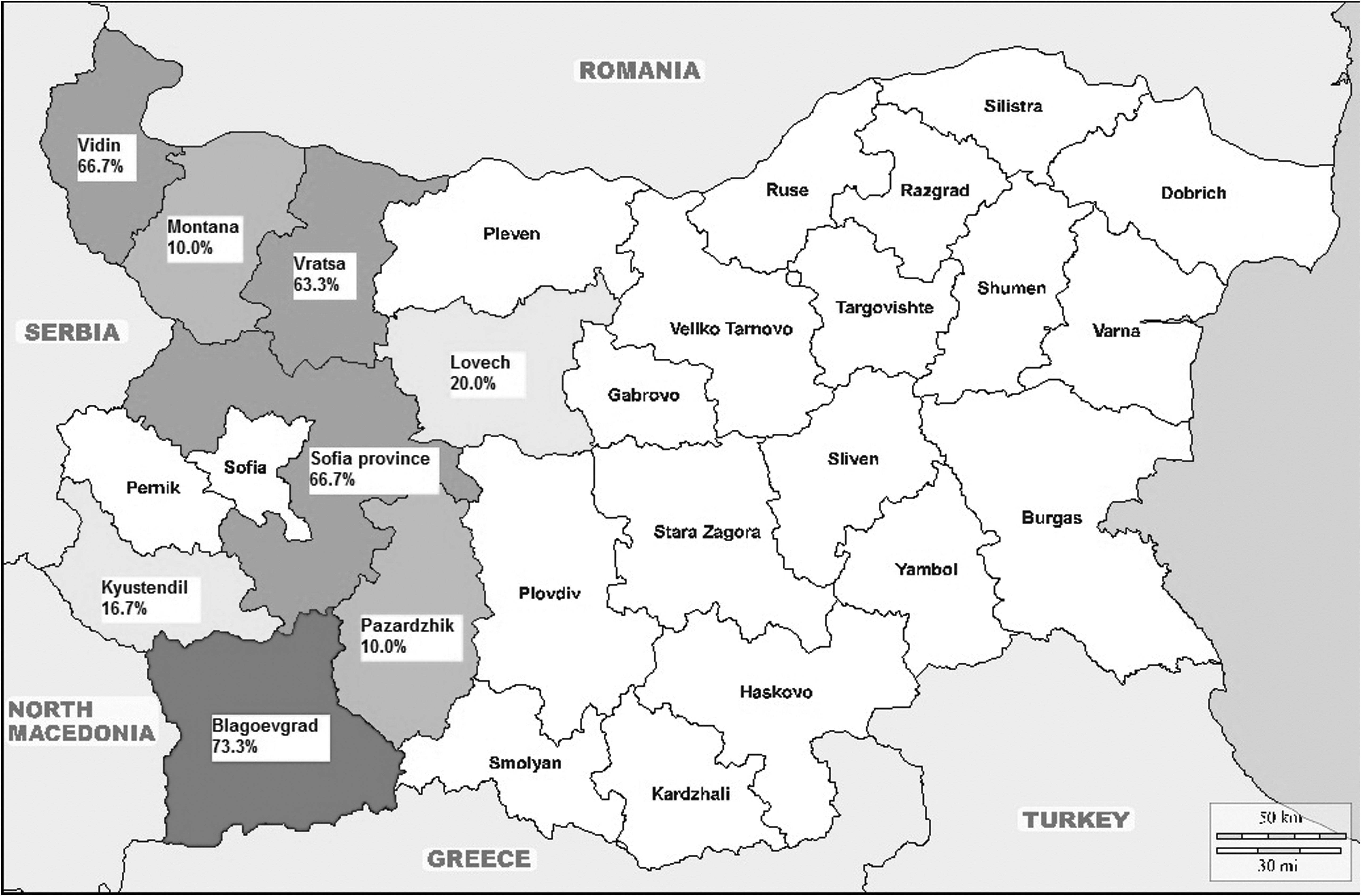
Geographic distribution of HEV infection in wild boars from western Bulgaria. HEV, hepatitis E virus.
Seroprevalence of Hepatitis E Virus Infection by Districts in Wild Boars from Western Bulgaria
df, degrees of freedom; HEV, hepatitis E virus.
Logistic Regression Showing the Relationship Between Hepatitis E Virus-Positive Wild Boars and Geographic Region
CI, confidence interval; NA, not applicable; OR, odds ratio; PE, parameter estimate; SE, standard error.
Discussion
Mild and moderate HEV seropositivity among wild boars was observed in countries from the Balkan Peninsula. In northern Turkey, 100.0% anti-HEV IgG negative results (93/93 serum samples) were found (Albayrak et al. 2013). The Romanian authors presented total anti-HEV seroprevalence of 9.61% (5/52) among wild boars from eastern Romania (Porea et al. 2015). An original article from Slovenia reported 30.21% (87/288) HEV seroprevalance in wild boar serum samples (Zele et al. 2016). The Croatian researchers found 31.10% (311/1000; 95% CI: 28.31–34.04) HEV seropositivity among wild boars in 6 of the 16 counties from their country (Jemersic et al. 2017).
In the southwestern European countries, the seroprevalence rates of HEV infection among wild boars varied. In Italy was observed the following positivity: 4.9% (29/594; 95% CI: 3.3–6.9) (Caruso et al. 2015), 10.2% (226/2211; 95% CI: 9.0–11.5) (Martinelli et al. 2015), 40.7% (93/228; 95% CI: 34.4–47.1) (Montagnaro et al. 2015), and 49.7% (186/374; 95% CI: 44.6–54.8) (Bertelloni et al. 2020). In France was reported the following HEV seropositivity: 14.0% (59/421) (Carpentier et al. 2012) and 29.2% (101/346; 95% CI: 24.5–34.4) (Jori et al. 2016). Moderate HEV seropositivity among wild boars was observed in the Iberian Peninsula: 26.3% (248/942; 95% CI: 23.5–29.2) (Boadella et al. 2012) and 28.0% (42/150) (de Deus et al. 2008).
In the other western European countries, there was a varied distribution of HEV infection in wild boars. Low HEV prevalence was reported from Netherlands (12% of 1029 wild boar) (Rutjes et al. 2010) and Switzerland (12.5%; 38/303; 95% CI: 9.1–16.9) (Burri et al. 2014). Moderate and high HEV spread was observed in Belgium (34.0%; 130/383; 95% CI: 29.7–39.4) (Thiry et al. 2017) and Germany (45.0%; 81/180) (Anheyer-Behmenburg et al. 2017). Also in countries from eastern Europe, different HEV seropositivity has been reported among wild boars. In the Czech Republic was observed a positivity of 8.5% (31/366) (Strakova et al. 2018); Estonia, 17.2% (81/471) (Ivanova et al. 2015); Poland, 44.4% (116/261) (Larska et al. 2015); and Lithuania, 57.0% (178/312) (Spancerniene et al. 2016).
In the countries from other continents, low and moderate HEV seroprevalence has been found. In Japan was reported the following HEV seropositivity: 8.1% (41/507) (Sato et al. 2011) and 25.5% (100/392) (Michitaka et al. 2007), China: 24.5% (186/758) (Liang et al. 2019), and Uruguay: 22.1% (31/140) (Mirazo et al. 2018).
The seropositivity in this study (40.8%) is similar to the results found in Italy (40.7%) (Montagnaro et al. 2015), Poland (44.4%) (Larska et al. 2015), and Germany (45.0%) (Anheyer-Behmenburg et al. 2017). The data from our research showed a high HEV seropositivity among wild boars in some districts from western Bulgaria (Vidin, Vratsa, Blagoevgrad, and Sofia province), and low seroprevalence in others (Montana, Lovech, Kyustendil, and Pazardzhik). The reasons for this could be different. On one hand, it is well known that positivity is lowest among age group “juveniles” and highest among age group “adults” (Carpentier et al. 2012, Burri et al. 2014). Another reason for these variabilities in different districts could be the month of the year, when the wild boar samples were collected. Probably, at the beginning of the hunting season (November), the population of juveniles was large and, therefore, the seropositivity was low. Conversely, most likely at the end of the hunting season (February), the selected samples showed higher seropositivity, because the population of adults is larger than the population of juveniles. On the other point of view, the applied ELISA diagnostic test (PrioCHECK HEV Ab porcine; Mikrogen GmbH) could influence on positive results (40.8% in this study). We think it is unlikely because results from Germany and Italy found low total HEV seroprevalence of 11.5% (12/104) and 4.9% (29/594; 95% CI: 3.3–6.9), respectively (Caruso et al. 2015, Weigand et al. 2018).
This research has some limitations that need to be addressed. Information regarding gender and age among wild boars was not recorded. Moreover, the study included a relatively small number of pigs and confirmatory immunoblot test was not performed (and also HEV RNA testing). Despite these limitations, this is the first seroprevalence study on the HEV infection among wild boars in western Bulgaria, which gives new insights for this infection in our country.
In conclusion, we present the first serological research of HEV infection among wild boars in western Bulgaria. Our results of this study showed high HEV seropositivity (40.8%) among wild boars in this part of the country. Thus, national veterinary institutions should take proper measures for monitoring and control of this infection among wild boars in Bulgaria.
Footnotes
Authors' Contributions
I.T., M.B., and R.P. contributed to study design, data collection, data interpretation, article preparation, literature search, and funds collection. P.M. and V.P. were involved in data collection and funds collection. K.G. carried out laboratory analysis. T.K. carried out statistical analysis. All authors read and approved the final version of the article.
Acknowledgment
We thank Dr. Emilia Ivanova (National Diagnostic Science and Research Veterinary Medical Institute, Bulgarian Food Safety Agency) for collecting blood samples from wild boars.
Author Disclosure Statement
No competing financial interests exist.
Funding Information
This research was funded by Trakia University, 6000 Stara Zagora, Bulgaria, Grant Number 08/2020.