
Abstract
Background:
Assessing the potential for mosquitoes to transmit medically important arboviruses is essential for understanding their threat to human populations. Currently, vector competence studies are typically performed by collecting saliva using a glass capillary tube system which involves sacrificing the mosquito at the time of saliva collection allowing only a single data point. These techniques also require handling infected mosquitoes and glass capillaries, constituting a safety risk.
Materials and Methods:
To improve the efficiency and safety of assessing vector competence, a novel containment and saliva collection approach for individually housed mosquitoes was developed. The improved housing, allowing longitudinal tracking of individual mosquitoes, consists of a 12-well Corning polystyrene plate sealed with a three-dimensional printed lid that holds organdy netting firmly against the rims of the wells.
Results:
This method provides excellent mosquito survival for five species of mosquitoes, with at least 79% of each species tested surviving for more than 2 weeks, comparable to the carton survival rates of ≥76%. When the plate housing system was used to assess vector infection, replication of West Nile virus (WNV) in mosquito tissues was similar to traditional containment mosquito housing. Mosquito saliva was collected using either blotting paper pads or traditional glass capillaries to assay viral transmission. The blotting paper collection showed similar or better sensitivity than the capillary method; in addition, longitudinal saliva samples could be collected from individual mosquitoes housed in the 12-well plates.
Conclusions:
The improved housing and saliva collection technique described herein provides a safer and more informative method for determining vector competence in mosquitoes.
Introduction
Mosquito-borne arboviruses are endemic on six continents, and half of people on earth are at risk of contracting illnesses caused by arboviruses (Leta et al., 2018). Many arboviruses have the potential to cause future epidemics through range expansion of host mosquitoes, increased vector competence, or adaptation to additional vector species (Braack et al., 2018; Ruckert and Ebel, 2018). To understand potential risks for arboviral emergence, characterizing the ability of various species of mosquitoes to transmit priority agents is critical.
An important measure of vector competence is whether virus is expelled during feeding, which can be determined by collecting saliva (Hurlbut, 1966; Ruckert and Ebel, 2018). A common method is to artificially induce the mosquito to salivate by inserting the proboscis into a glass capillary tube filled with immersion oil, fetal bovine serum (FBS), cell culture media, or blood (Aitken, 1977; Anderson et al., 2010; Collins, 1963; Colton et al., 2005; Hurlbut, 1966; Ledermann and Powers, 2016). However, this method has inherent safety risks, including possible percutaneous hand puncture from the glass tubes or scalpels, creation of aerosols during mosquito dissections, and accidental mosquito release while handling. In addition, when using the capillary method, mosquitoes are sacrificed upon saliva collection, thus only a single data point is collected and timing of the sample collection may dramatically affect results (Ledermann and Powers, 2016; Ruckert and Ebel, 2018). While multiple cohorts of mosquitoes can be tested on different days, there is no potential to study how the salivary viral load varies longitudinally in individual mosquitoes.
There is growing interest in alternative saliva collection methods to increase efficiency for studies on extrinsic incubation periods, virus evolution, surveillance, and vector competence. Methods have been developed to exploit mosquitoes' attraction for sugar to entice them to salivate onto a sugar-soaked substrate that is subsequently analyzed for viral RNA (Burkett-Cadena et al., 2016; Burkhalter et al., 2018; Flies et al., 2015; Fourniol et al., 2021; Fynmore et al., 2021; Grubaugh et al., 2017; Hall-Mendelin et al., 2010; Honorio et al., 2020; Koh et al., 2018; Melanson et al., 2017; Ye et al., 2015).
Experiments performed by Hall-Mendelin et al. (2010) described the use of field deployed CO2 traps fixed with honey-soaked FTA cards as a method to detect circulating arboviruses for surveillance. In laboratory settings, FTA cards or drops of sucrose solution have been used to collect saliva from individual flavivirus-infected mosquitoes for longitudinal studies (Grubaugh et al., 2017; Koh et al., 2018). Fourniol et al. (2021) individually housed Aedes aegypti and Aedes albopictus mosquitoes in 50 mL conical tubes and collected chikungunya virus-infected saliva using filter paper. However, this format becomes cumbersome as the number of mosquitoes increases. Dyed, honey-soaked University-Storage and Transport Optimized Platform (U-STOP) cards have also been used to aid in detecting ingestion of a sugar meal (Burkhalter et al., 2018).
Many of these studies used FTA cards which contain sodium dodecyl sulfate and ethylenediaminetetraacetic acid (EDTA) to preserve RNA (Burgoyne, 1996). These chemicals are toxic to Anopheles and Aedes genera mosquitoes eliminating the possibility of further saliva collections and allowing only a single time point assessment (Supplementary Fig. S1) (Alto et al., 2017). However, Grubaugh et al. (2017) were able to obtain longitudinal saliva samples from Culex genus mosquitoes using FTA, perhaps due to differences between genera or sugar meal preparation.
To facilitate longitudinal collections of arbovirus-infected mosquito saliva to understand critical components of vector competence, an improved system was developed. This method individually houses infected mosquitoes in a 12 well cell-culture plate using a three-dimensional (3D) printed lid to hold netting flush against the wells. During regular sugar feeding, the mosquitoes deposit saliva in blotting paper pads placed on the exterior of the netting each time they take a sugar meal, which can then be used to detect virus from infected mosquito saliva. Changing both the housing and the saliva collection method from the capillary technique enhanced biosafety and allowed for longitudinal monitoring of virus transmission in individual mosquitoes significantly increasing the data that can be collected from a single infection study.
Materials and Methods
Culture plate housing
Mosquitoes were housed individually in single wells of Costar 12-well plates (Corning 3512) with fine organdy netting held firmly against the tops of the wells by 3D printed lids and rubber bands (Fig. 1). The lids were designed in SketchUp (Google) and exported as .stl files (Supplementary Files S1 and S2). The main body of the lid, 6.0 mm thick, provides a flat surface to hold organdy netting firmly against the wells along with 2.0 mm legs that fit into small recesses of the plate to prevent shifting. Grooves, 4.0 mm wide, on top of the plate and around the edge, fit the rubber bands that hold the lid to the plate and the organdy to the lid. Holes in the lid (14.4 mm diameter) were centered above each 22 mm diameter well of the 12-well plate. The lid fits entirely within the outer lip of the plates. The lids were printed on a MakerBot Replicator 2X with MakerBot ABS (acrylonitrile butadiene styrene; MP01970) plastic filament with 10% diamond infill. The lids were 120.0 × 78.5 × 8.0 mm and weighed 19 g when printed. Additional information on experimental variables investigated is available in the Supplementary Table S1 and Supplementary Figs. S3–S9.

Twelve-well plate individual mosquito housing. Photograph of the disassembled and assembled housing setup, including Corning 12-well plate, 3D printed lid (120.0 × 78.5 × 8.0 mm outer dimensions, 14.4 mm diameter holes), organdy fine mesh, rubber bands, and 5 × 5 mm GB003 blotting paper squares. The organdy is clamped between the plates and secured by a horizontal rubber band. The lid is secured by two transverse rubber bands. Blotting paper is deposited into the holes in the lid and rests on top of the organdy. 3D, three-dimensional.
Safety assessment
A comparative safety analysis of the original glass capillary procedure and the new procedure for mosquito saliva collection in blotting paper in the novel housing was performed by Centers for Disease Control and Prevention (CDC)'s Division of Vector-Borne Diseases Safety and Occupational Health Specialist. Both methods were presented simultaneously with live mosquitoes under noninfectious conditions and were evaluated to identify any hazards associated between the researcher, the procedure, equipment utilized in the task, the arthropod, and the potential virus being collected.
Virus isolate
The virus used for laboratory mosquito infections, West Nile virus (WNV) strain R98432a, was obtained from the Arbovirus Reference Collection at CDC, Division of Vector-Borne Diseases (DVBD), Fort Collins, CO. This strain was isolated in 2012 from a human residing in Colorado and was passaged twice in Vero cells.
Mosquito species
Colonized mosquito species used were Ae. albopictus (Lake Charles [LC], LA) (Nasci et al., 1989), Ae. aegypti (Rexville-D) (Mitchell et al., 1987), Culex tarsalis (Kern National Wildlife Refuge [KNWR], CA) (Danforth et al., 2015), Culex quinquefasciatus (Sebring) (Kent et al., 2010), and Anopheles gambiae (G3) (BEI Resources, 2019).
Mosquito survival experiments
To assess adult survivorship inside the individual housing system, 1- to 3-day-old adult females of each of the five mosquito species were either individually contained in two 12-well plates per species, totaling 24 mosquitoes, or as cohorts of 30 individuals in pint-sized cartons. Each was held for 30 days at 28°C and 80% humidity with 12-h light/12-h dark photoperiods.
The mosquitoes in the plates were fed from squares (5 × 5 mm) of Whatman blotting paper grade GB003 (GE cat. no. 10427804) soaked in 10% sucrose solution placed on the organdy netting above each individual well. This blotting paper has a superior ability to retain the sugar solution compared with other tested products (Supplementary Table S1). Twenty-five microliters of a 10% sucrose solution was applied daily to the blotting squares, and the squares were replaced every 2–3 days. Carton-housed mosquitoes were provided 10% sucrose solution on 10 × 10 cm gauze pads, which were refreshed daily and replaced at the same time as the blotting paper pads.
Mortality from each housing system was recorded on a daily basis. Kaplan–Meier survival curves were plotted with Excel. MStat 6.1.2 software was used to perform two-tailed log-rank test to evaluate the survival curves (Drinkwater, 2011).
Vector competence studies
Three-to-four-day-old adult Cx. tarsalis (KNWR) mosquitoes held in cartons were allowed to feed on bloodmeals containing WNV (R98432a) at either 7.2 or 8.1 log10 plaque-forming units (pfu)/mL. The bloodmeals contained one part virus diluted in Dulbecco's minimal essential medium (DMEM), one part FBS with 10% sucrose, and one part chicken erythrocytes (Colorado Serum Co.) washed with phosphate-buffered saline and packed by centrifugation (Brault et al., 2004; Ledermann and Powers, 2016). A feeding system (Hemotek) was used to deliver the heated bloodmeal (37°C) to the mosquitoes for 1 h. After the feeding period, the mosquitoes were cold anesthetized, and fully blood-engorged females were collected and separated individually into wells of three 12-well plates per condition or grouped into cohorts of 30 in pint-sized cartons.
Females that had engorged on a noninfectious bloodmeal and nonengorged females were placed into 12-well plates to serve as untreated controls. The respective housing containers were placed in a humidified environmental chamber (80% relative humidity) and held at 28°C for 8 or 12 days until processing. The blotting paper sugar meal for individuals or gauze pad sugar meal for the cartons was replaced and/or refreshed every 24 h. Beginning on 3 or 4 days postinfection (dpi), the blotting papers were collected daily.
After the allotted incubation period of 8 or 12 dpi, mosquitoes were cold anesthetized for ∼5 min, legs and wings were removed, and the mosquitoes were induced to salivate into immersion oil in glass capillaries as previously described (Ledermann and Powers, 2016; Ledermann et al., 2017). For the mosquitoes housed in the plates, 2–3 additional min at 4°C were required to sedate the insects, as the plate setup insulated them slightly better than the carton housing. After salivation, heads and bodies were separated. Tissues were ground with micropestles in 400 μL of DMEM (Gibco) supplemented with 10% FBS, 100 U/mL of penicillin and streptomycin, and 1 U/mL of fungizone and gentamycin and filtered as previously described (Ledermann and Powers, 2016; Mossel et al., 2013). Samples were stored at −80°C until processing for virus testing.
Viral nucleic acid extraction and detection
Capillary saliva collections were centrifuged at 10,000 g and 4°C for 1 min before RNA extraction to excise the contents of capillary tubes into 500 μL of supplemented DMEM. Total RNA was extracted from 140 μL of each mosquito homogenate and capillary saliva using the QIAamp Viral RNA Kit according to the manufacturer's instructions before elution from the columns using 100 μL of elution buffer. The RNA was stored at −80°C. To elute virus from the blotting paper, each pad was placed into a 1.7 mL Sarstedt tube with 100 μL of Tris-EDTA buffer (10 mM Tris, 1 mM EDTA, pH 8.0; FisherBiotech BP1338-1). The sample was held at 28°C for 30 min and vortexed every 10–15 min. From this eluate, 10 μL was directly analyzed by quantitative reverse transcription polymerase chain reaction (RT-qPCR).
The QuantiTect Probe RT-qPCR Reagent Kit (Qiagen) was used to detect WNV nucleic acid from all sample types. A previously described WNV ENV specific primer and probe set was used with an experimentally determined detection threshold of cycle threshold (Ct) ≤39.7 (Lanciotti et al., 2000). Known standards were included, and a curve correlation coefficient of ≥0.950 and 90–110% PCR efficiency were used to validate each detection assay (Ledermann et al., 2011). Fisher's two-tailed exact test was performed in GraphPad Prism 7.05 to compare WNV positivity between plates and cartons and compare the sensitivity of saliva collection methods.
Results
Comparison of housing methods
To determine if the individual plate housing induced stress on the mosquitoes that might lead to increased mortality, the 12-well plate mosquito housing was used to house 5 species of mosquitoes representing 3 genera for 30 days (Fig. 2). The survival of mosquitoes in plates (2 plates per species) was compared with the standard carton housing method in which 30 mosquitoes were housed together in a pint carton sealed with organdy.
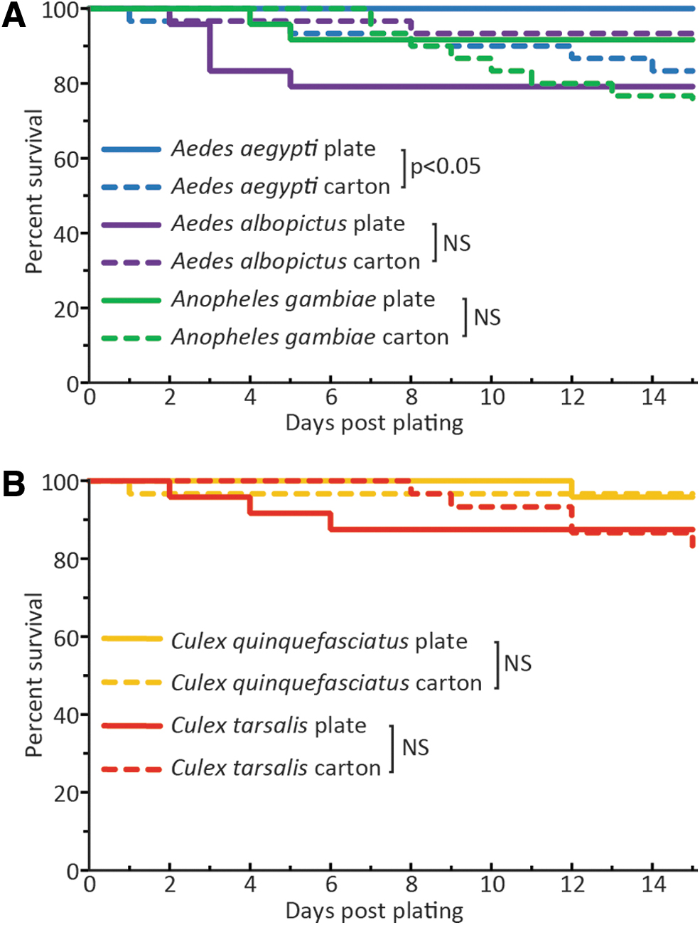
Mosquito survival in plates versus cartons for experimental study period. Mosquitoes representing three genera [Aedes and Anopheles
For the first 15 days (the typical length of a mosquito infection experiment), there was no significant difference between survival in plates versus cartons for Ae. albopictus, An. gambiae, Cx. quinquefasciatus, or Cx. tarsalis. Ae. aegypti in plates survived better than those in cartons (p = 0.04). Less than 25% of mosquitoes died in either housing system over 15 days. Across 30 days, there was considerably higher mortality, and the results were more variable (Supplementary Fig. S2). Across this longer time course, most mosquito species had no significant difference in survival between cartons and plates, although Cx. tarsalis survived significantly longer in plates (p = 0.003) and Ae. albopictus survived significantly longer in cartons (p < 0.001).
Safety assessment
A risk analysis review performed by a CDC safety specialist found that both the capillary and blotting paper saliva collection procedures had a low to moderate risk of percutaneous injury to the hands with the use of fine tip metal forceps during the initial selection of engorged mosquitoes (Supplementary File S3). However, in the newly described procedure, this risk was mitigated by the absence of manual mosquito manipulations to remove mosquito wings and legs with metal forceps or scalpels. In addition, the blotting pad method has fewer steps on open bench tops, which lack secondary containment measures. The original capillary collection procedure also was found to include a low to moderate risk of aerosolization of potentially infectious media or saliva and the possibility of injury by broken capillary tubes.
Conversely, the new procedure incorporated additional engineering controls designed to physically prevent injury or exposure of the operator. With no glass, the blotting paper method eliminated the risk created by capillary tubes which, if longer than 35 mm, must be snapped off to fit into a microfuge tube to expel the contents, a process that can generate micro shards of glass. In addition, the assembled plate served as primary containment for both the mosquitoes and the blotting paper with potentially infectious saliva. The assembled plate housing allowed for handling and collection of the mosquito saliva within a class II A2 Biological Safety Cabinet where capillary saliva collections could not feasibly be performed.
Detection of WNV in plate versus carton-housed mosquitoes
Because the plate housing may have affected viral infection patterns within the mosquito, infection and dissemination rates were compared between those housed in the plates with those in the traditional cartons. Three to four-day-old Cx. tarsalis were given a bloodmeal containing either 7.2 or 8.1 log10 pfu/mL of WNV. The heads, bodies, and capillary-collected saliva were assayed for the presence of detectable viral RNA at 8 and 12 dpi by RT-qPCR (Fig. 3A, B). Both the plate and carton method showed that the 8.1 log10 bloodmeal, which was consistent with titers found in birds (Komar et al., 2003), produced substantially higher WNV infection and dissemination rates than the 7.2 log10 group at both timepoints, as expected (Fig. 3C, D) (Reisen et al., 2006). The housing method made no statistically significant difference in the percent of heads or bodies infected or capillary-collected saliva at either time point or with either infectious dose.
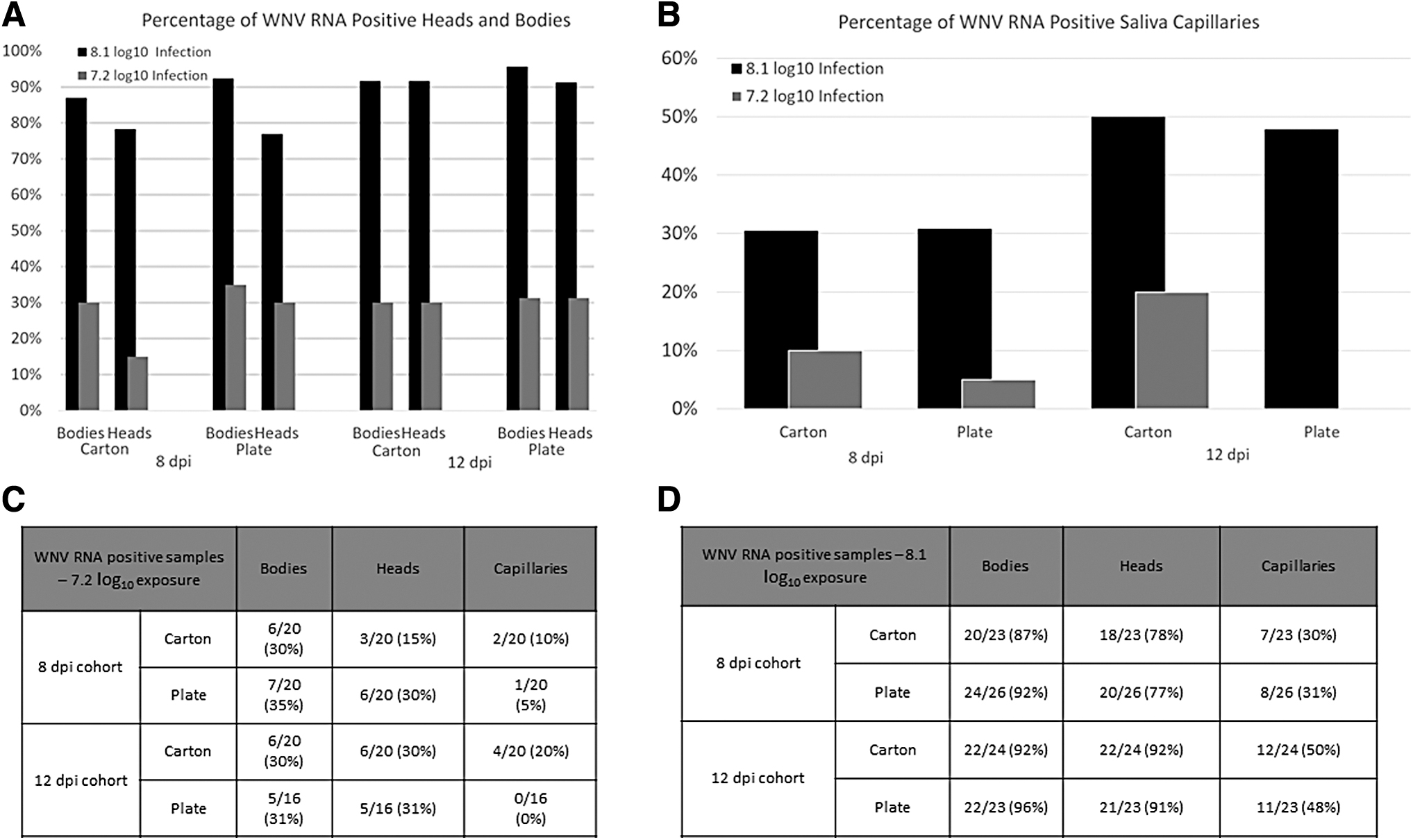
Infection rates for Culex tarsalis adult mosquitoes held in either 12-well culture plates or pint-sized cartons. Mosquitoes were exposed to either 7.2 or 8.1 log10 pfu/mL of WNV.
Comparison of WNV RNA detection in saliva from capillary and blotting paper collection
To assess if viral RNA in saliva could be detected in blotting paper at least as well as from capillary tubes, blotting paper pads from the plates were collected daily on days 4–12 postinfection during the experiments described above (Fig. 4A). In the 7.2 log10 pfu/mL bloodmeal group, the one mosquito in the 8 dpi group that had WNV RNA positive saliva by capillary collection also produced a series of WNV RNA positive blotting paper samples (Fig. 4B). One mosquito in the 12-day cohort provided a series of WNV RNA positive blotting papers without producing a positive capillary sample (Fig. 4C).

WNV RT-qPCR positive samples from mosquitoes individually housed in 12-well plates.
Mosquitoes infected with the 8.1 log10 pfu/mL bloodmeal had 8/26 (31%) WNV RNA positive saliva samples by the capillary collection method in the 8 dpi cohort (Fig. 4D). Comparably, blotting papers collected on day 8 from these same mosquitoes had a positivity rate of 7/26 (27%), which was not significantly different (p > 0.9999). The capillaries and blotting papers collected from the 8.1 log10 pfu/mL bloodmeal and 12 dpi plated mosquitoes showed similar trends with 11/23 (48%) and 12/23 (52%) positive, respectively, demonstrating no significant difference (p > 0.9999) in detecting WNV RNA in saliva samples due to capillary or blotting paper collection (Fig. 4E).
Sugar papers were collected daily from the 12-well plan housing which allowed for observation of longitudinal data from individual mosquitoes (Fig. 4B–E). Of the papers collected from the high dose, 8 dpi cohort, 12/26 (46%) were PCR positive on at least 1 collection day between days 3–8 dpi (Fig. 4D). All the mosquitoes in the 8 dpi cohort that had WNV-positive blotting paper samples had disseminated infection in their heads as well. In addition, four mosquitoes produced WNV RNA positive blotting papers on multiple days. When examining data from the high dose, 12 dpi cohort plates, 20/23 (87%) mosquitoes produced at least one WNV RNA positive blotting paper sample, which is significantly more than the 11/23 (48%) positive by capillary collection (p = 0.01) (Fig. 4E). Sixteen mosquitoes were positive on multiple collection days (Fig. 4E). Furthermore, over half of the mosquitoes (12/23) produced WNV RNA positive blotting paper pads on 3 or more consecutive days, suggesting the range of days in which the individual mosquitoes could transmit this virus.
Discussion
Based on work demonstrating that sucrose solution-soaked paper substrates could be used to identify virus-transmitting mosquitoes (Alto et al., 2017; Fourniol et al., 2021; Grubaugh et al., 2017; Hall-Mendelin et al., 2010; Honorio et al., 2020; Ledermann and Powers, 2016), a saliva collection method for use in studies on vector competence, extrinsic incubation period, and virus population genetics that would improve upon the traditional glass capillary mosquito saliva collection method was developed. This study provides optimized conditions to save each individual laboratory the trouble of doing it separately. The biggest innovation is in the improved housing that increases safety and capacity for housing infected mosquitoes, by allowing more mosquitoes per cage in an incubator and greater speed in the setup.
Most of these studies used FTA card substrate, which we (Supplementary Fig. S2) and others (Alto et al., 2017) have found to be lethal to Anopheles and Aedes genus mosquitoes, although Grubaugh et al. (2017) were able to get longitudinal data from Culex genus mosquitoes with it . Alto et al. (2017) utilized filter paper, whereas the blotting paper, Whatman GB003, that was selected for this study was optimized for liquid retention and handling characteristics (Supplementary Table S1). In addition, in Alto et al., cohorts of mosquitoes were used, and so the overall setup was substantially different from the combination of high-density individual housing, blotting paper, and longitudinal collection reported here.
The experimental design in Fourniol et al. (2021) was most similar to that described above, but used 50 mL conical tubes for housing and honey solutions on a different type of filter paper. Over the course of a typical 2-week experiment, there was no significant decrease in survival for 5 species of mosquitoes in the 12-well plates compared to carton housing. Similar WNV infection rates were detected in heads, bodies, and capillary-collected saliva from mosquitoes housed in plates and cartons indicating that there was no undue stress that affected the overall viral infection and dissemination patterns.
To evaluate the utility of the blotting paper technique in comparison with traditional capillary collection of saliva, WNV-infected Cx. tarsalis were tested using RT-qPCR at multiple collection times using both systems. RT-qPCR was selected for its high sensitivity and specificity. The results in this study suggested that both methods of saliva collection are comparable in their sensitivity at a single terminal timepoint.
However, when analyzed longitudinally, the blotting paper method demonstrated a higher inherent sensitivity by expanding the range of detection days. This temporal resolution could be of substantial benefit in studies on the extrinsic incubation period of arboviruses in mosquitoes. When assaying the blotting paper samples longitudinally, by 8 dpi, 50% more infectious saliva samples were detected than by the capillary collection (12 vs. 8 of 26), and nearly twice as many by 12 dpi (20 vs. 11 of 23). No positive blotting papers came from mosquitoes lacking disseminated infection.
Other researchers also report moderate sensitivity from paper substrates (Alto et al., 2017; Burkhalter et al., 2018; Fourniol et al., 2021; Grubaugh et al., 2017; Honorio et al., 2020) but those systems do not have the high throughput capacity and convenience of the compact 12-well plate housing. Transmissibility of WNV from Cx. tarsalis peaked at 9 dpi with the 8.1 log bloodmeal (Fig. 4E), consistent with previous reports (Danforth et al., 2015; Reisen et al., 2006). Distinct virus-vector combinations may behave differently when using this housing and blotting paper method compared with capillary collection of saliva so preliminary testing with other species would be warranted. In addition, recent work suggests that capillary collections may underestimate transmission rates, so comparing the sensitivity of longitudinal collections to other proxy measurements is warranted (Gloria-Soria et al., 2022).
The safety advantages of this compact housing approach are significant. Including the experiments described here and throughout the optimization phase, about 1000 mosquitoes were housed in the 12-well plate system and not a single mosquito escaped from its appointed well. The transparent plates allow for immediate visualization of the security of each mosquito. Live infectious mosquitoes do not need to be handled after they are housed in the plates, glass capillaries are eliminated from the procedure, and blotting paper can be changed in a biosafety cabinet. While the WNV is not inactivated by the blotting paper (so viable virus may remain present in the filter paper), the mosquitoes can be sampled daily which is not possible if using the lethal FTA paper.
In addition to the safety improvements, the 12-well plate housing is faster, easier, more compact, and cheaper than using 50 mL conical tubes. Use of the methods described above will aid in collecting longitudinal data on transmission dynamics for a variety of mosquitoes and arboviruses providing more data per experiment than can be collected with the traditional capillary saliva collection method thus increasing the value of critical laboratory vector competence studies.
Footnotes
Acknowledgments
The authors thank Chris Sexton of the Bacterial Diseases Branch of the DVBD, CDC, for 3D printing work. The authors also thank Sean Masters and the animal care staff at DVBD, CDC who maintained the mosquito colonies and Kevin Beggs for performing the safety risk assessment. We appreciate the staff at the Poudre River Public Library District who printed test lids and sparked the initial idea of using 3D printing. The findings and conclusions in this report are those of the authors and do not necessarily represent the official position of the CDC.
Author Disclosure Statement
No conflicts of interest.
Funding Information
The US CDC funded this work with additional support provided by LaSSI grant number 201902. N.M.B. was supported by an ASM/CDC Postdoctoral Research Fellowship and an ORISE Research Participation Fellowship (CDC). P.L.B. and L.P. were supported by ORISE Research Participation Fellowships (CDC).
Supplementary Material
Supplementary File S1
Supplementary File S2
Supplementary File S3
Supplementary Figure S1
Supplementary Figure S2
Supplementary Figure S3
Supplementary Figure S4
Supplementary Figure S5
Supplementary Figure S6
Supplementary Figure S7
Supplementary Figure S8
Supplementary Figure S9
Supplementary Table S1
References
Supplementary Material
Please find the following supplemental material available below.
For Open Access articles published under a Creative Commons License, all supplemental material carries the same license as the article it is associated with.
For non-Open Access articles published, all supplemental material carries a non-exclusive license, and permission requests for re-use of supplemental material or any part of supplemental material shall be sent directly to the copyright owner as specified in the copyright notice associated with the article.