
Abstract
Background:
Autochthonous human West Nile virus (WNV) infections were notified in the infectious disease surveillance system in Germany in 2018 for the first time and every year since then. Since clinically apparent infections are infrequent, we conducted two studies to investigate subclinical infections of this emerging disease in Germany in 2019 to detect infections not visible to surveillance based on symptomatic infections: limited-scope blood donor testing and a serosurvey among employees at two Berlin zoos with a history of demonstrated WNV infections in animals.
Methods:
For the zoo study, employees of the two zoos in Berlin were invited to participate in the study in late 2019. Blood samples were drawn and tested for the presence of antibodies (immunoglobulin M [IgM] and immunoglobulin G [IgG]) against WNV, and two other flaviviruses present in Germany: Usutu virus and Tick-borne encephalitis virus (TBEV). For the study in blood donors, four blood establishments with collection sites in regions with documented WNV-infected animals in 2018 and 2019 participated in the study. All donations in these regions were tested for WNV genome from July to November 2019.
Results:
In the enzyme-linked immunosorbent assay, none of the 70 tested zoo employees were WNV IgM-positive, 8 were WNV IgG-positive, additional 2 participants had equivocal results. All 10 were negative in the virus neutralization test (VNT) for WNV, but positive in the VNT for TBEV. None of the 4273 samples from blood donors tested in areas with WNV-infected animals was positive for WNV-RNA.
Conclusion:
Our results indicate that WNV circulation in Germany, though clearly documented in animals in 2019, apparently affected very few humans. Still areas with WNV-positive animals remain risk areas for human infection as well.
Introduction
West Nile virus (WNV) is an arthropod-borne virus (arbovirus). It circulates seasonally between ornithophilic mosquitoes as vectors and reservoirs and avian host species for amplification. Humans, horses, and other mammals are dead-end hosts. Most vector-borne human WNV infections remain asymptomatic, about 20% present as self-limited flu-like illness, ∼1% evolve into severe and sometimes fatal neuroinvasive disease, which most frequently occurs in the elderly or otherwise immunologically compromised persons. Human WNV cases have been identified in Southern and Eastern Europe for more than 20 years, but until 2018, Germany was considered to be a nonendemic country. This changed in 2018 when conditions for viral replication in mosquitoes and transmission improved, and the first WNV cases among mosquitos, birds, and horses were identified in specific areas in Central Eastern Germany (Ziegler et al., 2019). In 2018, a single first autochthonous human case in Germany was likely due to direct contact to the carcass of a WNV-deceased bird. Human WNV infections are notifiable according to the Infection Protection Act IfSG. In early 2019, no prior autochthonous mosquito-borne human infections had been noted. But due to the often mild or inapparent human WNV infections, detection by laboratory and physician-based surveillance is challenging and the presence of such infections could not be excluded at this time.
In addition to vector-borne infections, WNV can also be transmitted through blood transfusion and cell, tissue, or organ transplantation (Frank et al., 2022). A substantial proportion of recipients of these substances of human origin are immunocompromised. Therefore, such WNV transmissions often result in severe or fatal disease in these recipients (Pealer et al., 2003). Since WNV infections may compromise transfusion safety, an emerging circulation of WNV should prompt risk-minimizing measures. Since 2003, in accordance with Commission Directives 2004/33/EC and 2014/110/EC, potential donors of whole blood or blood components have to be deferred for 28 days after leaving an area with ongoing transmission of WNV to humans unless an individual nucleic acid test (NAT) is negative (Montgomery et al., 2006). Thus, sentinel studies in affected areas appeared necessary to identify infections (including asymptomatic infections) that would require the adaptation of blood safety measures. We therefore decided to investigate two populations at risk for WNV infections.
In zoological parks, WNV infected/deceased animals are more readily found and diagnosed compared to wild animals. To investigate mortality among valuable specimens (e.g., rare and exotic species of birds), dead birds are collected in zoological parks and submitted for WNV testing. Also, circulation may be comparatively intense because of the presence of highly susceptible bird species and anthropogenic mosquito habitats such as gutters, water troughs, and man-made bodies of water, even in dry periods. Mosquitos are clearly present: A survey in 2016/2017 in one zoo in Berlin demonstrated a wide range of Aedes, Anopheles, and Culex species, but at the time did not find WNV in any of the 16 taxa analyzed (Heym et al., 2019). Zoo Berlin hosts 1200 different species of animals including many birds on two sites in Berlin (zoo A and B), and WNV was identified in birds in zoo A and zoo B and in several mosquito pools in zoo B in 2019 (Ziegler et al., 2020). We therefore decided to perform a serosurvey among employees at the two Berlin zoos.
Blood donor specimens are readily available in large numbers and have been subject to WNV-RNA testing in several endemic countries (Busch et al., 2005; Cameron et al., 2005; Pisani et al., 2016). In Germany, WNV testing was not mandatory in 2019, but we performed limited-scope blood donor testing by NAT in areas with infected animals and thereby potentially exposed blood donors.
Methods
For the zoo study, employees of the two zoos in Berlin were invited to participate in the study in late 2019 (convenience sample). They were asked to fill in a brief questionnaire on illnesses, potential exposures, and vaccinations in 2019 after they gave informed consent.
Blood samples were drawn (serum) and tested for the presence of antibodies (immunoglobulin M [IgM] and immunoglobulin G [IgG]) against WNV, and two other flaviviruses present in Germany: Usutu virus (USUV) and Tick-borne encephalitis virus (TBEV) (WNV Dx Select IgM Capture/IgG enzyme-linked immunosorbent assay [ELISA], Focus Diagnostics, USA; Usutu IgG and Tickborne encephalitis (TBE) IgG ELISA, Euroimmun, Germany). All samples (n = 70) were submitted to virus neutralization test (VNT) against WNV, TBEV, and USUV run in parallel as described previously (Di Gennaro et al., 2014; Linke et al., 2007). Briefly, serum samples were analyzed using microneutralization assays against WNV and USUV and the highest dilution to achieve an infection of 50% was considered the NT50 titer, whereas 90% plaque reduction neutralization assays were used for TBE, using Vero E6 cells in 24-well plates. A cut off neutralization titer ≥1:20 was used to consider samples positive for USUV, whereas a neutralization titer ≥1:10 was used as cut off for WNV and TBEV VNT. The zoo study was approved by the ethics commission at the Charité - Universitätsmedizin Berlin (EA1/318/19).
For the study in blood donors, four Red Cross blood establishments (BE) with collection sites in regions with documented WNV-infected animals in 2018 and 2019 participated in the study, covering the vast majority of affected counties: Bavarian Red Cross Blood Service (BRC) German Red Cross Blood Service Niedersachsen, Sachsen-Anhalt, Thüringen, Oldenburg, Bremen (NSTOB) German Red Cross Blood Service Baden-Württemberg–Hessen (GRC-BH) German Red Cross Blood Service North-East (GRC-NE)
All donations in these respective areas (Fig. 1) were tested for WNV genome from July to November 2019 in pools of 8–94. Tests used with their respective limits of detection (LoD, copies/mL) are shown in Table 1.
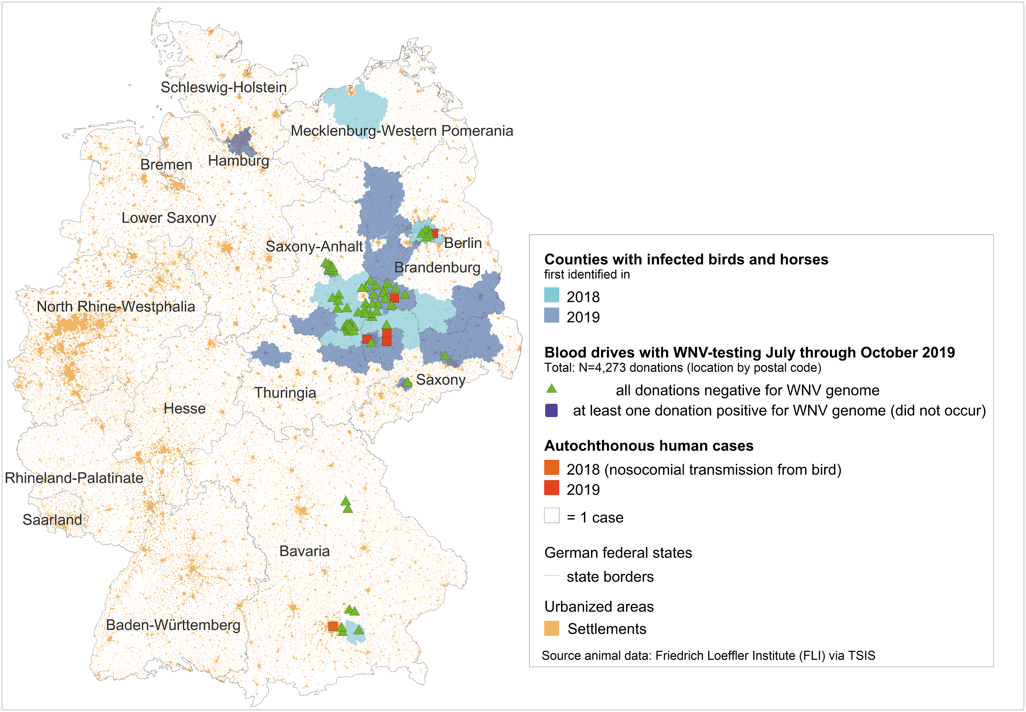
Results of the blood donor screening. The figure shows the counties in Germany with infected birds and horses in 2018 (light blue) and 2019 (dark blue). Autochthonous human WNV cases are shown as squares in 2018 (nosocomial transmission from bird, orange) and probably mosquito-borne in 2019 (red). Blood drives where all samples from donors were tested negative for WNV are shown as triangles (green). Source of animal data: Friedrich Loeffler Institute via TSIS. WNV, West Nile virus.
West Nile Virus-Nucleic Acid Test with Limits of Detection for Lineage 1 and 2 with Respect to the Individual Donation
BE, blood establishments; BRC, Bavarian Red Cross Blood Service; GRC-BH, German Red Cross Blood Service Baden-Württemberg–Hessen; GRC-NE, German Red Cross Blood Service North-East; LoD, limits of detection; n.a., not available; NSTOB, Niedersachsen, Sachsen-Anhalt, Thüringen, Oldenburg, Bremen; WNV, West Nile virus.
Data reported for the final analysis included the location of the blood drive, the date, and the number of donations screened and the test results. No additional data were collected from the donors. Data for the blood donor study were collected anonymously in compliance with the IfSG. As the data collection adhered to the legal requirements of the IfSG, no written informed consent was required.
Results
The zoo study managed to include 70 participants out of a combined 526 employees (13%, but 22% among animal keepers), 43 were male and 27 female. Seventeen participants worked closely with birds (i.e., entered the birds' indoor or outdoor habitats and thus shared mosquito exposure with the birds).
In the ELISA assay in the zoo study, none of the employees were WNV IgM-positive. Eight (11.4%) were WNV IgG-positive (one of them an employee with fever), additional two (2.9%) participants had equivocal results. All 10 were negative in the VNT for WNV (Table 2) but positive in the VNT for TBEV. Additionally, 4 of the 10 participants tested positive and 2 equivocal in VNT for USUV. Three participants reported a TBEV-vaccination in 2019, two of these participants tested positive in the VNT for TBEV. One employee reported having had a yellow fever vaccination in 2019 and had equivocal results for WNV-IgG and TBEV-IgG and no employee reported a Japanese Encephalitis Virus vaccination. Four employees (two very likely had contacts with birds) reported having had a fever between July and September 2019. One of the employees with fever tested positive in the VNT for TBEV. Another four employees (one had contacts with birds) reported having had a rash between July and September 2019. All four tested negative for all pathogens analyzed in this study.
Results of the Zoo Serosurvey
All sera yielded negative results in the WNV and USUV neutralization assays.
ELISA, enzyme-linked immunosorbent assay; TBEV, Tick-borne encephalitis virus; USUV, Usutu virus; VNT, virus neutralization test.
The participating BE collected 2.3 million donations in 2019 in their respective catchment areas. A total of 57.6% of donation were made by male donors and more than half of the donors were 45 years and older. The study included 4273 analyzed blood samples at 38 sampling sites in areas with WNV-infected animals. Specimens were collected from July 29 to November 29, 2019. None of the samples was positive for WNV-RNA (Fig. 1).
Discussion
When WNV circulation became evident for the first time in Germany in 2018, no information of the impact on the human population was available. Only five clinical WNV cases were diagnosed in 2019, all of which in areas with demonstrated WNV circulation in birds and horses. In parallel to these cases identified in the surveillance system, we initiated two sentinel studies to detect unidentified human WNV infections in affected regions. The blood donors were screened for detection of WNV viremia during the transmission season in a similar way WNV screening was performed in endemic countries, the zoo employees for antibodies after the season. But despite the well-documented WNV circulation, neither survey identified evidence for human infections. Taking the serological differentiation between cross-reactive flaviviruses into consideration, only evidence for past USUV infections and TBEV infections/vaccination was identified among the zoo employees.
Zoos are special in the study of WNV circulation because captive birds indicate local WNV circulation early (Levine et al., 2013). A study in Nashville, USA, demonstrated higher WNV infection rates in mosquitos in Nashville zoo compared to the wider city (Moncayo et al., 2023). WNV in North America was first demonstrated 1999 in zoo animals and anti-WNV-antibodies in zoo animals in Spain were early indicators of wider WNV circulation in the area the following year (Caballero-Gomez et al., 2020; Ludwig et al., 2002). Given the documented circulation of WNV at the two Berlin zoos in 2019, the lack of WNV-specific antibodies in employees with broad exposure to the animals (and sick zoo animals) is reassuring, showing that there was no large-scale WNV transmission to zoo personnel. By extension, the likelihood of infection for zoo visitors exposed for much shorter time periods was even lower.
The zoo survey was small, but included staff of both sexes and a variety of occupations including those with close contact to birds. We were able to identify past USUV infections in zoo employees. USUV is common in birds in all of Germany. Autochthonous asymptomatic human USUV infections have been demonstrated in blood donors (Cadar et al., 2017) and thus antibody evidence of USUV exposure is not surprising in zoo staff. We were also able to detect anti-TBEV-IgG, and the frequency of detection indicates TBEV-vaccinations or natural infection in the past. Even though Berlin is not considered endemic for TBEV, vaccination is recommended for those visiting endemic areas including large parts of Germany.
Our studies have limitations. The blood donor testing was performed using commercially available WNV NAT in pools with LoD of 22.2–193.5 copies/mL for lineage 1 and 93–99.5 copies/mL for the circulating lineage 2. WNV-viremia is only detectable in a short period of time after infection, and therefore, the WNV NAT cannot completely exclude WNV infections in the tested donor population. Also, donations with very low viral loads have been implicated in cases of transfusion-related transmission of WNV (Pealer et al., 2003). However, the minimum analytical sensitivity of 250 copies/mL of WNV RNA required by the German competent authority, the Paul-Ehrlich-Institut, was clearly met by all participating BE, and it can be assumed that most viremic donors would have been identified. In addition, the high number of samples included during the entire season in affected areas compensates for this limitation and allows us to conclude that the risk of transmission through blood products was negligible.
With respect to the serological analyses, the antigenic similarities between flaviviruses result in the presence of both species-specific and flavivirus cross-reactive antibodies following infection with a flavivirus. In addition, prior exposure to flaviviruses (either by natural infection or vaccination) may in some cases make it difficult to unambiguously identify the flavivirus responsible for the detected immune response. To mitigate this, we have included VNT since they are considered to be the more specific serological assay for identifying flavivirus infections.
WNV circulation in Germany was clearly documented in animals in 2019. It apparently affected very few humans, as during WNV season 2019 only five autochthonous human WNV-associated illnesses were notified. Another study in Berlin supports this: only one case of WNV infection in 2020 was identified in cerebrospinal fluid of over 600 screened cases of encephalitis/meningitis of unknown etiology from the summers of 2019 and 2020 (Schneider et al., 2022).
Areas with WNV-positive animals remain risk areas for human infection, and WNV continues to be found, including in zoo environments (Gunther et al., 2023; Rau et al., 2023; Ruscher et al., 2023). The degree to which WNV circulation affects humans depends among other factors on the presence of bridge vectors taking blood meals from birds and mammals. In areas with WNV circulation, reducing vector density for example, by larviciding with products based on Bacillus thuringeniensis israelensis can reduce the likelihood of human infection. In addition, people more likely to experience severe WNV infections (e.g., older and immunosuppressed people) in affected areas should protect themselves against mosquito bites.
Conclusions
To ensure blood safety, in Germany since 2003, blood donors had to be deferred for 28 days after visiting endemic areas during the WNV season. This measure was amended in 2014 with deferral or testing for the presence of WNV-RNA after visiting any endemic area. Until 2020, Germany was considered nonendemic, and thus the measure only applied for donors with relevant international travel history. As a consequence of the documented clinical human cases in 2019, in 2020, the measures to ensure blood safety (28-day deferral or testing) became mandatory in Germany also regarding exposure in affected areas within Germany.
Our study indicates that the risk of transmission through blood transfusion in 2019 was insignificant as we did not detect any positive samples among blood donors in the areas affected by animal and/or human WNV cases. The zoo study demonstrates the possibility of retrospectively assessing potential WNV transmission to humans on a very local level. The blood donor study highlights that blood donor specimens are accessible and should be used to aid surveillance efforts. This is particularly important during the emergence of infections like WNV, and the need for human data is urgent.
Footnotes
Acknowledgments
We thank the employees and management of Berlin Zoological Garden, Berlin Tierpark and Landesamt für Gesundheit und Soziales Berlin. We also thank Felix Reichert, Felix Moek and Kirsten Pörtner for their support in the zoo study.
Authors' Contributions
Conceptualization: R.L., K.P., R.O., S.E. Data curation: R.L., R.O., K.P. Methodology: R.O., K.P., S.E., L.B., A.S., C.D. Investigation: R.L., M.W.S., M.S., T.H.M., T.T., S.E., A.P., A.O., L.B., A.S., C.D. Resources: M.W.S., M.S., T.H.M., T.T., A.P., A.O. Writing—original draft preparation: R.L., C.F., R.O., C.D. Writing—review and editing: R.L., C.F., K.P., R.O., M.W.S., M.S., T.H.M., T.T., A.P., A.O., L.B., S.E., C.D. Project administration: C.F., R.O. All authors have read and agreed to the published version of the manuscript.
Author Disclosure Statement
All authors declare no conflict of interest. The funders had no role in the design of the study; in the collection, analyses, or interpretation of data; in the writing of the manuscript; or in the decision to publish the results.
Funding Information
The study received no external funding.