
Abstract
Detection of Helcococcus kunzii and Ignatzschineria ureiclastica/larvae has been made possible by recent advancements in microbiologic diagnostics. We report the first described case of polymicrobial bacteremia secondary to these two unique pathogens, and only the third case of I. ureiclastica/larvae bacteremia described in the United States. Myiasis has historically been thought of as an infestation. This case adds to the growing body of evidence that myiasis is potentially a vector for bacteria, and bacteremia, and the potential for the spread of other vector-borne diseases.
Introduction
Myiasis, or maggot-infested wounds, has historically been thought of as an infestation. Rarely is myiasis associated with systemic disease. Rather, medical maggot therapy has long been used for improved wound debridement and faster healing (Zubir et al., 2020). Myiasis is an infiltration of fly larvae on human tissue, usually decomposing flesh, and seldomly results in bacteremia (Le Brun et al., 2015; Do et al., 2021; Johnson et al., 2023; Reed et al., 2021). Rarer still is polymicrobial bacteremia secondary to the pathogens Helcococcus kunzii and Ignatzschineria ureiclastica/larvae. H. kunzii and I. ureiclastica/larvae are unusual pathogens whose detection have been made possible by recent advancement in molecular diagnostics and mass spectrometry (Nadrah et al., 2021). Myiasis has historically been thought of as an infestation. This case adds to the growing body of evidence that myiasis is potentially a vector for bacteria, and bacteremia, and the potential for the spread of other vector-borne diseases.
Case Report
A 56-year-old male with a history of alcohol dependence and homelessness presented to the emergency department complaining of a dorsal right foot wound infested with maggots. He was unsure of the duration of the wound but noted he had been wearing wet shoes and socks for a prolonged period before presentation. He denied any associated fevers or chills. Physical examination was notable for an open wound of the dorsum of the toes on his right foot, infested with hundreds of maggots. Blood cultures were obtained. The wound was copiously cleaned and irrigated unfortunately before any photographic documentation or species-level identification of the maggots.
Initial labs were notable for a white blood cell count of 6500/µL and platelet count of 123,000/µL. Erythrocyte sedimentation rate (ESR) was 90 mm/h and C-reactive protein (CRP) 12.79 mg/dL. Plain film X-rays of the foot revealed mild soft tissue swelling about the toes with no discrete areas of osteolysis. He was hemodynamically stable and was admitted to the medical floor. Orthopedic surgery was consulted and the wound was noted to be superficial with no appreciable abscess, drainable fluid collection, or communication with bone. He was empirically initiated on intravenous (IV) piperacillin-tazobactam.
Blood cultures initially returned positive for gram-positive cocci, with molecular detection of Staphylococcus epidermidis, negative for mecA and mecC genes. On day 2 of admission, blood cultures were revealed additional growth of both gram-positive cocci and gram-negative rods, neither of which were identified with molecular detection. On day 4 of admission, the gram-positive isolates were identified as H. kunzii and methicillin-susceptible S. epidermidis via matrix-assisted laser desorption/ionization time-of-flight (MALDI-TOF). The gram-negative isolate was unable to be identified by MALDI-TOF and was sent to a reference laboratory, and on day 6 of admission, it was identified as Ignatzschineria ureiclastica/larvae by MALDI-TOF. Susceptibility testing was not performed. Repeat blood cultures obtained on day 2 of admission were negative at 5 days. He received aggressive wound care with skin debridement throughout his hospitalization. He was ultimately discharged after being treated with a 12-day course of IV piperacillin-tazobactam.
Discussion
To the best of our knowledge, this case presents the first known coinfection with H. kunzii and Ignatzschineria, and only the third case of Ignatzschineria larvae/ureiclastica bacteremia in the United States to date (Table 1). Identification of both pathogens has been made recently possible with developments in microbiologic diagnostics, particularly MALDI-TOF. Both H. kunzii and Ignatzschineria are not commonly identified by routine microbiological lab testing.
Case Reports of Bacteremia due to Ignatzschineria and Helcococcus
published date of case, unknown date of clinical presentation.
H. kunzii was first described in 1993 (Collins et al., 1993). H. kunzii is a gram-positive coccus found in pairs or clusters (Lotte et al., 2015; Vergne et al., 2015a) and part of the skin flora (Woo et al., 2005). It is most often associated with lower extremity wound infections, although it has been described pathogenic in the setting of IV drug use (Woo et al., 2005). Detection of H. kunzii can be challenging, as it is usually isolated in polymicrobial specimens and overgrown by other pathogens (Vergne et al., 2015a). To date, H. kunzii has been rarely associated with systemic infections (Lotte et al., 2015; Park et al., 2014; Woo et al., 2005). In a case review of 39 isolates of H. kunzii in Europe, only a single case of bacteremia was noted (Vergne et al., 2015a). In that case, the culture was polymicrobial (Vergne et al., 2015a), as was seen in our case.
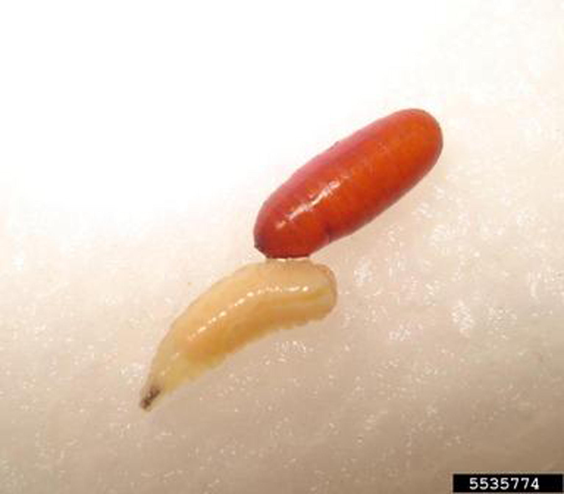
Ignatzschineria, alternatively, is not an inhabitant of the human skin flora. It is an aerobic gram-negative rod rarely described in human infections (Le Brun et al., 2015; Nadrah et al., 2021). It is a commensal of parasitic fly/maggot gastrointestinal tracts, first identified in Wohlfahrtia magnifica (Do et al., 2021; Nadrah et al., 2021; Reed et al., 2021). W. magnifica is also known as the spotted flesh fly. In the United States, it has been isolated in infections associated with Lucilia sericata (Le Brun et al., 2015; Reed et al., 2021), formerly of the genus name Phaenicia, a common green bottle fly found worldwide (Figs. 1 and 2). The species of maggot in our case was not known. As has been described in other cases of myiasis, fly larvae found are typically discarded in the emergency department following wound debridement before they can be sent for identification (Nadrah et al., 2021).
Common green bottle fly (Lucilia sericata). Photo courtesy of Joesph Berger, bugwood.org.
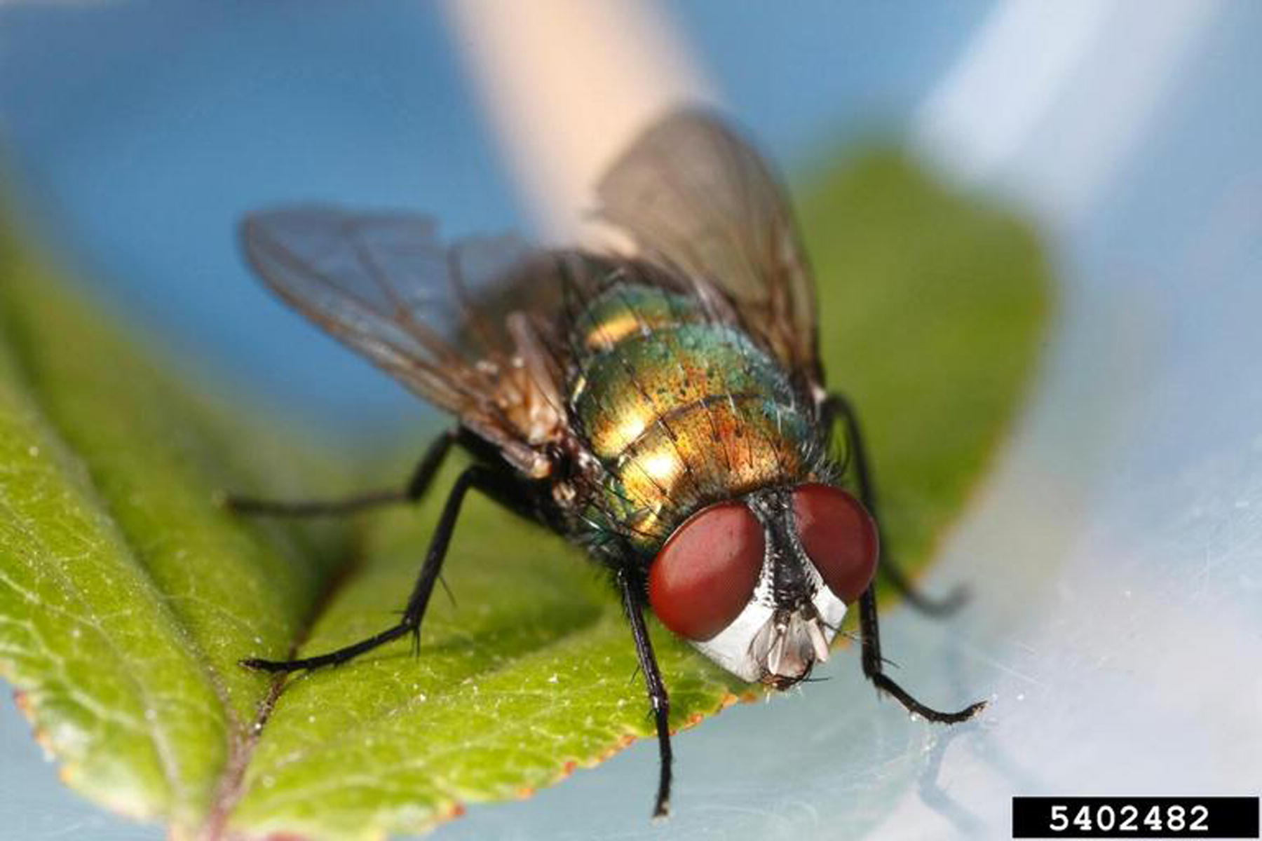
Common green bottle fly (Lucilia sericata). Photo courtesy of Mohammed El Damir, bugwood.org.
Ignatzschineria bacteremia is rare, with the few reported cases most frequently speciated to I. indica (Do et al., 2021; Reed et al., 2021). I. ureiclastica/larvae are infrequent enough that PCR sequencing can sometimes not distinguish between the two species (Do et al., 2021; Johnson et al., 2023; Nadrah et al., 2021; Reed et al., 2021). Species identification is likely not of clinical consequence at present time, as clinical manifestations and treatment are the same. To date in the United States, there have only been two other case reports of Ignatzschineria ureiclastica or larvae (Johnson et al., 2023; Reed et al., 2021), with only one having been sent for formal identification as I. larvae (Johnson et al., 2023).
Antimicrobial susceptibility for H. kunzii and Ignatzschineria was not performed in our case. Our patient clinically improved on therapy with piperacillin-tazobactam and repeat blood cultures documented clearance. Isolates of H. kunzii are generally susceptible to beta-lactams (Lotte et al., 2015; Vergne et al., 2015b), as well as glycopeptides and linezolid (Vergne et al., 2015b). Notably, H. kunzii resistance to macrolides and clindamycin has been reported (Park et al., 2014; Woo et al., 2005). Agents commonly active gram-negative agents, such as most beta-lactams, have demonstrated activity against most isolates of Ignatzschineria (Do et al., 2021; Johnson et al., 2023). Suspected susceptibility to piperacillin-tazobactam of both H. kunzii and Ignatzschineria could be presumed in our case given clinical improvement. However, it is possible that a more narrow-spectrum beta-lactam could have been equally effective.
Another case report of Ignatzschineria bacteremia in the setting of myiasis has been described and was postulated to have occurred secondary to wound debridement (Nadrah et al., 2021). However, in our case, blood cultures were drawn upon initial presentation to the emergency department and before any wound debridement. The timing of wound debridement in myiasis should be considered on a case-by-case basis, although it does not appear to have been correlated with this case of bacteremia. More likely it is associated with the size and extent of tissue breakdown leading to blood stream invasion.
Myiasis is a condition that disproportionately affects the socially disadvantaged. Those who are homeless and suffer from alcoholism seem to be at higher risk (Sherman, 2000). Interestingly, there is also a male preponderance (Sherman, 2000). Most cases of myiasis that occur in the United States are imported from Mexico and Central and South America and most commonly involve Dermatobia species (Safdar et al., 2003). Of the cases of myiasis that originate in the United States, the most common species involved is Lucilia sericata (Sherman, 2000). This case highlights the importance of obtaining blood cultures in all cases of myiasis, even in patients who do not appear to bacteremic or septic. Medical use of maggot therapy for wounds is not associated with risk for bacterial infection, as the maggots are grown in sterile environments (Do et al., 2021). However, community-acquired fly larvae infestations are at risk for polymicrobial infections with unusual pathogens, as seen in this case. This case adds to the growing body of evidence that myiasis can be a vector for bacteria, and bacteremia, and the potential for the spread of other vector-borne diseases. Our patient was afebrile, hemodynamically stable, and without leukocytosis, yet still was found to have polymicrobial bacteremia. Severe myiasis alone may be a signal for bacteremia.
Footnotes
Acknowledgments
Author Disclosure Statement
All authors report no conflicts of interest.
Funding Information
No funding was received for this article.