
Abstract
The induction of neutralizing antibodies specific for foot-and-mouth disease virus (FMDV) has been the central goal of vaccination efforts against this economically important disease of cloven-hoofed animals. Although these efforts have yielded much success, challenges remain, including little cross-serotype protection and inadequate duration of immunity. Commonly, viral infections are characterized by induction of cytotoxic T lymphocytes (CTL), yet the function of CTL in FMDV immunity is poorly defined. We developed an assay for detection of CTL specific for FMDV and reported that a modified adenovirus-vectored FMDV vaccine could induce CTL activity. This allowed us to determine whether FMDV-specific CTL responses are induced during infection and to test further whether vaccine-induced CTL could protect against challenge with FMDV. We now show the induction of antigen-specific CTL responses after infection of swine with FMDV strain A24 Cruizero. In addition, we developed a vaccination strategy that induces FMDV-specific CTL in the absence of significant neutralizing antibody. Animals vaccinated using this protocol showed delayed clinical disease and significantly suppressed viremia compared to control animals, suggesting a role for CTLs in the control of virus shedding. These results provide new insights showing induction of CTL responses to FMDV following infection or vaccination, and create the potential for improving vaccine performance by targeting cellular immunity.
Introduction
FMDV is a picornavirus with the genome consisting of single-stranded positive-sense RNA. This is translated as a single polyprotein that is subsequently cleaved into mature proteins by virus encoded proteases, especially 3C (3Cpro) (2,10,26,56). The leader protease (Lpro) (13) blocks host protein synthesis by cleaving initiation factor 4G (eIF4G) eliminating cap-dependent mRNA translation, and the combined actions of viral proteins 2B and 2C serve to disrupt vesicular transport (30,31). Virus RNA translation then co-opts the ribosomes as the 5′ UTR of the virus contains an internal ribosome entry site (IRES) which does not require intact eIF4G (4, 27).
Although many countries are largely free of FMD, the persistence of disease in parts of the world, and its highly infectious nature, require vigilance on the part of FMD-free countries. Agricultural concerns are focused primarily on cattle, swine, sheep, and goats. However, many wild ruminant and cloven-hoofed species are also susceptible to infection, though severity of clinical disease can vary from severe to inapparent (34). Routine control of FMD is maintained primarily through trade practices that limit the exportation of animal products from countries reporting FMD (18). Acute outbreaks have been controlled through vaccination or mass culling of susceptible animals (24,49,52,57). The adverse economic consequences of FMD outbreaks highlight the need for preventive measures, including effective vaccines.
Current vaccines consist of killed virus in adjuvant and are effective at generating protective neutralizing antibodies (14,45). However, the inability to differentiate infected from vaccinated animals (DIVA) makes deploying vaccines problematic for maintaining trade status. In addition, vaccine production requires the growth of large amounts of virus requiring expensive, high containment facilities for manufacture, thus providing an incentive for the exploration of alternative FMDV vaccines. A leading candidate DIVA vaccine for FMDV is vectored by a replication-defective human adenovirus 5 (Ad5). This construct delivers the coding regions for the P1 capsid precursor and 3Cpro to cells. Once expressed, 3Cpro cleaves the P1 polyprotein into the mature capsid proteins, which then form “empty capsids” (19). This approach to vaccination has been shown to protect both pigs and cattle (28,29,33,37,42). However, like the traditional killed virus vaccine, serotype and strain matching are required because the elicited neutralizing antibodies are type specific and cross-react poorly with other serotypes, and subtypes within serotypes (45).
Cytotoxic T lymphocytes (CTLs) are a CD8-expressing subset of T cells that can directly kill virus-infected cells in an antigen-specific manner. In contrast to antibodies, which recognize complex antigens, CTLs recognize processed antigens that are cleaved by the proteasome into short peptides of 8 to 12 amino acids, loaded into major histocompatibility complex (MHC) class I molecules in the lumen of the endoplasmic reticulum (ER), and then transported to the cell surface (43,58). The ability of CTLs to recognize peptides derived from all regions of the viral proteome makes their induction one potential way to bridge the gap of cross-serotype protection. Variation between serotypes of FMDV is located within the structural proteins forming the capsid (7), which is likely a consequence of immune selective pressure exerted by neutralizing antibodies specific for the capsid (23).
In contrast, the nonstructural proteins are very highly conserved (7), which increases the likelihood of different serotypes containing identical peptides that can be recognized by CTLs. However, in vitro, FMDV infection inhibits synthesis of new class I MHC proteins (47), which would block induction of CTLs if the same process occurs within virus infected cells in vivo. Further, the relatively rapid resolution of disease likely contributes to a dampened CTL response due to insufficient duration of antigen exposure. Despite their prominent role as part of the adaptive immune system and considering the lack of MHC expression predicted for infected cells, analysis of CTL responses to FMDV infection or vaccination has seldom been reported. This lack of data has led to the opinion that CTL do not play a role in the response to FMD (14).
CTLs are one component of cellular immunity, which refers to the more general response of multiple T cell subsets and the multiple functions those T cells exert during immune responses. Analysis of the response in cattle following vaccination with the commercial, killed virus vaccine has demonstrated CD8 T cells producing interferon gamma (IFNγ) (22). The peptide specificity of the response to vaccination has also been reported, analyzing proliferation and IFNγ secretion by CD4 T cells (16) or IFNγ secretion by CD8 T cells (16, 21). We have also reported IFNγ production by both CD4 and CD8 T lymphocytes following vaccination of cattle with the Ad5-FMDV vaccine (32). These results illustrate that there are multiple cell populations capable of producing IFNγ, especially within the CD8 T lymphocyte populations studied. Thus, these previous studies highlight the need to analyze CTL killing of virus-infected target cells and not assume that detected production of IFNγ is from virus-specific CTLs.
In part, the limited knowledge of CTL responses to FMDV is due to the technical difficulties of establishing reliable assays for CTL responses for this virus. FMDV is a highly cytopathic virus and therefore the experimental target cells for a CTL response cannot be infected with live virus (9,22). Further, the assay requires MHC class I matching of the responding animals and the target cells and intact expression of those MHC proteins. We previously reported the establishment of a CTL killing assay that utilizes the Ad5 expression system for the production of FMDV proteins that induces little cytopathic effect. Using this assay, we showed that vaccination of swine with an Ad5 construct lacking efficient processing of the P1 polyprotein resulted in the induction of anti-FMDV CTLs and that multiple vaccinations were needed in order to generate significant neutralizing antibody titers. The presence of CD8+ MHC-restricted CTLs was confirmed by swine leukocyte antigen (SLA)-1 tetramer staining. The companion parental vaccine construct, that processes the polypeptide into components that assemble into capsids, failed to induce a CTL response and required a single vaccination to induce a significant B cell response exhibited by neutralizing antibody titers (33,41). We refer to these vaccine constructs as Ad5-FMDV-T and Ad5-FMDV-B, respectively, for convenience and to signify the arm of the adaptive immune system preferentially stimulated.
These results provided an experimental system with which to test the contribution of CTL immunity to protection against infection with FMDV following challenge with live, virulent virus. This design was solely to address the experimental question of whether each can contribute to overall immunity to virus. Clearly, this is not an optimal vaccine design as maximizing both responses is the goal for an effective vaccine.
We now report the induction of CTL responses in naïve, infected pigs, the first demonstration of antigen-specific class I MHC-mediated killing of virus-infected target cells by CD8+ CTLs from FMDV-infected livestock. Further, we tested the efficacy of the CTL-inducing vaccine to protect swine against live virus challenge in the absence of significant neutralizing antibodies. Swine that received the T cell vaccine mounted a CTL response and developed delayed clinical disease with reduced/no viremia. Although insufficient to protect against challenge with FMDV under these designed experimental conditions, induction of CTL immunity under more optimal conditions, specifically while also inducing the well characterized humoral response to the virus, is likely to improve the performance of the Ad5-FMDV vaccination approach.
Materials and Methods
Cell lines
Porcine kidney [PK(15)] cells (ATCC CCL-33) and PK(15)-EGFP cells (41) were maintained in minimal essential medium (MEM) (Invitrogen, Carlsbad, CA), and baby hamster kidney (BHK-21) cells (ATCC CCL-10) were maintained in basal medium Eagle (BME) (Invitrogen). All media contained 10% fetal bovine serum (FBS) (Thermo Fisher Scientific, Waltham, MA) along with other supplements, as described previously (41).
Viruses
Viruses Ad5-A24-3C-mut (Ad5-FMDV-T), Ad5-A24 (Ad5-FMDV-B), and Ad5-VSV-G (Ad5 expressing the glycoprotein of vesicular stomatitis virus) have been described previously (33,41). Ad5-FMDV-T and Ad5-FMDV-B differ in that the 3C protease of the former is a mutant that is inefficient at P1 processing. For in vitro assays, FMDV, strain A24 Cruzeiro, was propagated in BHK-21 cells. A swine-derived isolate of the same strain was used for animal inoculation, prepared as previously described (40).
Animals
All animal experiments were performed in a secure biosafety level three laboratory on Plum Island Animal Disease Center (PIADC) following the protocol approved by the Institutional Animal Care and Use Committee following the guidelines of the United State Public Health Service, Department of Health and Human Services and the Animal Plant Health Inspection Service (APHIS) of the US Department of Agriculture (USDA). Sixteen National Institute of Health miniature (NIH mini) pigs, homozygous for MHC haplotype 4a.0/4a.0 (*0401 for all class I genes), also published as NIHd/d (51), were generously provided by Scott Arn and David Sachs (Transplantation Biology Research Center, Massachusetts General Hospital, Boston, MA). Animals ranged in age between 10 and 14 months, and weighed approximately 100 lbs.
Vaccination and challenge
Twelve NIH mini pigs were divided into three groups and vaccinated with either Ad5-FMDV-T (seven pigs), Ad5-FMDV-B (two pigs), or Ad5-VSV-G (three pigs). Animals were separated by gender with the Ad5-FMDV-T vaccinated animals (all male) kept in a separate room. The naïve animals (Ad5-VSV-G) were female, and the two animals vaccinated with Ad5-FMDV-B were a male and a female. The vaccine was delivered intradermally using the Derma-Vac® needle-free system (Merial Animal Health, Duluth, GA) at four sites on the neck for a total dose of 5×10⁹ plaque forming units (pfu)/pig. All animals were boosted on the same day with 5×10⁹ pfu of their respective immunogen 10 weeks (FMDV-B and VSV-G) or 12 weeks (FMDV-T) postvaccination. Eight days following the boost, all animals were challenged with 105 TCID50 of FMDV, strain A24 Cruzeiro, by intradermal needle inoculation in the heel bulb (39).
PBMC preparation
Vacutainer® tubes containing heparin were used to collect porcine blood. After dilution with PBS, the blood was underlain with Lymphoprep (Axis-Shield, Oslo, Norway), and then centrifuged at 1400 g for 20 min at room temperature. The resulting band of peripheral blood mononuclear cells (PBMCs) was harvested, washed with PBS three times, and then suspended Roswell Park Memorial Institute (RPMI)-1640 medium (Invitrogen, Carlsbad, CA).
Cytotoxic T lymphocyte killing assay
CTLs were sorted, and killing activity was measured as described previously (41). Briefly, PBMCs were cultured in the presence of PK(15) cells infected with Ad5-FMDV-T for 3 days, after which cells were purified on Lymphoprep and positively sorted for CD6 expression in order to reduce MHC unrestricted killing (12). CD6+ cells were sorted by incubating with anti-CD6 antibody (clone MIL8, AbD Serotec, Raleigh, NC) and then goat anti-mouse microbeads, followed by passage through LS columns using a MidiMACS magnetic separator (Miltenyi Biotec, Gladbach, Germany).
Following CD6 purification, effector cells were incubated for 4 h with PK(15)-EGFP target cells that were infected with Ad5-FMDV-T, Ad5-VSV-G, or mock-infected at E:T ratios of 50:1, 25:1, and 12:1 in the presence of 7-amino-actinomycin D (7-AAD) (BD Biosciences, San Jose, CA). The percent cell killing was determined by flow cytometry using a FACSCalibur flow cytometer with a high-throughput sampler (HTS) (BD Biosciences, San Jose, CA) by gating on the EGFP+ cell population and determining the percent 7-AAD+ cells. Background lysis of target cells alone was subtracted from all samples. In order to eliminate noise of day-to-day variability in the CTL killing assays, the greatest mean lysis of control target cells (mock infected or Ad5-VSV-G infected) was subtracted from mean lysis of the antigen-specific target cells at each time point (see Fig. 2A–C). Unadjusted data are shown in Supplementary Figure S1 (supplementary data are available online at
Serum neutralization assay
Four-fold dilutions of heat inactivated serum were incubated with 100 TCID50 FMDV A24 for 1 h at 37°C, and then incubated for 3 days with BHK-21 cells, which were scored for cytopathic effect. Endpoint serum neutralization titers (SNT) were calculated as previously described (38) and comparison of induction of neutralizing and total anti-FMDV antibody titers has been reported (29).
Serum virus titration
Frozen serum samples were thawed and used to make a series of 10-fold dilutions. Each dilution was added to a microtiter plate in quadruplicate, after which 104 BHK-21 cells were added to each well. Following 3 days of incubation at 37°C, wells were scored for presence of CPE and the TCID50 was calculated.
FMDV RNA detection
RNA was extracted from 50 μL of serum as previously described (1), and then detected by real-time RT-PCR according to the method of Callahan and co-workers (6). Cycle thresholds <45 were considered positive and converted to RNA copies/mL by comparison to a standard curve generated by real-time RT-PCR of known concentrations of in vitro synthesized FMDV RNA, as described (1).
Clinical evaluation
Scoring of clinical disease was based on appearance of vesicles, as previously described (39). A maximum score of 20 was possible based on the presence of vesicles on each digit, the snout, tongue, lower lip, or carpal/tarsal area of at least one leg. Clinical scores were cumulative through the period of evaluation.
Statistics
Statistical significance of CTL killing activity based on comparison between target cell groups was determined using a one-tailed, paired Student's t test. Statistical significant elevation of antigen-specific CTL killing compared to baseline was determined using repeat-measures one-way ANOVA with Dunnett's post test. CTL killing was considered statistically significant only if both comparisons gave p values of 0.05 or less. Statistically significant differences in serum antibody and viremia were determined using one-tailed unpaired Student's t test.
Results
Titration of challenge virus in genetically defined swine
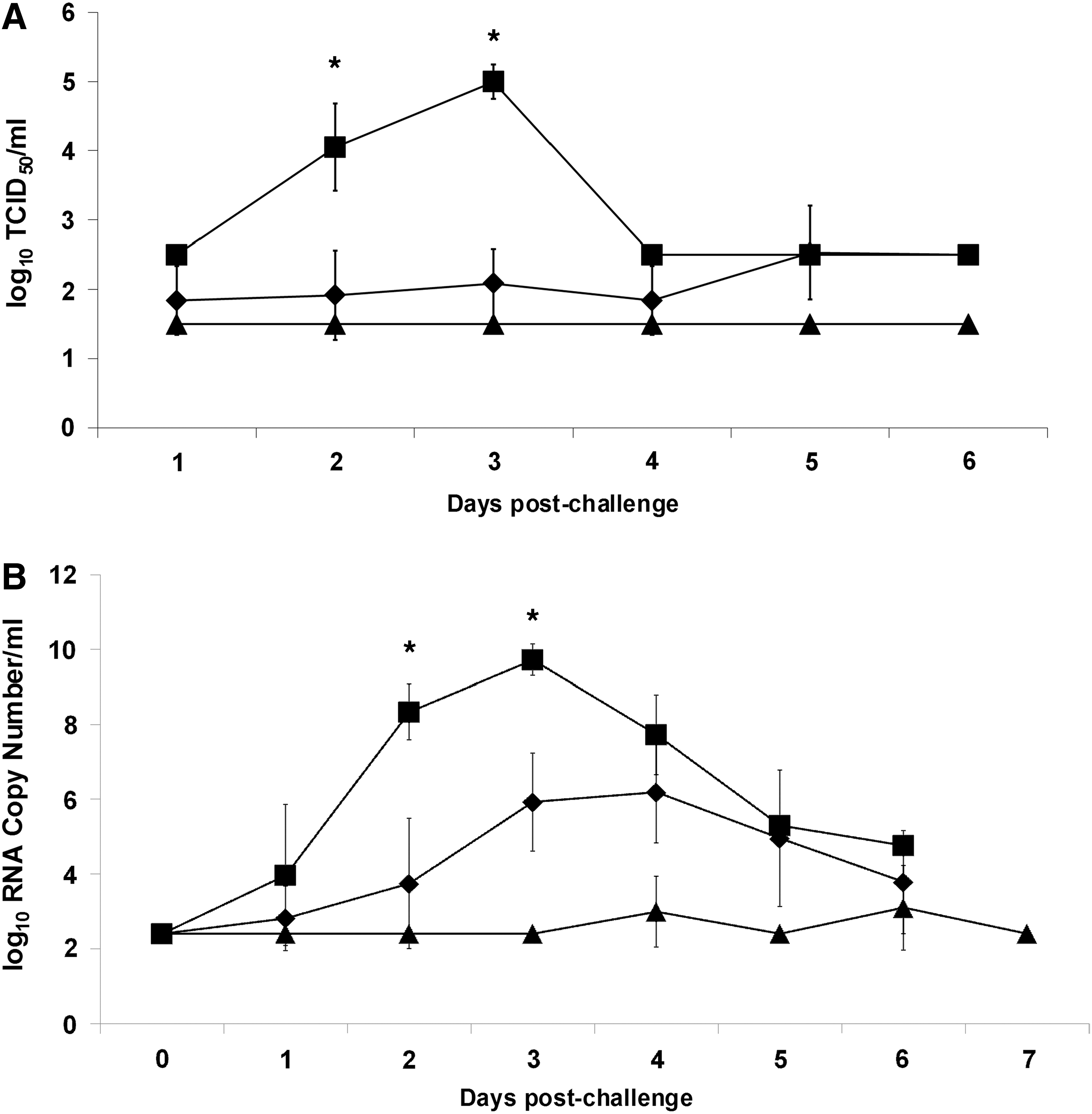
Analysis of CTL function requires an assay to measure lysis of virus-infected target cells in an antigen-specific, MHC-restricted fashion. This requires MHC class I matching between the effector T cells and target cells. We had availability of National Institutes of Health (NIH) miniature pigs (NIH mini pigs) homozygous for MHC for such studies. However, infection of this strain of pigs with FMDV, particularly animals that are 1 year old and weighing 100 lbs, had not been demonstrated. Therefore, we conducted a preliminary trial to determine an appropriate challenge dose of FMDV by inoculating four pigs with 10-fold dilutions of virus ranging from 104 to 107 TCID50, and then evaluating them for clinical signs on days 1, 2, 3, and 7 following infection. Viremia was confirmed by real-time RT-PCR. All pigs developed clinical disease and detectable viremia (Fig. 1). Severity of clinical disease increased with challenge dose such that the pig that received the highest dose, 107 TCID50, required euthanasia 2 days post challenge. The pigs with the two lowest doses had comparable clinical signs and viremia. Based on these data, we chose a challenge dose of 105 TCID50, assuring inoculation would result in clinical disease in the naïve animals.
Clinical evaluation of pigs used to determine challenge dose. Individual pigs were challenged with the indicated dose of FMDV and evaluated for clinical signs on days 1, 2, 3, and 7 (bars), as described in Materials and Methods. Although no clinical evaluation occurred on days 4 through 6, scoring is cumulative over the course of disease, which implies a score no lower than that of day 3. Sera were collected daily and viremia was measured using real-time RT-PCR (lines). Severe disease necessitated euthanasia of pig 73.
Humoral and cellular immune response following vaccination with Ad5 FMDV vectored vaccines
In order to test the ability of FMDV-specific CTLs to protect against disease, seven pigs were vaccinated with Ad5-FMDV-T, which expresses unprocessed P1 polyprotein. An additional two pigs were vaccinated with the standard empty capsid vaccine, Ad5-FMDV-B, the original vaccine vector containing the wild-type 3C protease. This virus-vectored vaccine has been shown to protect against homologous virus strain challenge of pigs with virulent FMDV as early as 7 days and for as long as 42 days (11,33). An irrelevant Ad5 vectored vaccine, Ad5-VSV-G (expressing the glycoprotein of vesicular stomatitis virus, VSV-G) was used to vaccinate three pigs and these animals served as FMDV naïve controls. Prior to vaccination and at several time points following vaccination, peripheral blood mononuclear cells (PBMCs) from all pigs were isolated and tested for FMDV-specific CTL killing activity as described in Materials and Methods. For pigs vaccinated with Ad5-FMDV-T, we observed an increase in CTL activity (p<0.01) 21 days after vaccination that returned to background levels by day 31 (Fig. 2A). There was minimal, statistically insignificant specific CTL activity detected using cells isolated from either the Ad5-FMDV-B or Ad5-VSV-G control vaccinated pigs (Fig. 2B and C). The lack of a VSV-G specific CTL response in the Ad5-VSV-G vaccinated animals (Supplementary Fig. S1C; supplementary data available on website
CTL killing activity induced by vaccination and challenge. PBMCs were collected from pigs at various time points relative to vaccination with
The presence of neutralizing antibodies was detected 21 days post-vaccination in sera from pigs vaccinated with Ad5-FMDV-B, but not in either the Ad5-FMDV-T or Ad5-VSV-G vaccine groups (Fig. 3). This initial dichotomy between induction of CTLs and neutralizing antibodies is consistent with our previous observations and allowed for analysis of the relative role of humoral and cellular immunity in protection against disease.
Serum neutralizing antibody titers. Sera were collected at indicated times relative to prime, boost, and challenge. Dilutions of sera were tested for neutralization of 100 TCID50 FMDV A24, as described in Materials and Methods. Geometric -mean neutralization titer is shown for vaccination groups (▴, Ad5-FMDV-B (n=2); ♦, Ad5-FMDV-T (n=7); ▪, Ad5-VSV-G (n=3)) with standard deviation. Asterisks indicate statistically significant difference (p<0.01) between Ad5-FMDV-B and Ad5-FMDV-T groups.
All animals were simultaneously boosted, 10 weeks following vaccination for the FMDV-B and VSV-G groups and 12 weeks following vaccination for the FMDV-T animals. PBMC from all animals were analyzed for CTL responses before challenging all animals with virulent FMDV, strain A24. We observed elevated CTL killing activity by cells derived from pigs vaccinated with Ad-FMDV-T (p<0.01), compared to 2 days before the boost (Fig. 2A). We also observed a small rise in CTL activity by cells derived from pigs vaccinated with Ad5-FMDV-B (Fig. 2B), although it did not reach statistical significance. Evaluation of serum samples taken 7 days following the boost (1 day before challenge) showed an increased neutralizing antibody titer in Ad5-FMDV-B-vaccinated pigs compared to levels prior to the vaccine boost (Fig. 3). Although neutralizing antibodies were detected in some of the pigs vaccinated with Ad5-FMDV-T, the titers were uniformly lower than Ad5-FMDV-B-vaccinated pigs (Fig. 3 and Supplementary Fig. S2A and B). The highest individual titer in this vaccine group was 1.8 log10 and three pigs from this group had no detectable serum neutralizing antibodies (Supplementary Fig. S2A). Control pigs vaccinated with Ad5-VSV-G showed no evidence of FMDV-specific CTL activity (Fig. 2C) or serum neutralizing antibodies (Fig. 3 and Supplementary Fig. S2B), as expected.
Clinical disease following challenge of vaccinated animals with FMDV
All animals in the study were challenged with 105 TCID50 of FMDV strain A24 Cruzero, and animals were evaluated for clinical disease on days 4 and 7. One of the animals vaccinated with Ad5-FMDV-B remained completely clear of vesicles, while the second developed a single vesicle on one digit by day 7 (Fig. 4). Animals vaccinated with Ad5-FMDV-T exhibited clinical signs of disease. However, with the exception of one animal, the clinical scores at day 4 were lower than the naïve controls, suggesting a delay in disease progression. By day 7 after challenge, clinical scores of the group vaccinated with Ad5-FMDV-T were very similar to the naïve controls (Fig. 4).
Clinical scores of individual animals challenged with FMDV. All animals were scored for presence of vesicles on days 4 and 7 postchallenge, as described in Materials and Methods. Scoring is cumulative over the course of disease, which implies a score no lower than that of day 4 on subsequent days.
Detection of viremia by virus isolation was greatly reduced in pigs vaccinated with Ad5-FMDV-T by a mean difference of almost 3 logs compared to the naïve controls (day 3 following challenge, Fig. 5A). The highest viremia detected in the Ad5-FMDV-T-vaccinated pigs was in animal 65, which had peak viremia on day 5 of log10 3.75, 1 log lower and 2 days delayed from the peak for all naïve animals (Supplementary Fig. S3A and B). The balance of the Ad5-FMDV-T-vaccinated pigs had either no detectable viremia following infection (log10 1.5 being the lower limit of the assay) or viremia that was 2 to 3 logs lower than controls. No viremia was detected in pigs given the standard vaccine, Ad5-FMDV-B (Fig. 5A).
Quantification of viremia. Daily sera were collected from animals following challenge.
Real-time RT-PCR detection of FMDV RNA present in serum yielded similar results (Fig. 5B). Again, pig number 65 showed FMDV RNA levels intermediate between the rest of the vaccine cohort and the naïve controls. In this assay, pig 68 also showed higher presence of FMDV RNA than the rest of the cohort but still an order of magnitude less than the naïve controls (Supplementary Fig. S4A and B). All other animals vaccinated with the FMDV-T construct had lower FMDV titers using the PCR assay. The average peak titer of this cohort of animals was log 6.19 on day 4 compared to an average titer of log 9.72 on day 3 for naïve, Ad5 VSV-G vaccinated animals. As in the virus isolation assay, the PCR assay showed reduced FMDV titers peaking in a delayed manner (Fig. 5). This assay detects presence of RNA viral genome resulting in signal combining presence of live virus genome and residual RNA from killed virus, yet relative results between these three treatment groups are the same as that of virus isolation.
CTL responses following challenge of vaccinated animals with FMDV
PBMCs were isolated and tested for CTL killing activity beginning 10 days after challenge. Cells from pigs vaccinated with Ad5-FMDV-T displayed an increase in CTL activity 10 days post challenge relative to preboost (p<0.01) that returned to background levels by 17 days post challenge (Fig. 2A). We attempted to evaluate CTL activity on day 4 but failed to obtain enough cells for the assay. We attribute this to the lymphopenia and immunopathology induced by FMDV (3).
Although we had observed a slight elevation in CTL activity in PBMCs derived from pigs vaccinated with Ad5-FMDV-B during the vaccination phase of the experiment, no CTL activity was observed following challenge with FMDV (Fig. 2B). However, serum neutralizing titers of these pigs continued to rise following challenge (Fig. 3A), suggesting a low level of virus replication in vivo. This was also observed in the Ad5-FMDV-T vaccinated animals following challenge, although pig 68 continued to lack detectable neutralizing antibodies as late as 4 days following challenge.
The pigs given the irrelevant vaccine, Ad5-VSV-G, served as FMDV naïve controls. These animals showed a statistically significant anti-FMDV CTL response at 24 days postchallenge (p<0.05). CTL killing of Ad5-FMDV infected target cells was no longer detectable 31 days following challenge (Fig. 2C). This followed similar kinetics to that observed for vaccination of animals with Ad5-FMDV-T, where significant killing was observed 21 days after vaccination and no longer detectable by 31 days (Fig. 2A). This represents the first report of porcine CTL killing activity following infection with FMDV.
Discussion
Historically, analysis of immune responses to FMDV vaccination and/or infection has focused on measurement of serum antibody, yielding an informative and widely reported assessment of humoral immunity to the virus. Defining the role of cellular immune responses to FMDV in controlling this acute viral disease has been difficult as this highly cytopathic virus does not allow for classical analysis of the killing of virus infected target cells using in vitro assays. Previous reports have analyzed surrogate responses for CTL killing such as proliferation or production of IFNγ by CD8 expressing cells (9,21,22,25,44,46). These data have been informative, but far from definitive, as many CD8-expressing lymphocytes that are not CTLs produce IFNγ, including NK cells and subsets of γδ T cells (5,53,55). Further, none of these reports sufficiently define the cell populations being assayed as CTLs such as detecting activation of perforin/granzyme expression and secretion of these proteins by αß T cell receptor expressing (TCR+), CD8 T lymphocytes.
We previously tested the hypothesis that inhibiting proteolytic processing of FMDV capsid proteins would enhance induction of CTLs, presumably by increasing the pool of antigenic peptides available for class I MHC presentation and stimulation of cognate CTLs (41). In those studies, we developed methodology to analyze antigen-specific, virus-infected cell killing and confirmed our results by showing MHC tetramer staining of the CD8 T cells correlates to target cell killing. The response we measured was generated in the artificial setting created by infecting the MHC matched target cells with the same replication-defective Ad5 virus, Ad5-FMDV-T, as was used for the vaccination. In the present study, we now show FMDV specific CTLs are induced following infection with live, virulent FMDV. Thus, we confirm that the CTL killing assay used in all of these studies can detect cellular immune responses to FMDV following infection, not solely in the case of immunization with the same Ad5 vector that is used in the CTL assay.
FMDV infection of cells in vitro was reported to mediate inhibition of MHC class I cell surface expression (47). Data presented here now indicate such inhibition in vivo is partial at best, as MHC class I expression by virus-infected cells is required to induce the CTL response we detected. This novel observation confirms that a primary CTL response is a characteristic of swine immunity to FMDV infection. Further, the kinetics of this response are comparable to the primary response of animals vaccinated with Ad5-FMDV-T, where CTL activity was detected 21 days following vaccination. This naturally induced CTL response is likely not robust, an inference that is supported by the rapid loss of CTL killing that we observed under experimental conditions more favorable for CTL induction.
We were able to design an experimental strategy that resulted in anti-FMDV antibody mediated immunity in the absence of CTL induction and alternatively, anti-FMDV CTL immunity in the absence of a significant neutralizing antibody response. This allowed testing of whether these respective immune responses would protect swine against FMD. During the challenge phase of this study, space limitations required that the cohort of pigs vaccinated with the standard Ad5 vectored vaccine (Ad5-FMDV-B), one male and one female, be housed in the same room as the naïve animals, which were all female. The pigs were allowed to co-mingle, except for the male, who was sequestered by gating in a separate pen. Analysis of the CTL response of the two Ad5-FMDV-B vaccinated animals showed no statistically significant induction of anti-FMDV CTL, as we reported previously (41).
Following primary vaccination with Ad5-FMDV-B, the neutralizing titers were approaching the titer of antibody response that is predictive of protection, 2.0 log10, and were both over 2.0 log10 after the secondary application of this vaccine. The development of neutralizing antibodies to this vaccine following priming has been repeatedly observed and reported (28,29,33,42). Following challenge, the sequestered male animal showed no signs of disease throughout the experiment. The female pig that received the Ad5-FMDV-B vaccine developed a vesicle on one toe. We attribute this result to overwhelming secondary challenge from the naïve animals in the same pen because pigs are prolific shedders of virus (15,50).
Although little or no CTL killing activity was detected in animals following priming with the standard or irrelevant vaccines, a significant CTL response was detected 21 days following primary immunization in animals given the CTL targeting vaccine, Ad5-FMDV-T,. This response rapidly returned to background by day 31. This result is consistent with the general response window of 2 to 4 weeks for porcine T cells (8). A similar result has been observed in cattle, with CD8 T cells detected by ELISPOT measuring IFNγ at 14 days following vaccination and returning to undetectable levels by day 21 (21). After secondary vaccination of these animals with Ad5-FMDV-T, the recall CTL response was detected within 4 days, which is suggestive of a memory response. Anti-FMDV neutralizing antibody was detected after this vaccine boost in 4 of the 7 animals in this group. This is consistent with our previous observation (41), and is presumably a result of residual 3Cpro activity of the mutated 3Cpro gene product in this construct (20). Nevertheless, the Ad5-FMDV-T-vaccinated pigs were not protected from disease.
Although neutralizing antibodies may have played a role in clinical outcome in some cases, three of the seven pigs did not have measurable neutralizing antibodies prior to challenge and one of those (pig 68) remained negative for neutralizing antibodies until at least day 4 postchallenge. Although this pig had the highest level of serum viral RNA of animals in the cohort, it was still reduced compared to the naïve controls, and there was a marked reduction in viremia as measured by virus isolation. Thus, although the presence of low levels of neutralizing antibodies in this vaccine cohort precludes a definitive interpretation, these data suggest that CTLs played a role in controlling disease in these vaccinated animals. The duration of CTL activity was limited and waned by 17 days following challenge. This is comparable to previous results obtained following a third inoculation of Ad5-FMDV-T (41).
These results may be interpreted as evidence that CTL immunity is of limited value in protecting swine from FMDV infection. However, an important caveat should be considered. Because the Ad5 expression system is nonreplicating, the expression of P1 antigen and 3C protease is relatively limited in duration and this may not provide sufficient stimulation to generate CTL activity robust enough to withstand viral challenge under these conditions. Thus, this may be a limitation in the delivery of peptide antigens that stimulate CTL responses, not the ability of CTL-mediated immunity to protect against infection. Further, the present Ad5 construct only immunizes with structural proteins. CTLs specific for nonstructural proteins are likely to be induced following infection, and vaccination with constructs expressing the additional nonstructural proteins may provide more effective protection. Though these possibilities remain to be investigated, data presented here show that even under these suboptimal conditions, disease was reduced and delayed and viral spread in vivo controlled. These factors strongly indicate that the results reported here do not represent the maximum possible contribution of CTLs to controlling virus propagation and disease during an infection, and optimizing vaccination for induction of CTL could be a highly effective vaccine formulation.
In previous studies, Sanz-Parra and co-workers immunized swine with the FMDV P1 capsid precursor using an Ad5 vector and observed partial protection based on clinical evaluation, and similar results were obtained in cattle (46,48). However, the presence of a cell-mediated response was inferred by lack of neutralizing antibodies and by in vitro lymphocyte proliferation in response to stimulation with virus. The results described here support and extend those findings by demonstrating the presence of killing activity of CTLs at the time of challenge corresponds to reduction of viremia and delay of disease progression compared to control animals lacking FMDV-specific CTL responses.
A vaccine that induces both neutralizing antibodies and a robust CTL response may be predicted to approach sterile immunity, the ultimate goal for vaccine performance. Minimally, a strong CTL response could reduce the effective antibody titer required to protect against clinical disease. Combined humoral and CTL immunity may therefore be able to slow outbreaks of FMDV, with CTLs especially important when the infecting viruses are poorly matched serologically to the vaccine strains available. Thus, if CTLs can be induced without compromising the efficacy of neutralizing antibody responses in new vaccine vectors, targeting CTL immunity may play an important role in FMDV vaccine strategies.
Footnotes
Acknowledgments
The authors would like to thank Ms. Raisa Glabman, Ms. Mital Pandya, and Mr. Ethan Hartwig for their support and assistance with these studies. We thank Scott Arn and Dr. David Sachs of the Transplantation Biology Research Center, Massachusetts General Hospital, Boston, MA, for providing the NIH mini pigs used in these studies. We would also like to thank the staff of the Animal Resources Unit (Plum Island Animal Disease Center, Department of Homeland Security) for their professional work with the animals used in these experiments. This work was funded by CRIS #1940-32000-057-00D from the Agricultural Research Service, USDA (MJG and WTG).
Author Disclosure Statement
No competing financial interests exist.
References
Supplementary Material
Please find the following supplemental material available below.
For Open Access articles published under a Creative Commons License, all supplemental material carries the same license as the article it is associated with.
For non-Open Access articles published, all supplemental material carries a non-exclusive license, and permission requests for re-use of supplemental material or any part of supplemental material shall be sent directly to the copyright owner as specified in the copyright notice associated with the article.