
Abstract
In recent years, Chikungunya virus (CHIKV) reemerged and numerous outbreaks were reported all over the world. After screening CHIKV-positive sera, we had already reported many dominant epitopes within the envelope E2 protein of CHIKV. In the present study, we aimed at developing a highly sensitive immunodiagnostic assay for CHIKV based on a multiple antigenic peptide (MAP) approach using selective epitopes of the E2 protein. MAPs in four different E2 peptide combinations were screened with CHIKV-positive sera. The MAPs reacted with all CHIKV-positive sera and no reactivity was seen with healthy or dengue-positive sera. Our results indicate that MAP 1 seems to be an alternate antigen to full-length protein E2 for immunodiagnosis of CHIKV infections with high sensitivity and specificity.
Introduction
A
CHIKV is an alpha virus with a single-stranded positive polarity 11Kb RNA genome. A subgenomic positive-strand RNA of CHIKV is synthesized and translated into four nonstructural proteins (nsP1, nsP2, nsP3, and nsP4) and five structural proteins (capsid, envelope proteins E1, E2, and E3, and a small 6K protein) (40). The structural proteins are synthesized as a polyprotein and are then cleaved separately by capsid autoproteinases and signalases (35). Three E1 and E2 proteins combine as a heterodimer and are present as spikes on the virion surface and are considered as two major glycoproteins responsible for infection. The E1 glycoprotein is responsible for cell membrane fusion, while E2 is primarily involved in receptor binding and cell entry (39).
The envelope proteins of CHIKV have been reported to elicit a protective immune response (22,47). After screening 19 CHIKV-positive sera with 17 synthetic peptides of the E2 glycoprotein, four potential peptides (RAGLLVRTSAPCT, GHFILARC, HGHPHEILYYEL, and HGKELPSSTYVQSC) have been reported for serodiagnosis (27). Using La Reunion (IMT) or Singapore (SGP11) CHIKV strains, multiple linear B-cell epitopes covering the entire CHIKV proteome have been reported (17). More recently, several B-cell epitopes common to mice and humans and their role in anti-CHIKV immunity have been reported (24). Similarly, we also screened 123 CHIKV-positive sera from different geographical regions of India using envelope peptides of CHIKV. Our data revealed new potential epitopes throughout the length of envelope E2 protein. Thus, our study, along with those of others, establishes E2 as the most promising candidate antigen for diagnostic as well as vaccine purposes (18,45,46).
Generally, peptides have been reported to display less sensitivity when used as coating antigens in enzyme linked immunosorbent assay (ELISA) (4). Also, due to their small size, they showed variation in seroreactivity with the patient sera. However, these limitations have been overcome by applying the MAP approach (41). The MAP technology allows assembling of several peptides to make it a highly immunogenic/antigenic structure over a lysine backbone, which can be further exploited for generating high titer antisera and also for producing highly immunogenic peptide vaccines for pathogens (42). Studies utilizing MAP in immunoassays for serodiagnosis of diseases have been reported. Some of the notable examples are malaria (14), hepatitis C virus (37), infectious bronchitis (16), peste des petits ruminants (PPR) virus (8), and infectious bursal disease IBD virus (33).
Many studies regarding the diagnosis of CHIKV have been reported, including viral isolation (7), real-time polymerase chain reaction (RT-PCR) (5,38), real-time loop-mediated isothermal PCR (28), antigen capture ELISA (21), classical serological assays like IgM detection assay (13), IgG-based immunolateral flow assay (32), immunofluorescence (44), IgM rapid immunochromatographic tests (19), complement fixation, and hemagglutination inhibition assay (6). To improve the specificity of the existing serological method, we reported assay detecting antibodies to identify immunodominant peptides instead of using the whole protein (46). The current study is an extension of the previous reported work, and here we describe the preparation of highly immunogenic multiple antigenic peptides (MAPs) using established peptides of the E2 protein of CHIKV. We prepared MAPs in four different combinations of E2 peptides and screened them with 45 CHIKV-positive sera. Our data identify the highly sensitive and specific MAP 1 as an alternate to the E2 antigen.
Materials and Methods
Study subjects
This study was approved by the human ethics committee, AIIMS, New Delhi. A total of 90 samples (between 7 and 23 days after onset of infection) were selected from different regions of India, among which 45 samples were from the confirmed CHIKV-positive individuals (RT-PCR and IgM kit method), 20 sera were from the confirmed dengue-positive individuals (IgM kit method), and 25 were from the healthy individuals. The blood samples were collected from the Department of Microbiology, AIIMS, New Delhi, India; the Vector Control Research Centre (VCRC), Pondicherry, India; and the Virology Department, NIMHANS, Bangalore, India, from July 2011 to September 2012.
MAP synthesis, purification, and characterization
MAPs were synthesized on Fmoc–Gly–HMP–Tentagel (AnaSpec) resin using Fmoc chemistry (1,30). In the beginning of the synthesis for each branch, Fmoc-Lys (ivDde)-OH was anchored to serve as the branching point. At the N-terminal end, the first peptide sequence was synthesized. After the peptide sequence was completed, t-Boc protected amino acid was added terminally to prevent further chain elongation. Another lysine (ivDde) was then added, on the ɛ-amino group of first lysine, and the next peptide sequence was synthesized on its α-amino group. The same procedure was repeated for synthesis of three/four peptide sequences. The MAP was cleaved from the resin, and crude MAP was purified by high-performance liquid chromatography (HPLC), using GF-250 column (Agilent). The MAP was lyophilized and stored till further use.
Multiple antigenic peptide ELISA
MAP (100 ng/100 μL diluted in 0.05 M carbonate–bicarbonate buffer, pH 9.6) was coated on the wells of microtiter plates for 4 h at 37°C. After coating, the wells were washed thrice with PBS/0.5% Tween 20 (PBS-T) and blocked with 200 μL blocking solution (5% skimmed milk powder in PBS-T) overnight at 4°C. After washings, the CHIKV-positive, dengue-positive, and healthy individual sera (1:100 dilutions) were added in duplicate wells and plates and incubated for 2 h at 37°C. The antigen–antibody complexes were detected using goat anti-human IgM/IgG antibodies conjugated with horseradish peroxidase (HRP). The plates were washed as above and color was developed with a substrate solution (0.05 M phosphate–citrate buffer, pH-5.0, with 30% H2O2) containing o-phenylenediamine (OPD). The reaction was stopped by adding 100 μL of 1 N H2SO4, and the optical density was measured at 492 nm.
Diagnostic performance of ELISA
The diagnostic performance of ELISA has to be expressed in terms of sensitivity, specificity, and efficiency (11). True-positive (TP), true-negative (TN), false-positive (FP), and false-negative (FN) results were determined by comparing the results for CHIKV-positive and healthy individual samples (by RT-PCR and IgM kit method) with MAP-ELISA results. The CHIKV-positive sample was considered as TP if there was a positive MAP-ELISA result and FN if there was a negative MAP-ELISA result. Similarly, the healthy individual sample was considered as TN if there was a negative MAP-ELISA result and FP if there was a positive MAP-ELISA result. Sensitivity of the immunoassay was calculated as TP/(TP+FN)×100 and specificity as TN/(TN+FP)×100. Efficiency was calculated as (TP+TN)/total number×100.
Statistical analysis
The data were analyzed using GraphPad prism 5 software. p-Value of less than 0.05 was considered as statistically significant. The cutoff value for each MAP was calculated using the receiver operating characteristic curve.
Results
Selection of peptides for MAP synthesis
In our previous work, we have shown many antigenic peptides (epitopes) of E2 protein after screening CHIKV-positive sera (46). Out of the 17 synthetic peptides of E2 protein, nine peptides that showed the highest sensitivity (60–95%) were selected. With the aim to get the best combination of these peptides exhibiting maximum sensitivity and specificity, we assembled these peptides as MAP with four different combinations. These were MAP 1 (E2-p16, E2-p11, E2-p3, and E2-p17); MAP 2 (E2-p5, E2-p7, and E2-p3); MAP 3 (E2-p10, E2-p9, and E2-p11); and MAP 4 (E2-p16, E2-p17, and E2-p15) (Table 1).
MAP, multiple antigenic peptide.
Characterization of MAP
The molecular weight of the MAPs was approximately 6.0 to 6.5 kDa as determined by SDS-PAGE and GF-HPLC (Fig. 1). Amino acid analysis of MAP was in agreement with the expected number of amino acid residues and all four MAPs showed immunoreactivity with rabbit anti-E2 sera using direct ELISA (data not shown).
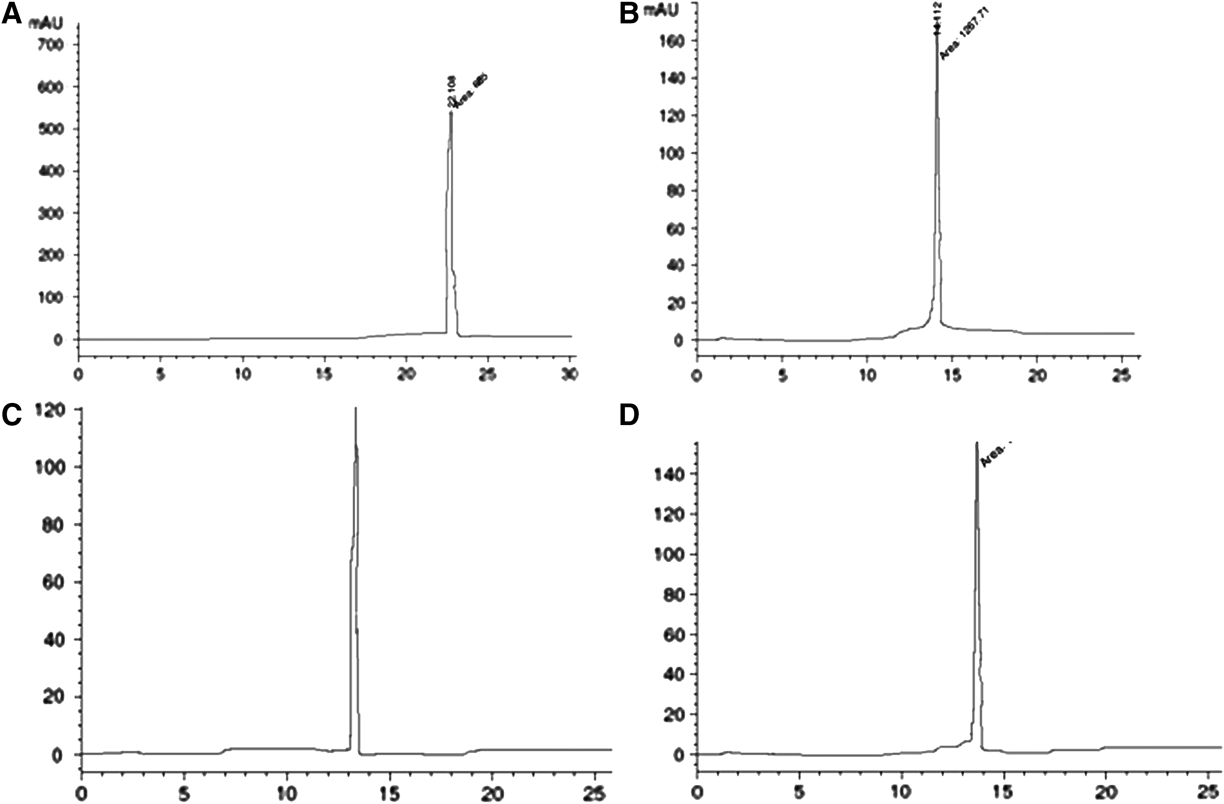
HPLC profile of all four multiple antigen peptides
Cutoff value calculation
A total of 45 CHIKV-positive, 20 dengue-positive, and 25 healthy individual sera were screened in ELISA using the E2 protein and all four E2 MAPs. Based on the seroreactivity of the 25 healthy sera tested for IgM immunoreactivity, the cutoff values for different E2 MAPs were as follows: 0.50 (MAP 1), 0.55 (MAP 2), 0.49 (MAP 3), and 0.54 (MAP 4). Based on the cutoff value for these E2 MAPs, we examined the seroreactivity of dengue-positive and CHIKV-positive sera. None of the 20 dengue-positive sera showed any significant seroreactivity with any of the E2 MAPs.
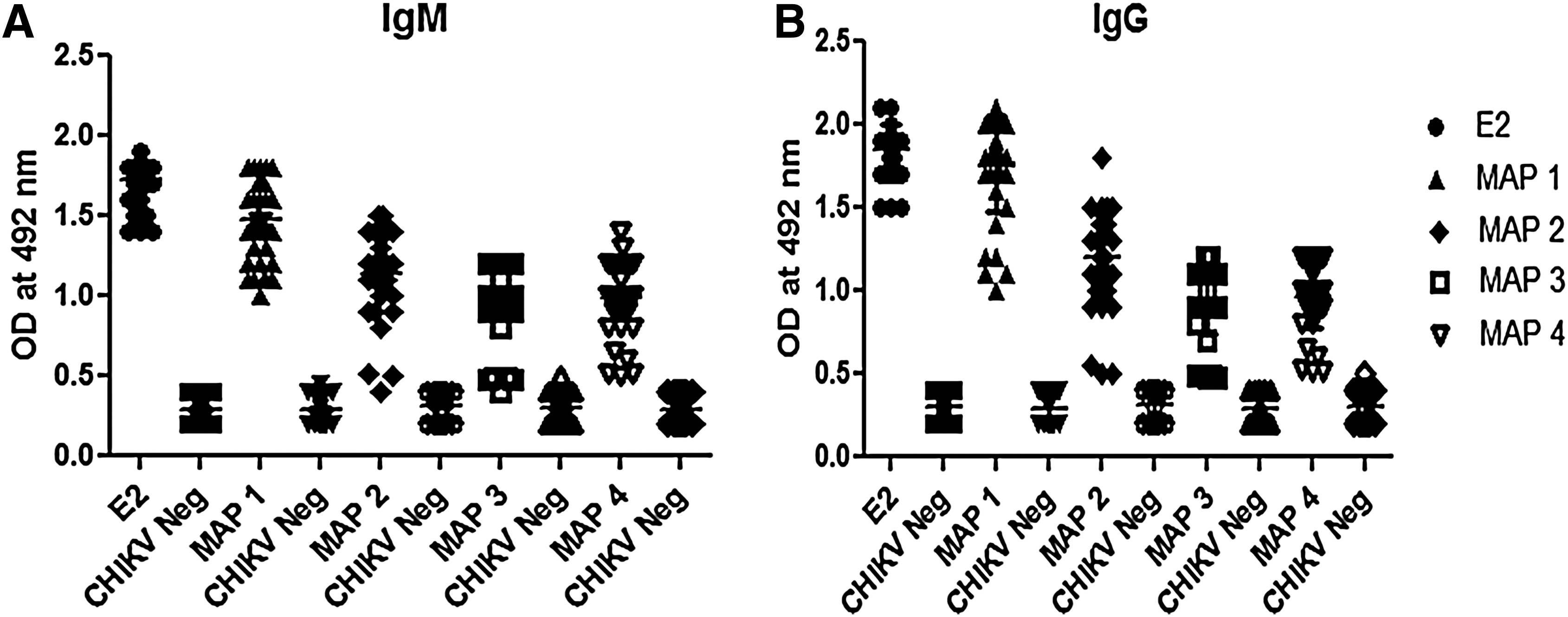
Reactivities of IgM and IgG antibodies with the combination of peptides MAP 1, MAP 2, MAP 3, and MAP 4 by ELISA
Evaluation of 45 CHIKV-positive sera with the calculated cutoff value revealed that the MAP 1 construct had the best combination of E2 peptides among the four MAPs studied. Figure 2 shows that IgM antibodies from all 45 patients (100%) reacted with MAP 1 and their positive value was higher than the calculated cutoff value (0.50). The mean±SD value of these positive sera is 1.83±0.17. These patients also showed 100% seroreactivity for IgG antibodies with a mean value of 2.0±0.15 (Fig. 2). The mean value of E2 antigen for IgM and IgG was 1.85±0.17 and 2.1±0.16, respectively. When the sera from the same patient was used to detect IgM antibodies using MAP 2, MAP 3, and MAP 4, a sensitivity of 97.7%, 91.1%, and 95.5% was observed, respectively (Table 1). With MAP 2, 3, and 4, the mean value of IgM antibodies observed was 1.3±0.12, 1.2±0.15, and 1.2±0.14, respectively, and the mean value of IgG observed was 1.43±0.12, 1.12±0.15, and 1.24±0.14, respectively. So far it is evident that the MAP 1 antigen showed a sensitivity equivalent to E2 antigen and thus it is best among all four E2 combinations studied for the diagnosis of CHIKV.
Discussion
In the present study, we describe a novel MAP-based immunoassay for the detection of CHIKV-reactive antibodies. Previous reports, including those from our own laboratory, had reported many peptides that are targets for antibody detection in CHIKV patients (46). These peptides showed significant sensitivity in immunoassay for CHIKV serodiagnosis. However, studies have reported that the sensitivity of an immunoassay based on antibody detection can be improved by using multiple peptides instead of using a single peptide (41). Thus, to determine the best performing group of peptides with maximum sensitivity and specificity, we synthesized four MAPs covering nine epitopes of E2 antigen of CHIKV.
MAP offers several advantages such as simple and convenient in design and synthesis (20), can be designed for multiple epitopes or multiple copies of a single epitope (36), highly immunogenic and antigenic, has a protein-like globular structure, noncytotoxic, biocompatible, resistant to proteolysis, easy standardization (2), and a multimeric nature, which provides reproducible result and increases surface binding. So MAP has been studied as an alternate source of antigen exploited for diagnosis (12). This study also indicates MAP as an antigen of choice for serodiagnosis of CHIKV.
Among all the antigens of CHIKV, the extensively studied envelope E2 is the most immunodominant viral protein, inducing a strong antibody response during acute phase of the disease (18). Also, the E2 protein generated a long-lasting immune response that is detectable even after 2 years of postinfection (17). In our previous study, we have identified many antigenic peptides that span the entire E2 protein. We observed that epitopes recognized by IgM, IgG, and IgG3 antibodies are almost consistent with the tested sera. Out of 17 synthetic peptides of E2 protein studied, E2-p3, E2-p11, E2-p16, and E2-p17 were found to generate both IgM and IgG antibodies with a sensitivity ranging from 91% to 95%. Along with these, other peptides like E2-p5, E2-p7, E2-p9, E2-p10, and E2-p15 also showed sensitivity in the range of 73% to 87% (22).
In the current study, we designed four different combinations of peptides to find out the best combination that gives the maximum sensitivity and specificity. The MAPs synthesized were as follows: MAP 1 (E2-p16, E2-p11, E2-p3, and E2-p17); MAP 2 (E2-p5, E2-p7, and E2-p3); MAP 3 (E2-p10, E2-p9, and E2-p11); and MAP 4 (E2-p 16, E2-p17, and E2-p15). These were used as coating antigens in ELISA for screening 45 CHIKV-positive sera collected from different epidemic regions of India. We observed that the epitopes recognized by individual patients are rather homogeneous and the use of four immunodominant peptides E2-p16, E2-p11, E2-p3, and E2-p17 together in the form of MAP 1 showed 100% sensitivity and specificity with all the patient sera.
Another combination of peptides E2-p5, E2-p7, and E2-p3 in the form of MAP 2 detected 97.7% of CHIKV-positive sera tested. The MAP 3 (E2-p10, E2-p9, and E2-p11) and MAP 4 (E2-p 16, E2-p17, and E2-p15) also showed high sensitivity of 93% and 97.7%, respectively. Interestingly, when all the four MAPs were mixed together and tested for antibody detection in the sera, a decrease in sensitivity (97.7%) was observed. The reason for the same is not known, but it could be due to masking of one MAP with the other when coated on the ELISA plate. This study includes patients from different epidemic regions of India, but whether the same MAP will be recognized by sera of chikungunya patients from other geographical regions needs to be studied in the near future.
To the best of our knowledge, this is the first study describing the use of MAP for the diagnosis of CHIKV. This study describes MAP 1 as a promising tool for the use in serodiagnosis of CHIKV.
Conclusion
We screened four MAPs with different combinations of antigenic peptides as a potential diagnostic marker for CHIKV infection in an ELISA system. The reactivity of same sera with all the four MAPs synthesized was tested, and interestingly, our results indicate that MAP 1 constitutes highly immunogenic peptides with a sensitivity close to the native E2 antigen. Our data established MAP 1 as the diagnostically most efficient reagent for CHIKV in terms of sensitivity and specificity as compared to peptides alone.
Footnotes
Acknowledgment
The authors are grateful to the Department of Biotechnology (DBT) and the Defense Research and Development Organization (DRDO), New Delhi, for financial support.
Author Disclosure Statement
No competing financial interests exist. There are no conflicts of interest among the authors.