




Abstract
This study describes an erythema multiforme (EM) in breeding sows, after their mixing in the group housing system. Sows at 30–35 days of gestation showed red and raised skin areas, depression, anorexia, fever, respiratory problems, and increased return to estrus. Blood and nasal samples from diseased sows were examined by quantitative polymerase chain reaction and enzyme-linked immunosorbent assay for respiratory pathogens. Hematological and biochemical analyses were performed on the blood samples. From diseased sows, vaginal swabs for microbiological examinations and samples at slaughterhouse for gross and microscopic examinations were collected. Samples from the complete gestation and lactation feed were examined for mycotoxins. All sampled sows were seropositive for Actinobacillus pleuropneumoniae (App) and porcine reproductive and respiratory syndrome virus (PRRSV). No viremia for PRRSV and porcine circovirus type 2 were detected. All nasal samples were positive for Streptococcus suis, one for Swine Influenza Virus and one for App, Hemophilus parasuis, and S. suis. In all vaginal swabs, Escherichia coli and Streptococcus spp. were detected. Diseased sows had moderate leukocytosis, mild anemia, and thrombocytopenia. No mycotoxins were detected in feed. Histopathological examination revealed increased vascularization of the superficial and middle dermis. EM was likely due to illness caused by viral and bacterial infections. This study suggests that stress caused by the sows' mixing might have triggered the problem.
Introduction
E
Internal diseases causing skin lesions or skin abnormalities in pigs such as abnormal color changes include bacterial (e.g., erysipelas, salmonellosis, pasteurellosis, pleuropneumonia, Glässer disease) or viral (e.g., classical swine fever and African swine fever, and dermatitis/nephropathy syndrome due to porcine circovirus type 2 [PCV2]) infections (2,28,30). It has been suggested that any septicemia or toxemia can cause erythema or cyanosis characterized by red to purple discoloration, especially on the extremities, and easily seen in white breeds (2).
Recently, a directive issued by the European Union (EU) requires stopping the use of individual sow stalls during the gestation period for animal welfare reasons, as from January 1, 2013 (4). Group housing of sows compulsory within the EU stipulates that sows and gilts be kept in groups from 4 weeks after service to 1 week before the expected time of farrowing (4). However, there are substantial data in the scientific literature on the impact of individual versus group housing of pregnant sows on reproduction, animal well-being, management, and health problems (e.g., injuries) (27). Sows housed in groups at weaning and regrouped after insemination experienced higher stress than sows housed in individual stalls at weaning and mixed in groups after insemination (22). It is known that physical and psychological distress can suppress immune function in animals, leading to an increased incidence of infectious disease. The innate defense mechanisms of an animal may not function optimally when the animal is immunosuppressed due to stressful conditions, a preexisting viral infection, immunotoxicants, or nutritional factors (23).
The present case study investigated EM in breeding sows of a commercial breeding herd (including gilts and sows of all parities), after mixing them at 30–35 days of gestation in a group housing system.
Materials and Methods
Description of the farm
The present study reports on a breeding stock of a farrow-to-finish commercial pig farm (commercial hybrids of Large White × Landrace, Topigs Norsvin). The capacity of the farm was 450 sows under production, located in Central Greece. A grandparent nucleus of 40 sows was kept in the farm for producing its own gilts. The farm facilities included 12 farrowing houses (10 pens), 18 flat-deck units (2 pens × 55 animals), growing houses (46 pens × 50 animals), one finishing house (4 pens × 40 animals), one mating-pregnancy (dry period) stable with 160 individual stalls (35th–105th day of pregnancy), two breeding stock house of group housing (18 pens × 10 sows—0–35th day of pregnancy), one breeding stock house of group housing for noninseminated gilts (5 pens × 25 gilts), a feed mill, and an artificial insemination (AI) laboratory. The herd practiced a 1 week batch production system. The weaning piglets were allotted equally according to the body weight and sex at random to 36 pens (22 piglets per pen) at flat-deck batteries for piglets in a climate-controlled post-weaning stable.
Weaned sows were housed in individual stalls, and they were inseminated twice with fresh semen from the same boar. Semen collection, dilution, and storage were performed in the farm (system “Do-it-yourself AI”). At 30–35 days of gestation, sows were removed to group housing rooms where they were housed with 10 animals per group. The sows were fed individually using separate feeding stalls. The gilts were also housed individually during the first month of gestation.
The vaccination scheme of breeding stock and weaners that was applied in the farm is shown in Table 1. All breeding females were treated with a single ivermectin injection 14 days prior to farrowing; the boars were treated twice a year.
ADV, Aujeszky's disease virus; Parvo, parvovirus; AR, atrophic rhinitis; Ery, erysipelas; PRRSV, porcine reproductive and respiratory syndrome virus; E. coli, Escherichia coli; Cl. perfringens, Clostridium perfringens; PCV2, porcine circovirus type 2; M. hyo, Mycoplasma hyopneumoniae.
The feed provided to the animals was self-prepared based on a corn/barley/wheat–soya meal, depending on the season. The breeding animals received a different feed during gestation and lactation (Table 2).
Drinking water was provided for ad libitum consumption by the animals. There was one drinking nipple per sow during lactation, and one nipple in each crate during gestation.
Housing facilities had fully automated temperature and humidity control system, as well as automated feeding. During the study, feed was manually provided for ad libitum consumption in order to monitor feed consumption.
The farm was seropositive to PRRSV and PCV2. One year before the study, the farm had experienced an outbreak of PRRSV infection in breeding stock and weaning to finishing stage. Diagnosis was based on clinical signs (mainly premature farrowings, decreased number of live born piglets, agalactia and anorexia in lactating sows, and increased secondary respiratory bacterial co-infections in weaners and growers/finishers) and subsequent real-time polymerase chain reaction (RT-PCR) in serum samples from pigs of all ages (PCR positive breeding stock 30%, weaners 90%, and growers/finishers 35%).
Case study
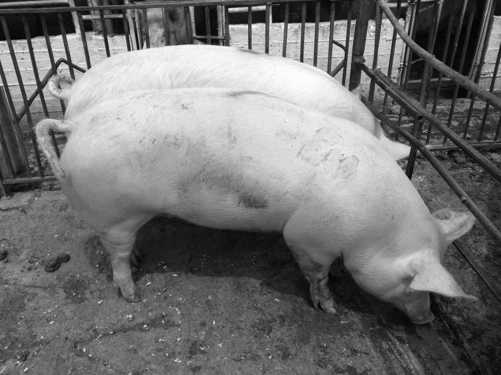
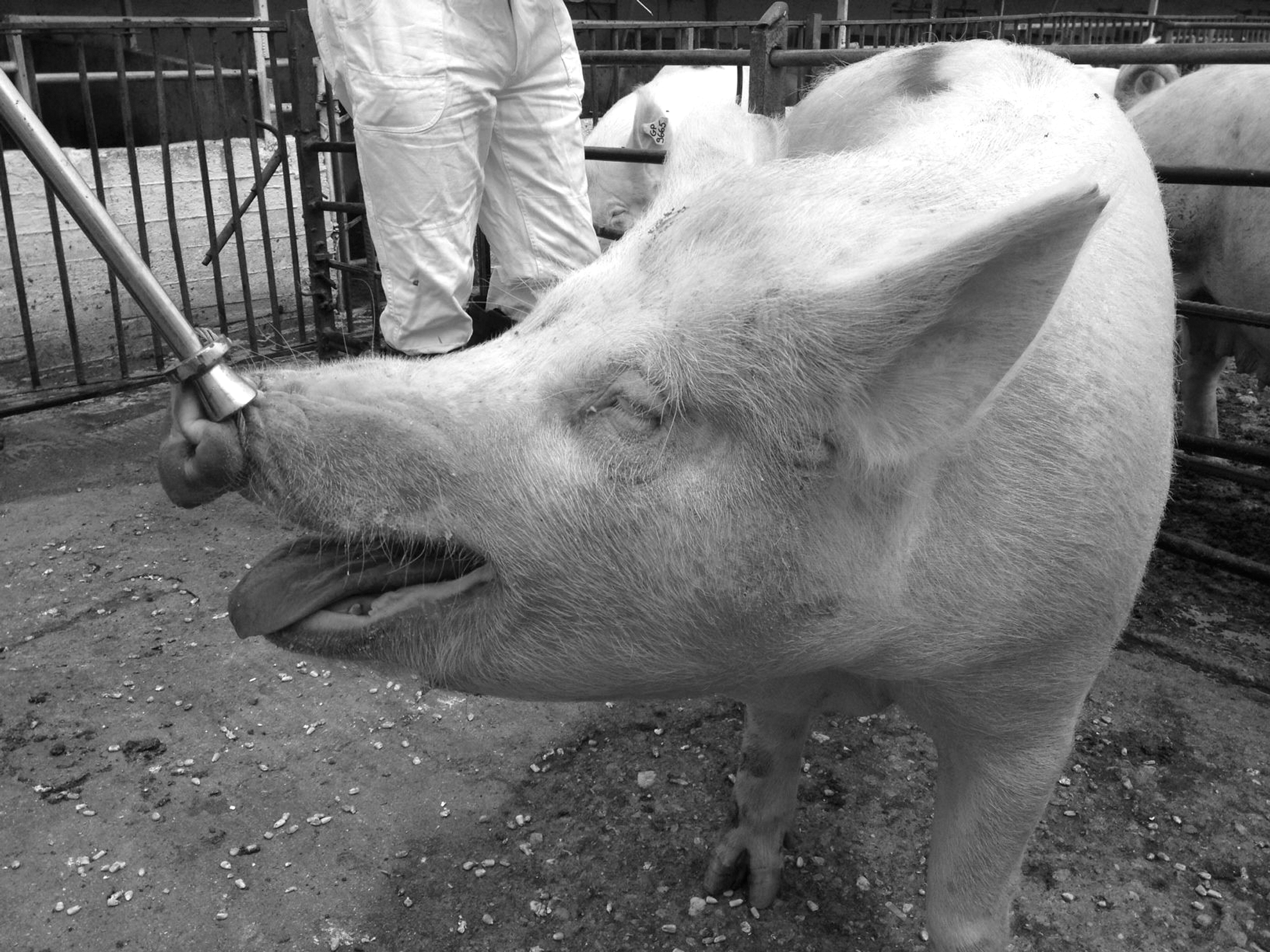
The current case study was manifested by EM in the majority (90%) of breeding stock, in combination with respiratory signs and appetence but without mortality. Since January 2014, up to this clinical examination in May 2014, the sows were moved after artificial insemination to a group housing, and after mixing (around 30–35 days of gestation), they developed EM, characterized by symmetrical, red, raised skin areas that appeared all over the body, although they seemed to be more noticeable on the neck and the face, especially around the eyes and ears (Figs. 1 –8). Moreover, the diseased sows showed depression, decreased appetite, high fever (40–41.5°C), stiffness and posture difficulties, as well as respiratory signs. The respiratory signs were characterized by moderate breathing difficulties, eye and nasal discharge, accompanied by mucus and blood in severe incidences (Figs. 6 –8). The above symptoms were not detected in lactating and recently weaned sows (during the first month of gestation) or in the building of newly introduced gilts that were not yet inseminated. This clinical picture was obvious in the majority of pregnant sows after mixing them in the group housing system (about 90%), and the symptoms remained for several weeks. The sows also showed depression and decreased appetite, and few of them had a fever for 1–3 days. The aforementioned symptoms were noticed in all breeding stock, including sows and gilts, and in sows or gilts of the grandparent nucleus.

Diseased sow in the dry period with EM, characterized by red, raised skin areas that appeared all over the body. EM, erythema multiforme.
Diseased sows with high fever (40–41.5°C) and EM, characterized by symmetrical, red, raised skin areas that appeared all over the body.
Diseased gilts in the dry period with EM, characterized by symmetrical, red, raised skin areas.
Diseased sow with EM, characterized by red, raised skin areas on the neck and the face, especially around the eyes and ears.
Diseased sow in the dry period with EM, characterized by red, raised skin areas on the neck and around the eyes and ears.
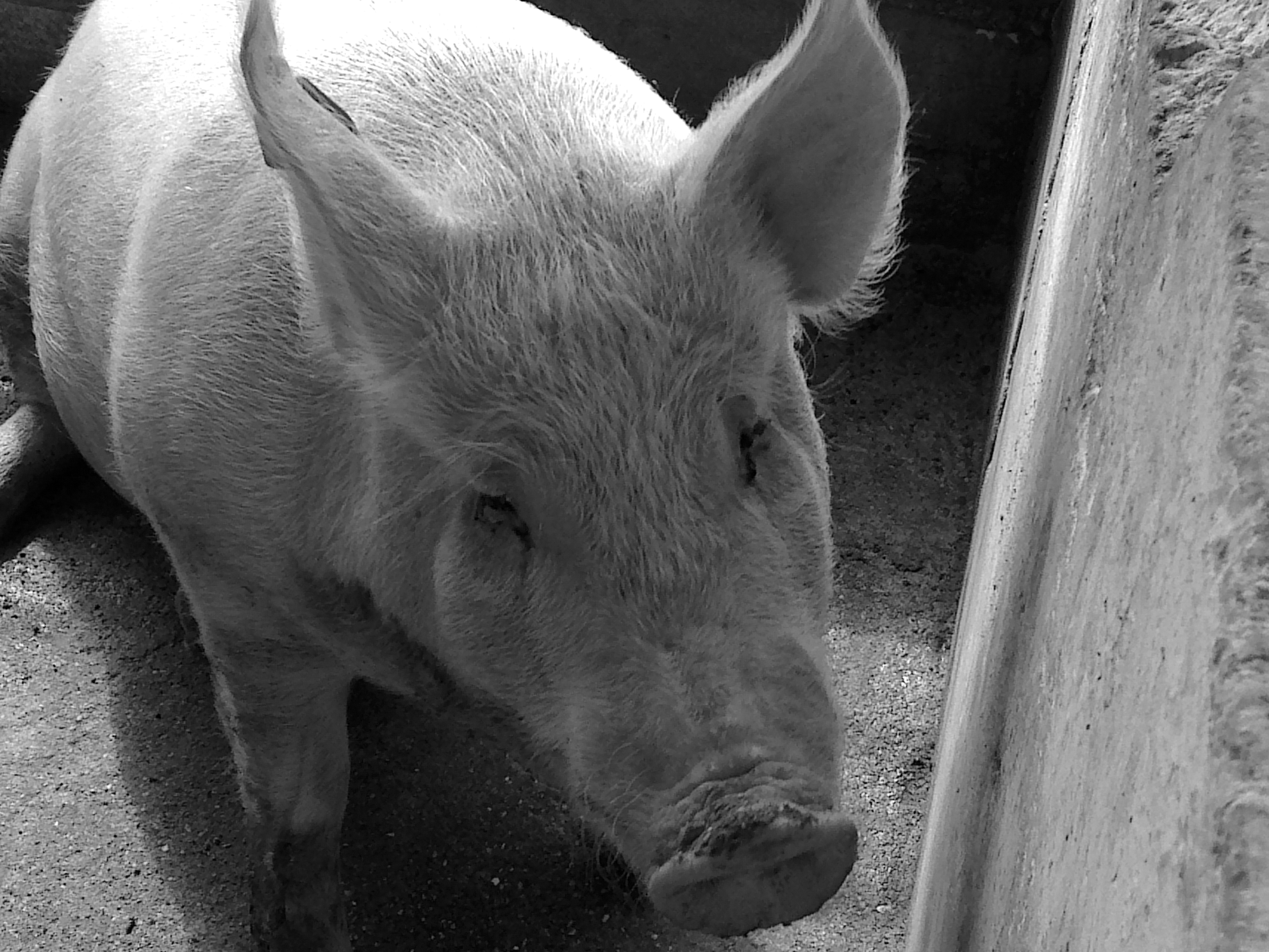
Diseased sow with depression, high fever (40–41.5°C), moderate breathing difficulties and eye and nasal discharge.
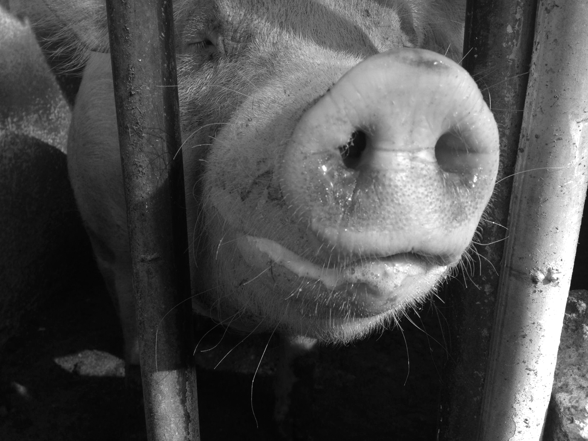
Diseased sow with high fever (40–41.5°C), and severe nasal discharge, accompanied with mucus and blood.
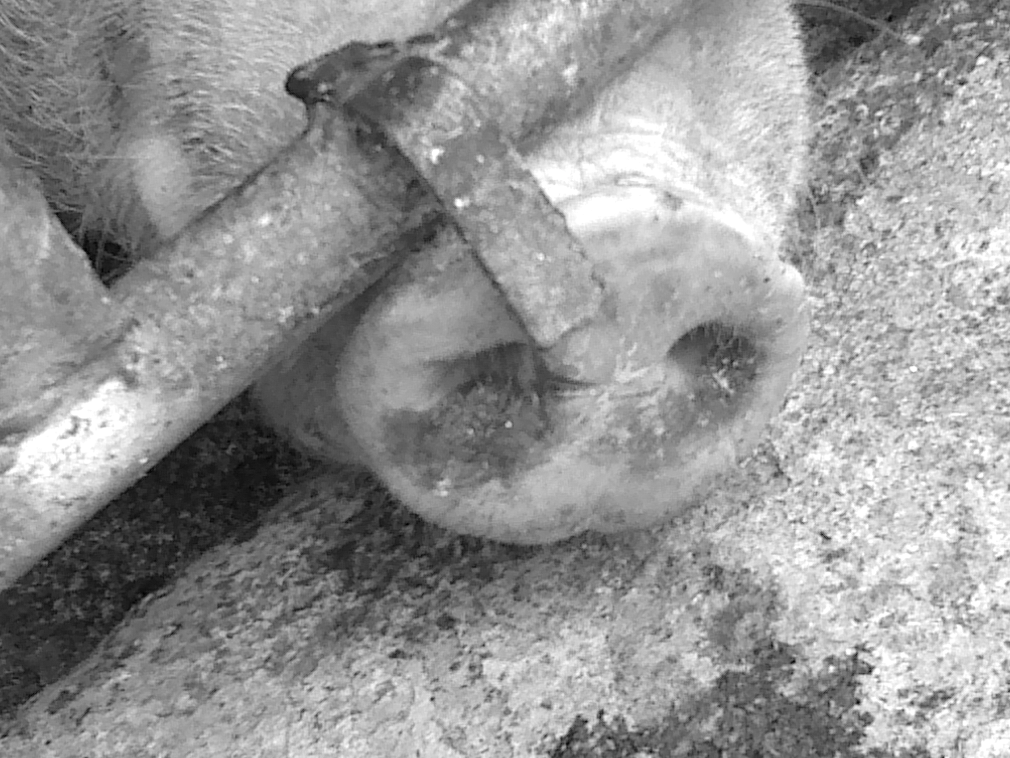
Diseased sow with depression, anorexia, high fever (40–41.5°C), and severe respiratory signs (breathing difficulties, eye and nasal discharge, accompanied with mucus and blood).
In addition, all inseminated gilts showed similar clinical signs, mainly after the first month of gestation, when they moved to the group housing room. Shortly after mixing them in the group housing system, 10–15% of inseminated sows and gilts returned to estrus (Table 3).
Sampling and laboratory examinations
In May 2014, blood samples from seven diseased sows and nasal swabs from four diseased sows other than those that were blood sampled were collected. The sampled sows were at 30–45 days of gestation and ranged from parity 1 to 5. In addition, in August 2014, nasal samples from seven diseased sows were collected. Moreover, in May 2014, vaginal swabs were collected from the same four diseased sows from whom nasal samples were collected.
Serum blood samples were examined by (a) quantitative PCR (qPCR) for PRRSV (European PRRSV type 1—PRRSV EU—and American PRRSV type 2— PRRSV US) and for PCV2; and (b) enzyme-linked immunosorbent assay for Actinobacillus pleuropneumoniae (App) antibodies (Abs), Erysipelothrix rhusiopathiae (Ery) Abs, PRRSV Abs, Aujeszky gE Abs (positive: field virus infected—antibodies against ADV gE), African Swine Fever Virus (ASFV) Abs, classical swine fever virus (CSFV) Abs, and Leptospira spp. Abs (L. Pomona, L. tarassovi, L. canicola, L. grippotyphosa, and L. Bratislava).
Nasal swabs were examined by PCR for apx-IV gen of App, Bordetella bronchiseptica, Haemophius parasuis, Pasteurella multocida, Streptococcus suis, porcine cytomegalovirus (PCMV, inclusion body rhinitis), and Swine Influenza Virus (SIV; H1N1, H3N2, H1N2).
In addition, two blood samples, (with anticoagulant) from sows were collected for blood culture, using strict aseptic technique and sterile equipment. The samples are inoculated into the blood culture bottles (Oxoid SIGNAL Blood Culture System) and mixed with the medium. The sampled sows had not received any antibiotic treatment.
Moreover, hematological and biochemical analyses were performed of the blood samples. At the slaughterhouse, samples from seven diseased sows with typical symptoms (skin, liver, kidney, lung) for gross and microscopic examinations were collected.
In addition, samples of the complete feed from gestation and lactation feed were examined for mycotoxines (Aflatoxines [B1, B2, G1, G2], Deoxynivalenol [DON], Acetyldeoxynivalenol [acetyl-DON], Nivalenol, Zearalenone [ZEN]), using high-performance liquid chromatography (HPLC; Varian 9010 and 9050). The limits of detection of each mycotoxin were: 0.4 μg/kg for aflatoxines (B1, B2, G1, G2), 16.3 μg/kg for DON, 22.2 μg/kg for acetyl-DON, 17.1 μg/kg for nivalenol, and 1.4 μg/kg for ZEN.
Results
The results of ELISA and PCR in serum blood samples are shown in Table 4. All sows were seropositive for App and PRRSV, but seronegative for ASFV, CSFV, ADV, and Leptospira spp. In addition, no viremia for PRRSV and PCV2 was detected.
PRRSV antibody ELISA results: <0.4 negative; 0.4–0.99 positive 1; 1.0–1.49 positive 2; 1.5–1.99 positive 3; 2.0–2.49 positive 4; 2.5–2.99 positive 5; ≥3.0 positive 6.
Positive, field virus infected (antibodies against AK gE); negative, not infected with field virus (no antibodies against AK gE).
L. Pomona, L. tarassovi, L. canicola, L. grippothyphosa, L. Bratislava: all samples were < 1:100.
PCR, polymerase chain reaction; ELISA, enzyme-linked immunosorbent assay.
The results of nasal swabs are presented in Table 5. B. bronchiseptica, P. multocida, and PCVM were not detected in any sample. S. suis was detected in all samples; one sample (No 11) was positive for App, H. parasuis, and S. suis. One sample was positive for SIV in the May 2014 sampling.
Examination of the vaginal swabs revealed E. coli and Streptococcus spp. infections in all samples. The sensitivity testing (according to CLSI1) of E. coli and Streptococcus spp. in vaginal swabs is presented in Table 6.
According to the manufacturer's guidelines.
Sensitive; **resistant.
Streptococcus spp. and Actinobacillus spp. were isolated from blood culture, while the antibiogram revealed that both of them were sensitive to the following antibiotics: penicillin, ampicillin, and a combination of amoxicillin + clavulanic acid (Table 6).
The results of hematological and biochemical analyses of the blood samples from diseased sows are shown in Table 7. The complete blood cell (CBC) count with differential revealed moderate leukocytosis with atypical lymphocytes and lymphopenia, possibly secondary to the depletion of CD4 lymphocytes. An eosinophil count >3,000/μL was also seen in two cases. Neutropenia occurred in the most severe case and may be indicative of poor prognosis. A severely elevated total white blood cell (WBC) count in two cases was consistent with infection. Mild anemia was also present, and thrombocytopenia was found in one case.
HCT, hematocrit; RBC, red blood cells; Hgb, hemoglobin; MCV, mean corpuscular volume; MCHC, mean corpuscular hemoglobin concentration; PLT, platelet count; WBC, white blood cell count; NEU, neutrophil count; LYM, lymphocyte count; MONO, monocyte count; EOS, eosinophil count.
Values out of reference range are presented in bold.
The histopathological examination of the skin revealed increased vascularization of the superficial and middle dermis mainly (Fig. 9). No remarkable lesions were found in the other examined organs.

Histopathological examination of the skin: increased vascularization mainly of the superficial and middle dermis.
The examination of completed gestation and lactation feed did not reveal mycotoxins at detectable levels in the diet of diseased sows. The detection of Aflatoxines (B1, B2, G1, G2) (<22 μg/kg), DON (<250 μg/kg), acetyl-DON (<250 μg/kg), Nivalenol (<250 μg/kg), and ZEN (<50 μg/kg) was below the contamination limits.
Management practices—treatments
In May 2014, based on the results from vaginal swabs for the treatment of E. coli and Streptococcus spp. infections of reproductive system, injectable amoxicillin + clavulanic acid was applied in all sows on the day of weaning, while in the first week of the introduction of sows/gilts to the group housing room, 400 ppm of amoxicillin were administrated in the feed. In addition, to reduce the negative effects of fever on the reproductive performance of sows (e.g., returns to estrus; Table 7), acetylsalicylic acid powder was administrated using top dressing for 3 days in diseased sows. Finally, to reduce fighting between sows, a commercial product (Anty—Gryz, Over Group Sp. Z.o.o. Sp. K., Poland) based on herbs (e.g., herb wormwood, marigold flowers, crumble chamomile, sage leaf) was used locally on the skin around the neck and upper back. This product has an extremely bitter taste, discouraging aggressive attack and aggression.
In August 2014, based on the laboratory results, vaccination against SIV and H. parasuis was included in the routine vaccination program of sows and gilts, and against App in the routine vaccination program of replacement gilts.
Discussion
Generally, EM presents with a skin eruption characterized by a typical target (iris) lesion, raised spots, or other lesions on the skin, and there may be mucous membrane involvement. The reason for EM is not known in animals and humans (17). It is a type of hypersensitivity reaction, caused by a reaction to an infection, certain medications, or illness, but oftentimes the cause is unknown (3,12,18,20,25). Animal EM is very different from 90% of human EM, which is herpes virus associated (32). EM has been reported in dogs after Canine parvovirus-2 infection (31), as well as in minipigs (9) after stress factors and in wild animals resulting from an aberrant immune response, such as after vaccination (7,10). Animal EM is often attributed to drugs, but this is rarely proven (32).
EM reported in the Gottingen minipig has a close resemblance to the syndrome called “Dippity Pig Syndrome” observed in Vietnamese potbelly pigs. Red streaks on the back of the pig develop rapidly. Soon, they start oozing, and the pig arches its back and may vocalize in pain. The condition appears to be stress related (9). In the present study, even if the clinical picture was not similar to the reported signs in the minipig, stress as a trigger factor is common point. A possible trigger for the incidence of the current clinical disease was probably the stress under the group housing conditions. Stress impairs immune functions (24), so infections are more likely to affect pigs under the stressful conditions of the group housing system. In addition, stress may cause a subclinical infection to be activated (23). In the present case, it is possible that inseminated sows or gilts pigs were subjected to “stress” at a time when their immunity status was low, making them more susceptible to becoming infected and developing disease. It is known that crowding and mixing may cause stress, negatively influencing the immune function (14) and immune responsiveness (23). In group housing conditions, fighting happens between sows, who may injure one another, as well as these conditions causing fear and anxiety (5,6,13,29). Aggression due to mixing of unfamiliar animals is inevitably associated with physiological and psychological stress in animals due to competition for access to limited resources, or to establish a social relationship between unfamiliar animals (19,26). However, it is impossible to eliminate aggression in the group housing system (27).
In the present study, 2 months after the start of vaccination against H. parasuis and SIV, the clinical signs characterized by EM—respiratory problems, appetence, and reproductive performance (Table 3)—decreased dramatically, and the general picture in the breeding stock was normal. Based on the current clinical picture of the farm, the vaccinations against H. parasuis and SIV and antibiotic treatment against S. suis infection led to the significant improvement of the health status in the farm, especially with regard to skin condition and reproductive parameters (Table 3). Sows are reservoirs of H. parasuis in infected herds, and the clinical disease is often associated with stress, especially during transport of pigs (21). Furthermore, S. suis can usually be found in the vaginal tracts of sows or gilts (8), and it has a very similar within-herd epidemiology compared with H. parasuis (21). In the present case study, aggression due to mixing sows in the group housing system after insemination was possibly a main trigger factor of decreasing their immune status, resulting in outbreaks of H. parasuis and S. suis infections, especially in gilts and young sows, who were immunologically naïve before the vaccination and then mixed with other enzootically infected older animals. As EM is a hypersensitivity reaction usually triggered by infections or illness, the possible explanation of the clinical manifestation of EM in the present case study could be fever (due to bacterial infections from S. suis, H. parasuis, and App) and possibly the stress after mixing. The possibility of SIV infection to be included in the etiology is low, as it had not spread to other sows, and normally it should not only have occurred in sows after mixing them in the group housing system.
Based on the clinical, clinicopathological, and pathological findings, a diagnosis of EM was documented. EM is a skin condition possibly mediated by deposition of the immune complex in the superficial microvasculature of the skin that usually follows an infection or drug exposure and other various triggers. In the present study, under stressful group housing conditions, a subclinical infection or an interaction of different respiratory pathogens seems to have been activated, affecting negative the health status and performance of the breeding stock.
Footnotes
Acknowledgments
The authors acknowledge the participating pig producer Thedoros Xiromeritis for his technical assistance.
Author Disclosure Statement
No competing financial interests exist.