
Abstract
For induction of an appropriate immune response, especially in the case of an inactivated vaccine, the use of an adjuvant is crucial. In this study, adjuvanticity effect of G2 dendrimer in veterinary rabies vaccine has been investigated. A nonlinear globular G2 dendrimer comprising citric acid and polyethylene glycol 600 (PEG-600) was synthesized and the toxicity was studied in vitro on the J774A.1 cell line. The adjuvanticity effect of the dendrimer was then investigated on rabies virus in NMRI mice as a model. Different concentrations of dendrimer were used to determine the best formulation for the survival of the mice after virus challenge. The rise of neutralizing antibody was also checked by rapid fluorescent focus inhibition test (RFFIT). The relative potency of the prepared formulation was finally calculated using standard NIH test and the results were compared (and discussed) with the commercially available rabies vaccine. The accuracy of dendrimer synthesis was confirmed using Fourier transform infrared (FT-IR), size, and zeta potential analysis. The in vitro toxicity assay revealed that no significant toxic effect is observed in cells when data are compared with the control group. The in vivo assay showed that a higher survival rate in the mice received a special formulation due to adjuvanticity effect of dendrimer, which is also confirmed by RFFIT. However, the relative potency of that formulation does not give expected results when compared with the alum-containing rabies vaccine. In the current investigation, the adjuvanticity effect of G2 dendrimer was demonstrated for the first time in rising of neutralizing antibodies against rabies virus. Our data confirm that nanoparticles can enhance immune responses in an appropriate manner. Moreover, engineered nanoparticles will enable us to develop novel potent multivalent adjuvants in vaccine technology.
Introduction
R
Currently, with the emergence of nanotechnology, various promising areas are introduced in medicine. Nanotechnology now becomes an effective tool for cell-specific delivery and sustained release, as well as codelivery of antigens in the field of vaccine development (17). Antigen-loaded nanoparticles are being investigated as vaccine delivery systems, as alternatives to the currently used alum, with an objective to develop better vaccine systems and minimize the frequency of immunization. Recently, several studies were conducted on the use of nanoparticles as adjuvants. It was shown that many inorganic nanoparticles such as silver (4,38), gold (10), calcium phosphate (13), and also biodegradable organic nanoparticles, including polymeric nanoparticles (2,12), enhance the immunogenicity of antigens. Dendrimers are classified as highly branched polymeric three-dimensional nanostructures with symmetrical shapes (1). The structure of these materials has a significant impact on their physicochemical properties. Due to the unique behavior of dendrimers, they have a wide range of biomedical and industrial applications (18). The unique features of dendrimers include (a) the ability to precisely control the size and shape, (b) high purity, (c) high capacity for particle loading, (e) low toxicity, (f) creation of branches with different degrees, (g) solubility in water, (h) presence of internal cavities, and (i) multifunctional terminal active groups (6,16, 21,39). The active groups and spaces between branches provide higher capacity and tolerability to encapsulate the guest molecules with different sizes (40). In this study, the adjuvanticity effect of G2 citric acid-based dendrimer in veterinary rabies vaccine has been investigated.
Materials and Methods
Materials
Polyethylene glycol 600 (PEG-600), citric acid, dicyclohexylcarbodiimide (DCC), dimethyl sulfoxide (DMSO), and alum were purchased from Merck. Dialysis membrane (100–500 cutoff) was purchased from Spectrum Co., and the inactivated rabies virus was obtained from Pasteur Institute of Iran (Production and Research Complex).
G1 and G2 dendrimer synthesis
First and second generations of dendrimers (G1, G2) were prepared according to Namazi and Adeli (30). Briefly, one mmole PEG-600, preactivated by 10 mmol DCC, was added to 2 mmol citric acid and the reaction continued for 2 days in DMSO. Dialysis was carried out using a dialysis bag (100–500 cutoff) in phosphate-buffered saline (PBS) solution for 24 h and the solvent replaced every 6 h. First-generation dendrimer was then subjected to the lyophilization process for further analysis and G2 synthesis. Similar to the first generation, 1 mmol of G1 dendrimers was activated with 10 mmol DCC and subsequently added to 6 mmol citric acid. The reaction continued for 4 days, purified using a dialysis bag, and finally lyophilized for in vitro and in vivo analysis.
Structural characterization of G1 and G2 dendrimers
Fourier transform infrared (FT-IR) spectra were measured on a Bruker Model Tensor-27 spectrometer (Kyoto, Japan) for each dendrimer generation. Size and charge distribution of G1 and G2 dendrimers were also investigated by the dynamic light scattering (DLS) technique (Malvern; Zetasizer Nano ZS, Worcestershire, United Kingdom) in double-distilled water at 0.1 mg/mL concentration.
In vitro toxicity
J774A.1 cells (ATCC® TIB-67™) were cultured in Dulbecco's modified Eagle's medium (DMEM) supplemented with 10% fetal bovine serum (FBS) and 1 × Pen-Strep (Invitrogen) at 37°C and 5% CO2. For MTT assay, 20,000 cells/well were cultured in a 96-well plate (Nunc) and incubated overnight at 37°C and 5% CO2. Different concentrations of dendrimer (10−3, 10−2, 10−1, and 1 mg/mL) were added to each well as sextuplicate and incubated for 24 h at 37°C; 100 μL (0.05 mg/well) of MTT (Sigma-Aldrich) was then added to each well and incubated for 4 h at 37°C. The supernatants were removed and 100 μL DMSO (Sigma) was added to each well and the reaction read at 570 and 630 nm.
The adjuvanticity of dendrimer
In sterile conditions, different amounts of G2 dendrimer (0.5, 1, and 1.5 mg) were added to 1 mL of inactivated rabies virus (Lot 92-1; Pasteur Institute of Iran) and the mixture was incubated under gentle stirring at 4°C overnight. Female NMRI mice (∼16 g, 4 weeks old, Pasteur Institute of Iran) were randomly divided into four groups (six mice in each group). Mice were kept at 21°C ± 3°C with 60% humidity under a standard 12-h light/12-h dark cycle. In this study, all animal experiment protocols were approved by the Review Board and Ethics Committee of Pasteur Institute of Iran and conformed to the European Communities Council Directive of November 1986 (86/609/EEC) in such a way to minimize the number of animals used and their suffering. Prepared vaccines (0.5 mL) were intraperitoneally injected on days 0 and 7 into mice. PBS and a commercially available veterinary rabies vaccine containing alum adjuvant (Lot 92-1; Pasteur Institute of Iran) were used as negative and positive controls, respectively. One week later, all groups were intracerebrally injected with 0.03 mL CVS-11 (12 LD50; Pasteur Institute of Iran) under anesthesia. The mice were then monitored for 14 days after 5 days (incubation period) and mortality confirmed by fluorescent antibody test (FAT) using antinucleocapsid FITC-conjugated polyclonal antibody (Bio-Rad).
Neutralizing antibody titration assay
The rising of neutralizing antibodies against rabies virus was determined as previously described (25). Briefly, the isolated serum samples were inactivated by incubation at 56°C for 30 min. Then, 50 μL of CVS-11 (50 FFD50) was incubated with 50 μL of threefold dilutions of the tests and reference for 1 h at 37°C; 5 × 10⁴ BSR cell suspension (a clone of baby hamster kidney [BHK] cells; Pasteur Institute of Iran) was then added to the wells and incubated overnight at 37°C in 5% CO2. The plates were rinsed and fixed by cold acetone. Finally, the plates were stained with 50 μL FITC-conjugated antinucleocapsid polyclonal antibody (Bio-Rad) and the percentage of infection was determined by fluorescence microscopy. The neutralizing antibody titers were calculated using the Reed and Muench method, a simple method for estimating fifty percent endpoints.
Determination of vaccine potency using NIH test
According to results of the adjuvanticity test, the concentration of 12.5 mg/kg (250 μg dendrimer per injection dose) was used for NIH test. Serial dilutions (1:5, 1:25, 1:125, 1:625) of the test and the reference (Rabipur, India, Lot No. 2078) vaccines were prepared and 0.5 mL of each dilution was intraperitoneally injected into 16 NMRI mice (∼13–16 g, 4 weeks old, Pasteur Institute of Iran) on days 1 and 7. On the 14th day, the mice were intracerebrally challenged with 0.03 mL of rabies virus (CVS-11, 20 LD50). For precise LD50 determination of challenge virus, 0.03 mL of each CVS-11 dilution (10−5, 10−6, 10−7, and 10−8) was injected intracerebrally into 10 mice and LD50 was determined using the Reed and Muench method. In addition, for inactivity test, reference and test vaccines were also injected intracerebrally into 10 mice. After a latency period of rabies disease in mice the mice were monitored and the number of sick (hunched back and incoordination), paralyzed (paralysis signs), or dead animals was recorded for 14 days. To confirm the mouse deaths due to rabies disease, the FAT method was performed on mouse brains. Finally, mouse survival was compared with the reference vaccine and relative potency was calculated using the Spearman-Karber method (3). The cutoff values of approved vaccines are at least 1 IU/dose for veterinary vaccines.
Results
FT-IR spectroscopy
In the first-generation spectrum, the peak is seen in the 3,000–3,500 cm−1 region, which shows the presence of a hydroxyl group related to citric acid in the structure of PEG and the peak seen in 2,800–2,900 cm−1 region is a sign of the presence of aliphatic carbon of citric acid and PEG. In the peak of the first generation, the 1,700 cm−1 region peak related to carbonyl citric acid has moved to the 1,600 cm−1 region, which indicates the low concentration of citric acid in the structure of dendrimers and also the conjugation of citric acid to PEG (Fig. 1a). In the second-generation spectrum, the reinforced peak in the 3,000–3,500 cm−1 region shows the presence of hydroxyl group related to citric acid, where its increase is a sign of continuing of conjugation and the appearance of second-generation structures in PEG. Furthermore, observing a reinforced peak in 2,800–2,900 cm−1 region shows the presence and increase of aliphatic carbon of citric acid and PEG due to continuance of conjugation (Fig. 1b). In the second-generation spectrum, a peak in the 1,700 cm−1 region is observed, which represents a reinforced ester bond due to the increase in citric acid, which also confirms the high concentration of citric acid, the dendrimer structure, and the conjugation of citric acid with PEG. With regard to the heightened aliphatic and hydroxyl peaks in the infrared spectrum related to the second compared with the first generation, it could be concluded that the infrared spectrums validate both dendrimer generations considering their functional groups and the peak intensity related to the added factor.
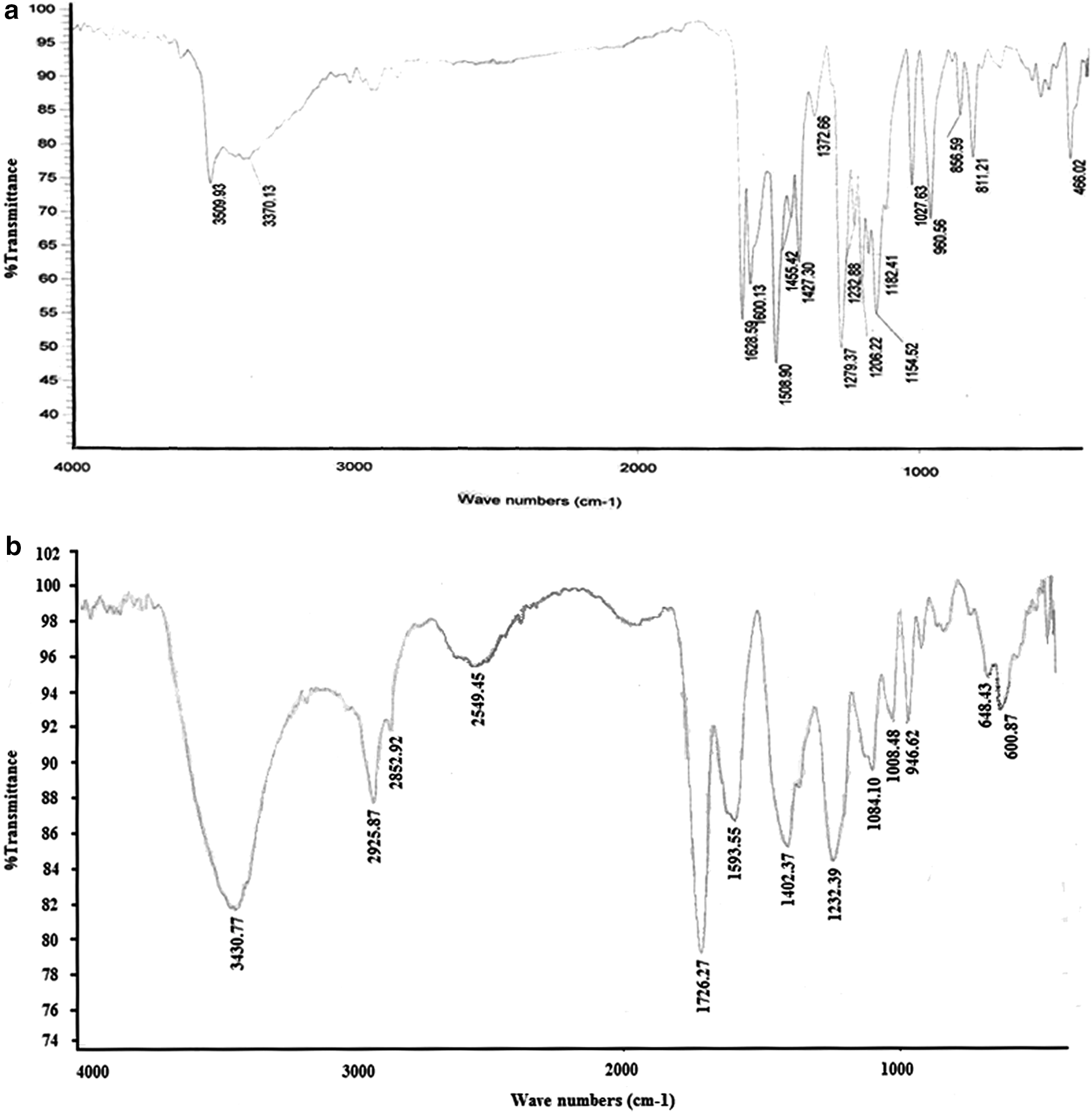
Infrared spectrum of first generation (G1)
Measurement of size and charge
The size and charge of the first generation were determined as 70 nm and −3.5 mV, respectively. This is while the data regarding the second generation showed the 90 nm size and −11.8 mV charge of this dendrimer, which could be due to the increase in the number of citric acid groups on the surface of the second-generation dendrimer as well as an increase in the negative charge (Fig. 2).

The measurement of size and zeta potential of first
Toxicity of dendrimer
In vitro toxicity of G2 dendrimer was investigated using MTT assay on J774A.1 macrophage-like cells. The assay elucidated that the nanoparticles have no more significant toxic effect at all concentrations than cells that received PBS as control (Fig. 3).
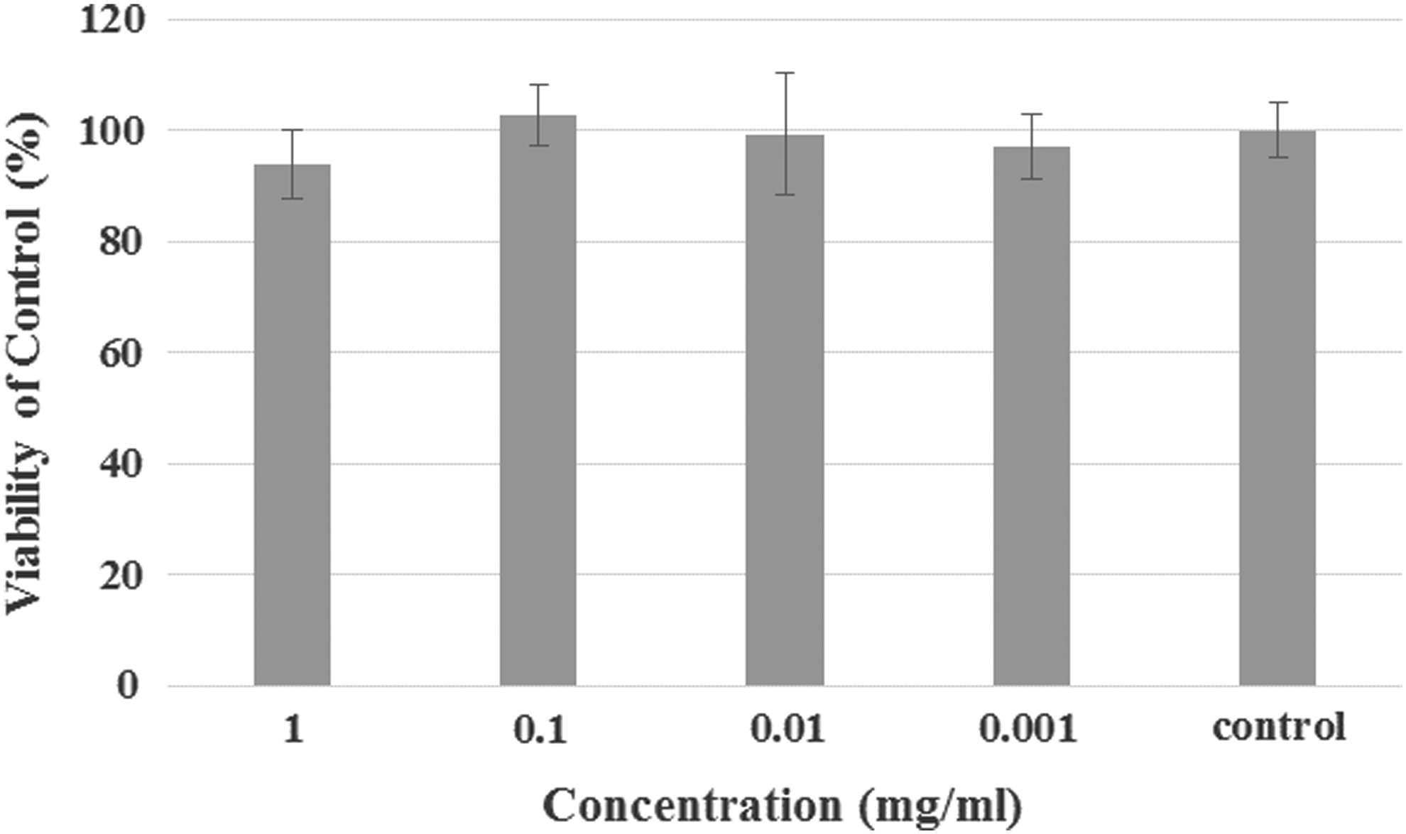
In vitro toxicity assay of dendrimer on J774A.1 cell line. The figure shows that the G2 dendrimer has no significant toxic effect on the cells when compared with the control.
The adjuvanticity of dendrimer
The mortality of mice at the different concentration of dendrimer is shown in Figure 4 and Table 1. The highest percentage of viability was seen at 12.5 mg/kg compared with the PBS group when analyzed with chi-square test (p < 0.05). However, at higher concentrations, there was no significant difference in the numbers of live and dead mice compared with the PBS group. Rabies-neutralizing antibodies were confirmed by neutralizing antibody titration assay (Table 1).
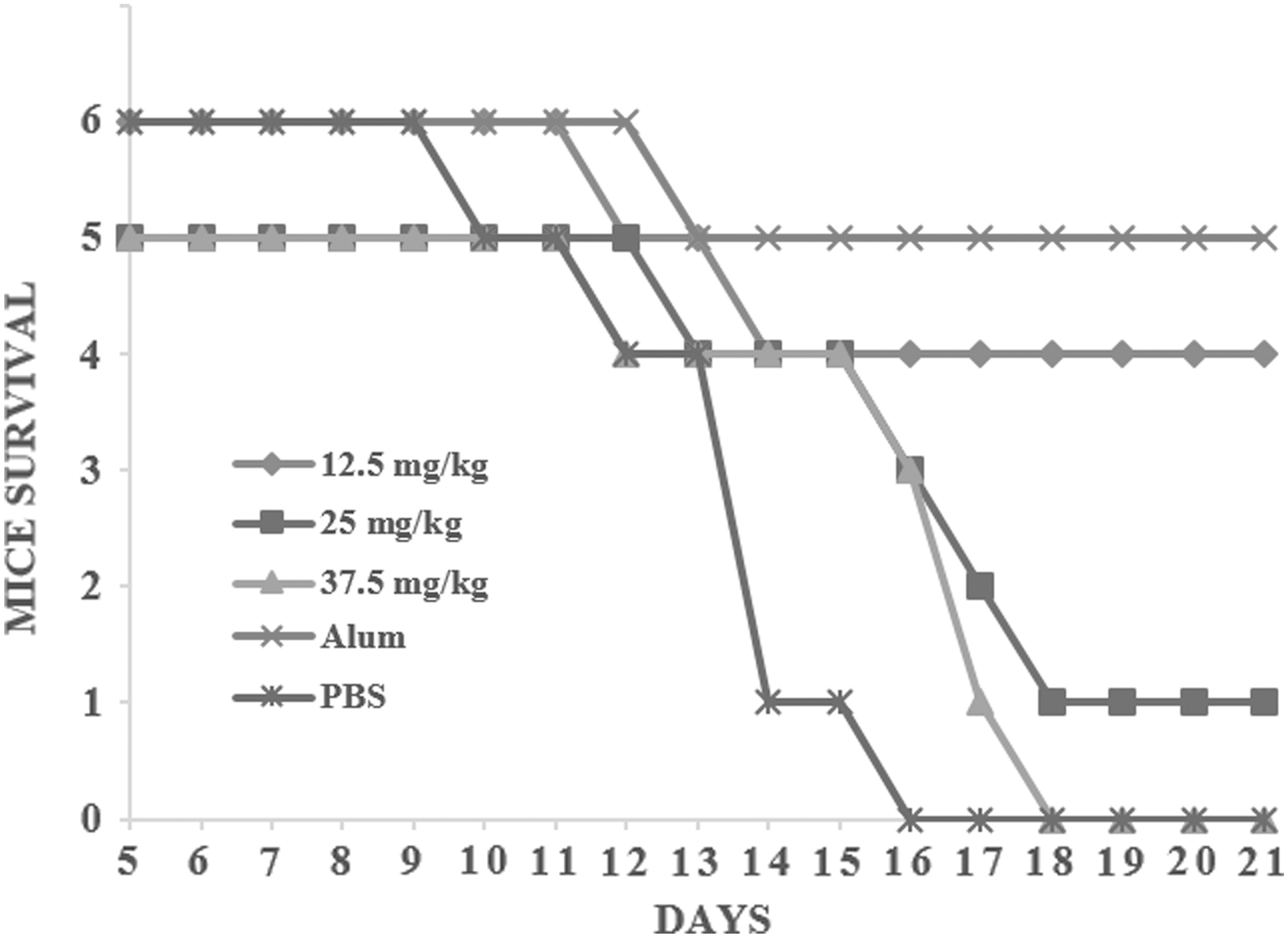
Kaplan–Meier plot of mouse survival at different concentrations of dendrimer-containing vaccines. PBS and alum-containing vaccine were used as negative and positive controls, respectively. The highest survival rate was seen at 12.5 mg/kg, which is comparable with the alum group. alum, aluminum hydroxide; PBS, phosphate-buffered saline.
Commercially available vaccine containing alum and PBS used as positive and negative controls, respectively.
This group was not challenged with live virus.
alum, aluminum hydroxide; PBS, phosphate-buffered saline.
NIH test results
Table 2 shows the mortality of mice in the NIH test. Statistical analysis showed that the calculated potency for alum adjuvant vaccine is 1.757 and, for the dendrimer-containing vaccine, is 0.293.
The potency was calculated using the Spearman–Karber method.
Discussion
Subcellular, killed, or attenuated vaccines do not strongly trigger immune responses such as living microorganisms (32). Therefore, adjuvants are usually added to the formulation to increase the immunogenicity of vaccines and the strength of immune responses (14). The adjuvant must be biodegradable, stable in the formulation or after injection, able to stimulate the desired immune responses, easily produced, and low in cost without any adverse effects (28,31). Currently, only the aluminum salts are widely used as adjuvants and most of them have not received necessary licenses for use in humans. Although in the past decade, a wide variety of new compounds, such as oligonucleotides and emulsifiers, have been evaluated for their adjuvanticity effects, none of them have been commercially available so far. Recently, nanoparticles were evaluated for increasing immune responses against various antigens and the data showed promising results in in vitro or in vivo models (2). These natural or synthetic tiny particles can be micro or nano in size and enhance the quality of immune responses. Association of biomolecules with fine particles, as adsorption or entrapment, is a successful approach to controlling the presentation of antigens to the immune system (34). This provides an enhanced immune response due to a targeted introduction of antigen to specific immune cells such as antigen-presenting cells (APCs).
In this study, the construction of G1 and G2 dendrimers was investigated. The synthesis was carried out through a series of controlled steps, in which the number of chains was gradually and symmetrically increased up to 10 around a central PEG core. The external functional shell of dendrimer provides a multivalent surface or deep cavities, which can react with or entrap biomolecules, respectively. In other words, several antigens can be simultaneously attached to these functionalized surfaces and completely delivered to the immune system to have instantaneously immunological responses. This will lead to development of new multivalent vaccines, reduce the frequency of vaccine administrations, and decrease remarkably medication care costs in public health both for customers and vaccine producers. Reduction in the number of immunizations is more highlighted in livestock vaccination due to the huge number at a time, animal containment, and heavy costs in animal husbandry. In addition, with the use of appropriate chemical linkers between dendrimer and antigens, sustained release or controlled delivery of such immunogens can be achieved in the body.
In the next step, the adjuvanticity effect of G2 dendrimer was investigated by adding the inactivated virus rabies as a model vaccine. For this purpose, different concentrations of dendrimer were added to a constant amount of virus and the preparations were then injected into six mice. Mortality was evaluated after the virus challenge of vaccinated mice according to the mentioned protocol. The preliminary experiment revealed that the mouse group that received the concentration of 12.5 mg/kg elucidated the best adjuvanticity effect with a survival rate of 66%, while the value was 83% for mice that received the commercially available vaccine containing alum adjuvant. Adjuvant effects of nanoparticles have been observed in other studies. He et al. indicated that calcium phosphate nanoparticle plus HSV-2 induces protection against live HSV-2 infection. In addition, this nanoparticle induces very little inflammation at the site of entry and induces a higher IgG2a response and a lower IgE response relative to the responses induced by alum (13). Many different vaccine antigens encapsulated into PLGA nanoparticles were shown to induce broad and potent immune responses, including Plasmodium vivax with monophosphoryl lipid A as adjuvant (29), Bacillus anthracis (22), and hepatitis B virus (HBV) (7,36). Hepatitis B therapeutic vaccines were designed and formulated by loading the hepatitis B core antigen (HBcAg) into PLGA nanoparticles (300 nm) with or without monophospholipid A (MPLA) adjuvant (7). A single immunization with HBcAg encapsulating PLGA nanoparticles containing MPLA induced a stronger cellular immune response than those induced by HBcAg alone or by HBcAg mixed with MPLA in a murine model. More importantly, the level of HBcAg-specific immune responses could be significantly increased further by a booster immunization with the PLGA nanoparticles. These results suggested that codelivery of HBcAg and MPLA in PLGA nanoparticles promoted HBcAg-specific cellular immune responses. In addition, nanoparticle adjuvants derived from inulin such as Advax have shown enhancement of immune response in vaccines against various viruses, including influenza (15) and hepatitis B (35). In addition, carbohydrate-based (23) and polyphenylene-based (27) dendrimers have also been reported to be useful as carriers of peptides for immunization purposes.
In the present study, it was interesting that higher concentrations of dendrimer did not show expected responses and the survival rates in these groups were low. It may be because the best ratio of dendrimer/antigen required to carry or release the antigen for stimulating the immune response was not achieved at these concentrations. In other words, one of the reasons why the mortality of mice that received the 12.5 mg/kg dose is less than two higher doses (25 and 37.5 mg/kg) is that at this concentration, the ratio of antigen/nanoparticles is appropriate. It can be concluded that not only nanoparticle concentration but also the ratio of nanoparticles/antigen is very important in adjuvanticity function of dendrimer. This phenomenon was also observed in similar studies (4). In addition, the proliferation assay of synthesized G2 dendrimer revealed that the nanoparticle has no toxicity on murine macrophage-like cells when compared with the control group. Since G2 dendrimer only comprises citric acid branches and a PEG core, in which the first can be easily metabolized in Krebs cycle and the latter remains intact without any structural change, no toxic intermediates are generated after internalization to the APCs.
For determination of dendrimer-containing vaccine potency, the dendrimer/antigen ratio with the best protective effect was selected for final NIH test. However, the relative potency of the prepared vaccine was obtained equal to 0.3 IU/mL, which is not acceptable based on WHO requirements for veterinary rabies vaccines. It must be mentioned that the acceptable relative potency is at least 1 IU/mL for veterinary rabies vaccines (3). A deep view of detailed data in the NIH test revealed that the dilution, which was carried out on the test, significantly influences the survival rates in mice that received vaccines containing dendrimer as an adjuvant. The reason is most likely due to dissociation of antigen from dendrimer as a result of dilution. This leads to loss of ability of dendrimer to show its adjuvanticity effect at higher dilutions in the test. Since the interaction between dendrimer and the antigen is simple and noncovalent, the preparation process used in the study just provides the loading of antigen by entrapment or weak electrostatic interactions, which are easily affected by dilution in the test (41). This issue can be simply resolved by changing the type of interaction between the nanoparticles and the antigen. This aim can be easily achievable because the surface features can be changed by applying various groups at the last generation in the preparation process of dendrimers. In this way, the dendrimer can strongly couple to the antigen and efficiently be loaded with new performance features, which were not more affected by environmental or test conditions. The adjuvanticity feature of dendrimers, shown for the first time in this study, in addition to other advantageous features such as biodegradability, high purity, low toxicity, and controllable production variables, makes them power tools in the field of vaccinology.
Conclusion
In the current study, the adjuvanticity effect of G2 dendrimer was demonstrated in the stimulation of neutralizing antibodies against rabies virus. However, universality and interpretation of results are needed for further investigations such as injection of G2 dendrimer in different routes of administration in several animal models. In addition, the effect should be tested with a range of different antigens to clearly determine its mechanisms. Our results confirm that nanoparticles can enhance immune response than the antigen alone. Although more evaluations must be performed to clarify the various aspects of nanoparticles in this field, they promise a bright future for the vaccine industry.
Footnotes
Acknowledgments
The authors are grateful to the WHO Collaborating Center for Reference and Research on Rabies of Pasteur Institute of Iran for technical support and services. The authors declare this work was carried out in Pasteur Institute of Iran, which was not supported by any governmental funding.
Author Disclosure Statement
No competing financial interests exist.