
Abstract
The purpose of this case report is to describe for the first time concurrent porcine reproductive and respiratory syndrome virus (PRRSV) and porcine circovirus 2 (PCV-2) infections in a commercial farrow-to-finish pig farm in Albania, as well as the phylogenetical analysis of isolated PRRSV strain. The present study reports on a farrow-to-finish commercial pig farm, located in South Albania. In a percentage of about 40% of weaners in each batch (60–70 piglets per batch), clinical signs, including fever, severe respiratory signs, wasting, jaundice, rough hairy coat, palpable inguinal lymphadenopathy, and high mortality rate, were performed. The clinical signs of sows included sporadic premature farrowings (22%), with increased number of stillbirth (3.3%) and weak piglets (4.1%) based on the record system of the farm. Blood samples were obtained from 8 sows (4 lactating and 4 dry-period sows), 25 piglets of 5 different batches (5 at 15–20 days, 5 at 40 days, 5 at 50 days, 5 of 60 days, and 5 of 70 days), and 5 finishers of 130–150 days of age. Moreover, tissue samples were collected from five weaners at 20–70 days of age. Histopathological examination of lung and lymph node sections revealed findings compatible with PRRSV and PCV-2 infection. Pigs between 15 and 130–140 days of age were positive for type 1 (European) PRRSV and pigs between 50 and 130–140 days of age were positive for PCV-2. Blood serum samples were tested by real-time polymerase chain reaction (PCR) for PCV-2 and one real-time reverse transcription–PCR-positive sample was selected for subsequent complete ORF5 (Gp5) gene sequencing. The results of this case report confirm the detection of PRRSV and PCV-2 concurrent infection in an Albanian farrow-to-finish pig farm. The full-length ORF5 sequence of the detected PRRSV strain (named “Mursi/AL/15”) was successfully determined, revealing high nucleotide identity with other type 1 European isolates.
Introduction
P
Porcine circovirus 2 (PCV-2) is a member of the family Circoviridae (26). Since the discovery of PCV-2 in 1998 (4,32), this small, circular, nonenveloped DNA virus is recognized as one of the most important pathogens of the pig population worldwide. PCV-2 was first connected to the emergence of a condition designated as postweaning multisystemic wasting syndrome (PMWS) in Western Canada (15). Besides PMWS, PCV-2 has been also associated with other disease conditions, including porcine dermatitis and nephropathy syndrome, enteritis, reproductive failure, and porcine respiratory disease complex and proliferative and necrotizing pneumonia (36,39,40,45). Altogether, these conditions have been grouped under the term porcine circovirus diseases (15) or porcine circovirus-associated diseases (35, 39). A picture of PCV-2 as a pig pathogen commonly present in association with other viruses or bacteria became obvious (13,21), making a dramatic appearance toward the end of last century, and soon became recognized as the most important pig pathogen (18).
PRRSV and PCV-2 remain major causes of significant economic losses in pig production (2,33,37,40). During the past years, new pig farms were established in Albania, considering the increasing demand of the meat processing industry (12). No published data exist regarding PRRSV and PCV-2 infections in Albania. The aim of the present study is to report for the first time PRRSV and PCV-2 infections in a commercial pig farm in Albania.
Materials and Methods
Herd description
The present study reports on a farrow-to-finish commercial pig farm, located in South Albania (Mursi, coordinates: 39° 42′ 17″ North, 20° 4′ 36″ East). The capacity of the farm was 120 sows under production (commercial hybrids of Large White× Landrace), including gilts introduced from France and Greece. The farm's vaccination scheme for breeding stock and weaners did not include immunizations against PRRSV and PCV-2. The routine farm's vaccination scheme included vaccination of all sows/gilts against Aujeszky's disease (AD), parvovirus (PV) infection, atrophic rhinitis, and erysipelas (Ery). All boars were vaccinated every 6 months against Ery, AD, and PV, whereas weaners were vaccinated against Mycoplasma hyopneumoniae.
Case description
Based on the records of the farm, the last introduction of new animals in the farm was 3 months before the occurrence of the outbreak, including replacement gilts and PRRSV/PCV-2 unvaccinated weaned pigs. The evaluation of the applied biosecurity measures in this farm revealed that the low biosecurity level was the main risk factor for the introduction of pathogens. An evaluation of the main risk factors for the herd health status is shown in Table 1.
PCV-2, porcine circovirus 2; PRRSV, porcine reproductive and respiratory syndrome virus.
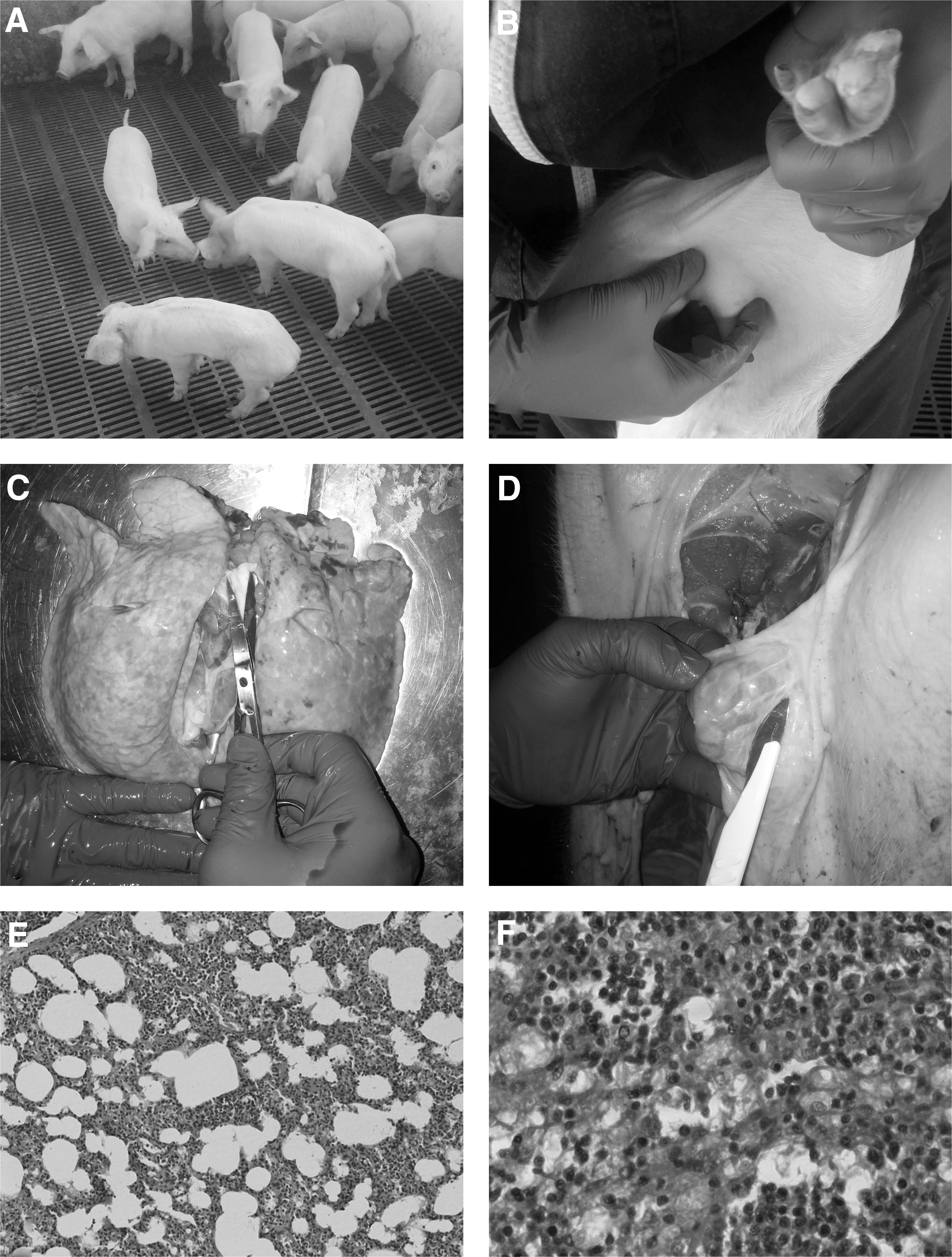
Based on clinical examinations, a percentage of about 40% of weaners in each batch suffered from high fever, poor growth performance, severe respiratory signs, as well as from an increased mortality rate. The acute respiratory disease in weaners included coughing, sneezing, increased respiratory rates, dyspnea (“thumping”), and nasal and eye discharges. In addition, depression, wasting, sporadic jaundice, rough hairy coat and palpable inguinal lymphadenopathy were noticed in pigs (Fig. 1A, B). The clinical signs of sows, based on the clinical examination and record system of the farm, included sporadic premature farrowing (22% of total gestating sows), moderate in appetence and anorexia, as well as increased number of stillbirth (3.3%) and weak piglets (4.1%). In addition, based on clinical examinations, a percentage of about 15% of finishers performed moderate-to-severe respiratory signs.
Clinical findings.
Sampling/laboratory exams
In November 2015, blood samples were obtained from 8 sows (4 lactating and 4 dry-period sows), 5 piglets of 15–20 days of age, 20 piglets of 40–70 days of age (4 of 50 days, 4 of 60 days, 4 of 70 days), and 5 of 130–150 days of age. In addition, tissue samples were collected from weaners (e.g., lymph nodes, lung, kidney, and liver) after euthanasia, for histopathological examination, through staining of tissue sections with Hematoxylin and Eosin.
Virological testing was performed at the Diagnostic Laboratory (School of Veterinary Medicine, Aristotle University of Thessaloniki). Blood serum samples underwent nucleic acid extraction using the PureLink® Viral RNA/DNA Mini Kit (Invitrogen, Carlsbad, CA). Extracts were tested for PCV-2 using a previously described TaqMan probe-based real-time polymerase chain reaction (PCR) (49) and for PRRSV (European type 1 and North American type 2), using a previously described TaqMan probe-based real-time reverse transcription (RT)-PCR (28). Reactions were carried out on a CFX96™ Real-Time System (Bio-Rad Laboratories, Hercules, CA). One real-time RT-PCR-positive extract was selected and reverse transcribed into complementary DNA (cDNA) using random hexamers, according to Dovas et al. (11). The complete PRRSV ORF5 (Gp5) sequence was amplified by PCR, employing the primer pair Gp5-SeqUp (5′-ATGAGGTGGGCYACAACCATYGC-3′) and Gp5-SeqDo (5′-AGGGCRTATATCATTATRGGTGTGTATGT-3′). The product was purified using the NucleoSpin® Gel and PCR Clean-up Kit (Macherey-Nagel, Düren, Germany). Sanger dideoxy sequencing was performed at Eurofins Genomics (Ebersberg, Germany), using an ABI 3730xl Genetic Analyzer (Applied Biosystems, Darmstadt, Germany). Phylogenetic analysis of the obtained sequence and additional European genotype ORF5 sequences, which were retrieved from GenBank, was conducted using the MEGA 6 software (44).
Diagnostic Results
The postmortem examination revealed that many lymph nodes (superficial inguinal, mediastinal, lung, mesenteric) were enlarged and pale on cut surface, whereas lungs failed to collapse and they were edematous and occasionally mottled (Fig. 1C, D). Histopathological examination of lung and lymph node sections according to Kiernan (20), revealed findings compatible with PRRSV and PCV-2 infection, such as lymphoplasmacytic interstitial pneumonia and mild histiocytic infiltration of lymph nodes (7) (Fig. 1E, F). The virological testing of the samples obtained from sows revealed negative results for both viruses. Pigs between 15 and 130–140 days of age were positive for type 1 (European) PRRSV. In addition, positive results for PCV-2 were obtained in pigs between 50 and 130–140 days of age. The full-length ORF5 sequence of the detected PRRSV strain, named “Mursi/AL/15” was successfully determined, and the obtained sequence was submitted into GenBank, under the accession number MF770711. Comparison of the obtained sequence with respective sequences of other European genotype strains available in GenBank, revealed nucleotide sequence identity of 90.8% to the strain “Spain 28/2003” (accession No. DQ345755), of 86.8% to “Lelystad virus,” that is, the prototype European genotype strain (accession No. M96262), and of 86.3% to the vaccine strain “Amervac PRRS” (accession No. GU067771) (Fig. 2).
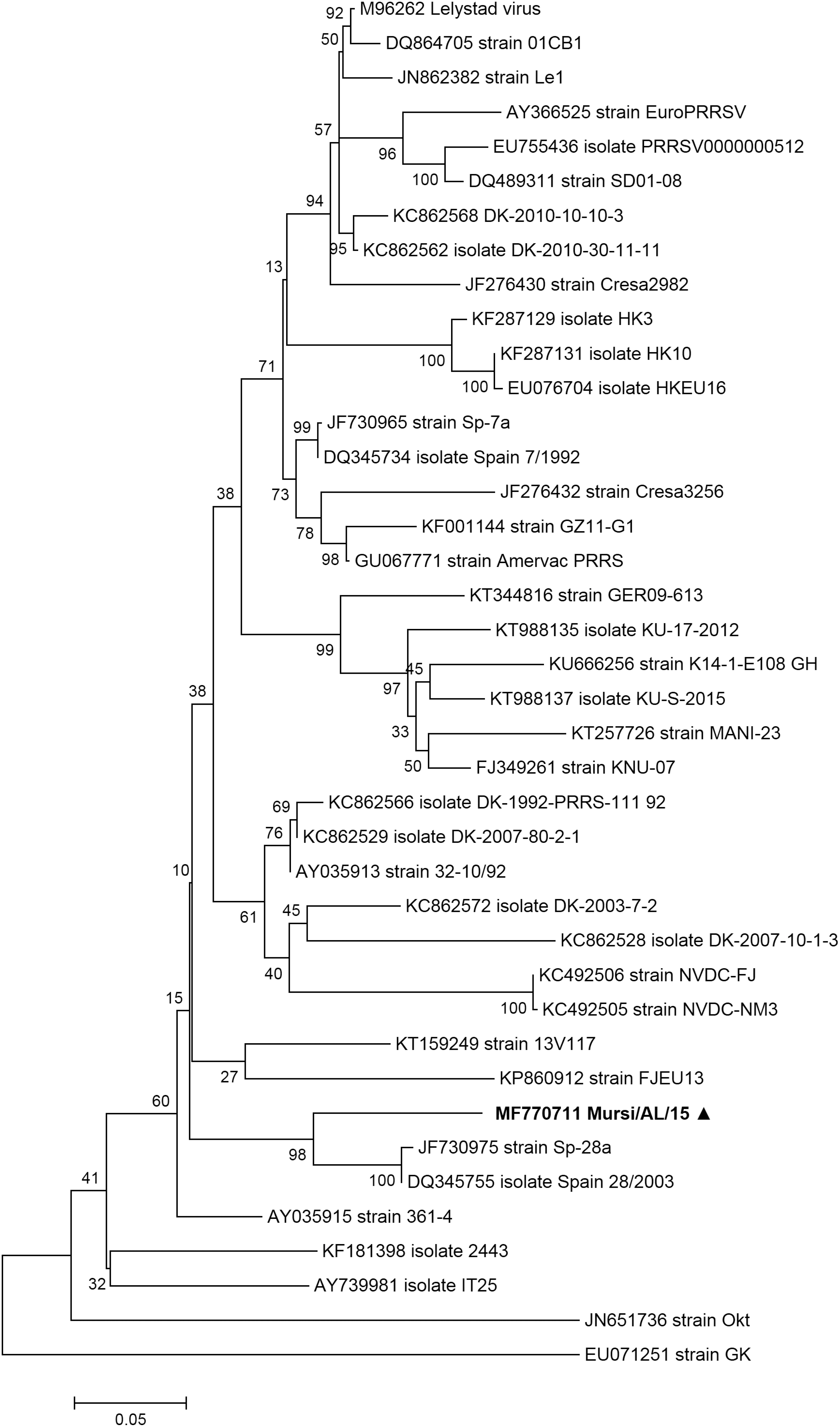
Phylogenetic tree inferred with ML analysis, based on Gp5 sequences of selected European genotype PRRSV strains. The tree was mid-point rooted. The Kimura 2-parameter model with gamma-distributed rates across sites (K2+Γ) was selected as the best fitting nucleotide substitution model for the dataset. The numbers indicated on branches are NPB probabilities. Scale bar indicates nucleotide substitutions per position. The sequence obtained from the Mursi/AL/15 strain is indicated in boldface and with a triangle. ML, Maximum Likelihood; NPB, nonparametric bootstrap; PRRSV, porcine reproductive and respiratory syndrome virus.
Interventions Applied and Follow-Up
It is suspected that PRRSV was introduced in the farm through purchase of replacement gilts and unvaccinated weaned pigs, 3 months before the occurrence of the outbreak. For this reason, inadequate external and internal biosecurity measures were proposed (e.g., maintaining quarantine program for newly introduced animals for up to 60 days, more frequent disinfections per batch) (6,50). According to Chae (8), if a farm is coinfected with PCV-2 and PRRSV, then PCV-2 vaccination is a priority. In this case report after the initial diagnosis of PRRSV and PCV-2 coinfection, a vaccination scheme was proposed, including vaccination of weaners against PCV-2 at weaning day and vaccination of sows against PRRSV with a modified live virus vaccine at 60th day of gestation and 6th day of lactation. Moreover, a treatment protocol with doxycycline and tiamulin in the feed for 14 days was proposed at the start of finishing stage to prevent secondary bacterial coinfections. The clinical picture was improved and the mortality was reduced in a period of 2 months after the application of the aforementioned vaccination scheme.
Discussion
This article constitutes the first report of PRRSV and PCV-2 infection in Albania. Based on the results of the records of the farm, it is suspected that PRRSV was introduced in the farm through purchase of replacement gilts and unvaccinated weaned pigs, 3 months before the occurrence of the outbreak. Purchase of gilts from PRRSV or/and PCV-2-positive herds, or movement of other types of infected animals between different stages of production, represents a known mechanism of virus transmission between production sites (24,30). Considering the numerous pathways of PRRSV transmission, internal and external biosecurity could be implemented to slow down the transmission of an endemic strain within or between herds (6,10). The introduction of young gilts into a herd of pregnant sows may not only cause problems for the gilts, but may also severely affect gestation in the sow population. This is the case when the gilts introduced act as subclinical carriers of pathogens, for which the sow population is seronegative. This risk may be especially high for PRRSV, because young gilts may be subclinical virus carriers for several months after infection (5,47), and viral shedding can be reactivated by transport stress and immunosuppression (3). Furthermore, the ability of the virus to persist in herds, and its wide biologic, antigenic, and genetic variability (50) may further complicate control plans in Albania. Many factors, including management practices and pig flow, the level of risk associated with local pig density, and the inherent characteristics of the specific PRRSV strains on farm level, contribute to a successful porcine herd health management program development (37,50).
In conclusion, the results of the present study suggest that PRRSV and PCV-2 are present in Albania. Awareness of veterinarians implicated with swine health should be further increased. Moreover, testing for PRRSV and PCV-2 should be included in the differential diagnosis of swine farms located in Albania, where increased mortality rates and poor growth performance are observed, along with piglets presenting with respiratory disease signs, and sows with reproductive disorders. Data obtained from the molecular characterization of PRRSV will be of use for epidemiological analyses of outbreaks, for evaluation of vaccine programs, as well as for the future design of diagnostic tests and vaccines, due to high degree of genome heterogeneity. In addition to diagnosis, knowledge regarding infected farms is important, so as to intensify biosecurity measures, given that the control of PRRSV and PCV-2 infections and minimization of economic losses in commercial swine farms are a challenge. Last but not the least, immunizations of animals could further assist in the control of the infections, by maximizing herd immunity.
Footnotes
Author Disclosure Statement
No competing financial interests exist.