
Abstract
This case study report describes a transmissible gastroenteritis coronavirus (TGEV) infection presented in a commercial pig herd. The clinical signs of infection appeared in newborn piglets, including medium morbidity and low mortality rates. Rectal swabs were collected from five different affected litters for laboratory examinations. Samples from two dead piglets and two euthanized affected piglets were collected for gross and histopathological examinations. All fecal samples were tested TGEV positive by real-time polymerase chain reaction (RT-PCR). Necropsy revealed nonspecific gross lesions. The histopathological examinations revealed villi fused with denuded tips and severe villus atrophy, leading to extensive epithelial flattening in middle and lower small intestine. The architecture pattern of villi presented columnar and cuboidal poorly differentiated enterocytes with mild subepithelial edema. In some enterocytes, pycnotic nuclei were detected. Microscopic examination of brain tissue revealed diffuse gliosis in the area of pia matter with mild congestion of the meningeal and parenchymal vessels and neuronal degeneration. In conclusion, this case study reported an epidemic TGEV infection in piglets, characterized by low mortality and medium morbidity rates accompanied by typical histopathological lesions in small intestine, as well as by coexisting brain lesions, that are described for the first time.
Introduction
T
On a herd basis, two epidemiological forms of TGE are recognized: epidemic and endemic. Typical clinical signs of epidemic TGEV infection in piglets are vomiting and diarrhea (profuse watery, yellowish), rapid loss of weight, dehydration, and high morbidity and mortality rates in piglets of <2 weeks of age. Clinical signs of epidemic TGE in finishing swine and in sows include inappetence, transient diarrhea, and vomiting (17). Typically, in endemically infected herds, TGEV appears as a mild diarrhea in piglets ∼6 days of age. The consistency of the feces ranges from clear and watery to white and creamy, and in late-stage disease it becomes gray and pasty (16). Low mortality (usually <10–20% in pigs from 6 days of age until 2 weeks after weaning) and concurrent infection are characteristic, although some litters may experience severe clinical signs. Severity of clinical signs is dependent on the age of pig, the management system, degree of exposure to the virus, and degree of passive maternal immunity (1). Infected sows usually do not show clinical signs.
The caused acute maldigestive/malabsorptive diarrhea and the dehydration in piglets are due to a marked reduction in enzymatic activity in the small intestine, resulting in a disruption of digestion and cellular transport of nutrients and electrolytes (15). The main caused lesion of TGE is markedly shortened villi of the jejunum and ileum (2). Villous atrophy is more severe in newborn pigs than in older pigs (15), as neonates are more susceptible to TGEV infection.
The aim of this case report is to describe a TGEV infection in newborn piglets in a commercial pig herd and to report the lesions observed.
Materials and Methods
Description of the farm
The present study is based on data from the breeding stock of a farrow-to-finish commercial pig farm (commercial hybrids of Large White × Landrace). The capacity of the farm was 550 sows under production, located in Central Greece. A grandparent nucleus of 40 sows was kept in the farm for producing its own gilts. The farm facilities included 12 farrowing houses (10 pens), 18 flat-deck units (2 pens of 55 animals), growing houses (46 pens of 50 animals), one finishing house (4 pens of 40 animals), one mating-pregnancy (dry period) stable with 240 individual stalls (0–35th day of pregnancy), two breeding stock house of group housing (18 pens of 10 sows/35–105th day of pregnancy), one breeding stock house of group housing for noninseminated gilts (5 pens of 25 gilts), a feed mill, and an artificial insemination laboratory. The herd practiced a 1-week batch production system. The weaning piglets were allotted equally according to the body weight and sex at random at flat-deck batteries for piglets in a climate-controlled postweaning stable.
The vaccination scheme of breeding stock and weaners that was applied in the farm is shown in Table 1. All breeding females were treated with a single ivermectin injection 14 days before farrowing; the boars were treated twice a year. The feed provided to the animals was self-prepared based on a corn/barley/wheat–soya meal, depending on the season. The breeding animals received a different feed during gestation and lactation. Drinking water was provided for ad libitum consumption by the animals. Housing facilities had fully automated temperature and humidity control system, as well as automated feeding.
ADV, Aujeszky's disease virus; AR, atrophic rhinitis; Ery, erysipelas; Parvo, parvovirus; PCV2, porcine circovirus type 2; PRRSV, porcine reproductive and respiratory syndrome virus.
Case study
During November 2017, the first observed clinical sign was vomiting, followed by diarrhea in 5 pens at one of 12 total farrowing houses. The age of newborn piglets in this farrowing house ranged from 1 to 4 days, and all sows were at first parity. At the beginning, diarrhea was very sparse, watery, ran down the hind legs, and dripped from the tail. As the disease progressed, diarrhea was more obvious, more specifically thickened, becoming yellowish and foamy. The skin of the rump was usually wet and soiled. Subsequently, the piglets became dehydrated and had eyes that were sunken. Their hair coat was also quite rough and presented significant weight loss. In a week interval, all pens (totally 10) of the farrowing house were affected by diarrhea. The morbidity rate was medium (∼30–35%), whereas the mortality rate was low (<8%), mainly due to severe dehydration. In the following weeks, diarrhea was observed in other two farrowing houses, where the 50% were at their first parity.
Sampling
A combination of different tests was carried out so that to have an accurate diagnosis of the cause of the infection. Fecal samples (rectal swabs) were taken from five different affected litters (five samples from live piglets 1–4 days of age/litter) for laboratory examinations (real-time polymerase chain reaction [RT-PCR], bacteriological testing). In addition, three of the aforementioned piglets with symptoms were euthanized, and sent to laboratory for necropsy and histopathological examinations.
Laboratory examinations
Fecal samples were examined by RT-PCR for TGEV and rotavirus infection. This method allows the rapid and sensitive detection of RNA of TGEV from samples purified from nasal swabs and feces (12). A specific RNA sequence of the TGEV genome is transcribed into cDNA and amplified by the RT-PCR, which is applied on Light Cycler 2.0 Roche®. Specific primers and probes are used to amplify and detect a part of the S region of TGEV. Using the Taqman probes principle maximizes the specificity of this method.
The euthanized piglets were autopsied to find any gross lesions. In addition, tissue samples from brain and all the anatomic regions of the gastrointestinal tract (stomach, small and large intestine) were collected for histopathological examinations. The tissue samples were fixed in 10% buffered formalin and embedded in paraffin routinely. Dewaxed 3–5 μm thick sections were obtained, and stained with hematoxylin and eosin (H&E) for histopathological evaluation.
Results
Fecal samples
The results of RT-PCR in fecal samples are shown in Table 2. All piglets were positive for TGEV but negative for porcine rotavirus. Moreover, the bacteriological testing was negative for Escherichia coli and other enteric pathogens.
TGEV, transmissible gastroenteritis coronavirus.
Gross and histopathological results
The necropsy revealed dilation of the intestine, which contained a small amount of yellowish fluid. No specific gross lesions were detected in any other organ.
The histopathological appearance of the small-intestine-examined samples was similar and characterized by villi fused with denuded tips. Severe villus atrophy led to extensive epithelial flattening. It was observed in the middle and lower small intestine (Fig. 1). The architecture pattern of villi was characterized by columnar and cuboidal poorly differentiated enterocytes with mild subepithelial edema. In some enterocytes, pycnotic nuclei were detected. No significant lesions were detected either in stomach or in the large intestine. The examined tissue sections from the brain revealed diffuse gliosis in the area of pia matter with mild congestion of the meningeal and parenchymal vessels and neuronal degeneration accompanied with neuronophagia (Fig. 2). The above-described lesions were observed mostly in the parietal and occipital lobe.
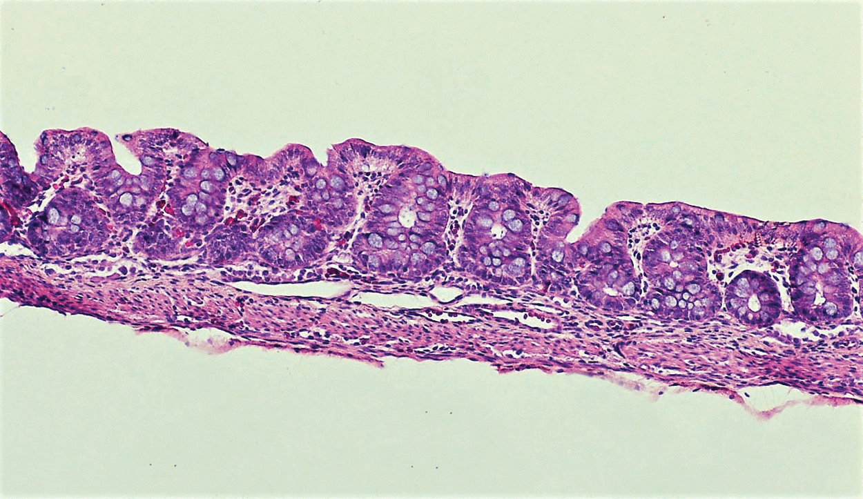
Ileum: Severe villus atrophy resulting in epithelial flattening. HE, × 20.
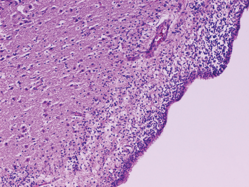
Brain, occipital lobe, pia matter: extensive gliosis with congested parenchymal vessels. HE, ×10.
Management practices
At first step, oral antibiotics were used in affected piglets for the prevention of secondary bacterial enteric infections. At the same time, oral feed supplements in liquid form based on plant extracts and herds (phytogenics) were used to support gut health and improve nutrient utilization. In addition, electrolytes and milk replacers were provided to piglets to reduce the negative effects of severe diarrhea and dehydration.
Based on the low mortality rate in piglets, the common practice of feedback control of TGE (feeding of sows with TGEV-infected minced intestines) was not applied to avoid the potential hazards (e.g., possible spread of other pathogens to pregnant sows and throughout the herd).
Discussion
This case report described a TGEV endemic infection in newborn piglets, characterized by low mortality rate and medium morbidity rate accompanied by typical histopathological lesions in middle and lower small intestine. Our findings on the lesions of small intestine are in agreement with those of previous studies (9,11,16). Furthermore, coexisting brain lesions were observed, indicating nonsupportive encephalitis, characterized mainly by diffuse gliosis in the area of pia matter. This finding suggests that it is possible that TGEV could also cause brain lesions beyond the known ones. The aforementioned finding also supports our initial hypothesis on possible effect of TGEV on brain function, causing vomit due to neural pathway. It is known that jejunal enterocytes undergo massive necrosis within 12–24 h after TGEV infection, resulting in an acute maldigestive and malabsorptive diarrhea and severe dehydration (15). Moreover, other mechanisms such as altered intestinal sodium transport with accumulation of electrolytes/water in the intestinal lumen and loss of extravascular protein are attributed to diarrhea caused by TGEV infection (4). Using different pathways, several viruses have been shown to be able to penetrate the central nervous system (CNS), and infect neurons and glial cells (8). The detected brain lesions as degeneration of neurons, reactivity of the glia, and perivascular inflammatory reaction are in accordance with the general hallmarks of viral infection of the CNS (13). These lesions, indicating no-supportive encephalitis, could result from viral infection of CNS as part of systematic infection. Coronaviruses are considered to be neuroinvasive, neurotropic, and occasionally neurovirulent in various hosts, including human, cats, pigs, rodents, and fowl, especially on susceptible individuals (3,7,18).
Hypoglycemia, caused by diarrhea and vomiting in newborn piglets, is considered the main cause of death of piglets infected with TGEV (5,13). As it is established, the brain requires glucose and oxygen for its energy production. So the microscopic effects of reduced glucose are similar to those of oxygen depletion (10). Hypoglycemia causes excitotoxicity (14), which is the term used for the neuronal death process induced by massive release of the excitatory amino acid l-glutamate into the extracellular space after lysis of neurons from the infarct core. So according to the above theory, the hypoglycemia in our case could be the reason of neuronal degeneration. However, more rigorous studies are required to investigate the pathogenesis of TGEV, especially if the TGEV has direct or indirect effect on brain function.
In conclusion, this case study reported a TGEV infection in newborn piglets, characterized by typical histopathological lesions in small intestine, as well as the typical pattern of viral brain lesions, suggesting that TGEV has neurotropic effect.
Footnotes
Author Disclosure Statement
No competing financial interests exist.