
Abstract
Bovine rotavirus (BRV) is one of main pathogens responsible for diarrhea, fever, and vomiting. In this study, we developed a colloidal gold immunochromatographic test strip for detecting BRV according to the principle of double-antibody sandwich. The monoclonal antibodies (mAbs) and polyclonal antibodies (pAbs) were prepared and purified. On the strip, the purified mAbs labeled with the colloidal gold were used as the detector, and the goat anti-mouse antibodies and purified pAbs were coated on the nitrocellulose membranes as the control line and the test line, respectively. We optimized different reaction conditions, including the amount of mAbs, the pH of colloidal gold solution, coating solution, blocking solution, sample pad treatment solution, antibody concentration in control line, and antibody concentration in detection line. In specificity assay, the strip had high specificity in detecting BRV. No cross-reaction was observed in detecting other viruses. The detection sensitivity of the strip was found to be 1 × 10³ TCID50/0.1 mL. Two hundred twenty clinical samples were detected with the strip compared to reverse transcription-polymerase chain reaction. No false-negative or false-positive results were found, and the results obtained by the two methods were similar. In conclusion, we developed a novel immunochromatographic strip to rapidly detect BRV. The strip developed exhibited high sensitivity and specificity for BRV detection. It could be a rapid, convenient, and effective method for the rapid diagnosis of BRV infection in the fields.
Introduction
Rotavirus infects a wide variety of animal species, and some of them are responsible for severe clinical diseases. Bovine rotavirus (BRV) is one of main pathogens responsible for diarrhea, fever, and vomiting (18,22,32). BRV persists in cattles and exhibits the durable and high infection. BRV is classified in the family Reoviridae and the genus Rotavirus. The genome of BRV contains 11 double-stranded RNAs and encodes 12 viral proteins. Six of them are structural proteins (VP1, VP2, VP3, VP4, VP6, and VP7) and the others are nonstructural proteins (NSP1-NSP6) (18,19,20,29).
VP6 gene is very conserved in group A BRV and suitable as a specific gene for BRV detection (21,31,32). The protein VP6 is a trimer formed by oligomers, which constitutes the icosahedral structure of the mid-layer capsid of BRV. Removing VP6 from virus-like particles, the virus will lose transcriptase activity due to the lack of VP6 (3). The VP6 protein is the group (subgroup)-specific antigen of virus, which is the basis for RV grouping. According to the difference in antigenicity of VP6 protein, RV is divided into seven groups, including A–G group (7). The protein VP6 is encoded by the sixth gene fragment of BRV, which consists of 387 amino acids with a molecular weight of about 45 kDa. The VP6 protein accounts for 51% of the total viral protein and 39% of the total structural protein. It has good immunogenicity and is considered to be the main diagnostic antigen of BRV (5).
The main transmission route of BRV is through the digestive tracts. The prevention of BRV infection mainly depends on vaccine immunization. Inoculation of the inactivated vaccine in 1–3 months before the delivery of the cow can make the newborn calf passively immune and obtain the protection by maternal antibodies (11,12,15,16). Traditionally, detection of BRV has been mainly based on serological approaches, etiological approaches, and molecular methods. Most of traditional methods need long time and require special equipment to diagnose in the laboratory (4,24). Therefore, a rapid, specific, and convenient method for the field detection has important practical significance for the prevention and control of BRV infection.
Immunological assays show better effects in detecting pathogen due to the specific interaction between antigen and antibody. For this reason, colloidal gold has been introduced into immunochemistry in recent years. Colloidal gold particles can be used to label antibodies instead of enzymes. The reaction of the gold-labeled antibodies with the corresponding antigens can result in a visible color reaction (17,30).
The distinctive advantages of colloidal gold particles are that they can be directly observed without staining and have high resolution for accurate positioning results. It has been introduced to field analysis, which simplifies the time-consuming and laborious traditional cultivation method (28). Colloidal gold immunochromatographic (GICG) assays not only speed up the detection process but also provide detection methods without using reagents (2). Their rapid analysis, high sensitivity, and simplicity of operation provide promising solutions to the need for pathogen diagnosis. They have been increasingly applied to more research fields (6,8,10,37).
In this study, a colloidal GICG strip for detecting BRV was developed by using purified monoclonal antibodies (mAbs) (against BRV VP6 protein) and polyclonal antibodies (pAbs) (against BRV). This method was simple, rapid, and specific for the detection of BRV, which is more suitable for pathogen diagnosis in the field.
Materials and Methods
Reagents and materials
Gold chloride and bovine serum albumin (BSA) were obtained from Sigma Corporation (St. Louis, MO). The hybridoma cells secreting antibodies against BRV VP6 protein were prepared and stored in our laboratory. Goat anti-mouse immunoglobulin G (IgG) was obtained from Beijing Zhongshan Jinqiao Limited Corporation (China). Nitrocellulose membranes, sample pad, glass–fiber conjugate pad, and absorbent pad were obtained from Millipore. All other chemicals were obtained from Sigma-Aldrich.
Preparation of pAbs
New Zealand rabbits were administered with purified BRV (500 μg) after emulsifying in complete Freunds' adjuvant for the first injection and same dose after emulsifying in incomplete Freunds' adjuvant for two boosters every 2 weeks. Sera were collected 7 days after the last booster. The immunoglobulins were precipitated with standard ammonium sulfate precipitation. In brief, an equal amount of saturated ammonium persulfate solution was dropped into the mixed serum, which was kept stirring on ice until the precipitation formed. The mixture was centrifuged at 12,000 × g for 30 min. The deposit was dissolved in PBS and then dialyzed against PBS. The amount of protein was determined. The Ig protein was analyzed by sodium dodecyl sulfate-polyacrylamide gel electrophoresis (SDS-PAGE) and BandScan software.
Preparation of monoclonal antibody
BALB/c mice were pretreated with liquid paraffin and then injected with the hybridoma cells secreting antibodies against BRV VP6 protein by intraperitoneal route. The mAbs were harvested after 2 weeks and purified with HiTrap™ Protein G HP according to the manufacturer's instructions (GE Healthcare, Milwaukee). The activity of the mAbs was analyzed by SDS-PAGE and enzyme-linked immunosorbent assay (ELISA).
Preparation of colloidal gold
Gold particles with an average diameter of 20 nm were prepared according to the method described by Wang (33). In brief, the suspension of gold particles was prepared as follows: under reflux conditions, 100 mL solution of gold chloride solution (0.01%) was heated to boiling. Approximately 1.0 mL of trisodium citrate solution (1%) was then added rapidly, while stirring. The solution was boiled for another 5–10 min until the color of mixture changed to wine red. The gold particles were detected by transmission electron microscope (TEM) after cooling. Finally, 0.05% sodium azide (as a preservative) was added to the gold particle solution and then stored at 4°C.
Conjugation of anti-BRV-VP6 mAb with colloidal gold
The complexes of mAbs conjugated with colloidal gold were prepared according to the previous method (26). In brief, the pH value of the colloidal gold solution was adjusted to 8.0–9.0 with 0.2 M K2CO3. To estimate the minimal amount of mAb to stabilize colloidal gold, the procedure was conducted as follows: 1,000 μL of colloidal gold solution was added quickly into 100 μL of serial dilutions (10–100 μg/mL) of mAbs at increasing concentrations, respectively. After 5 min, 100 μL of 10% NaCl solution was added to the mixture for another 2 h. The absorption at 520 and 580 nm was detected (A520–A580).
When the mAbs added exceeded the minimum amount to stabilize colloidal gold, the color remained unchanged or it would change from reddish to blue. The optimum concentration of mAbs added was 130% of the lowest concentration to label so that the color remained unchanged. The colloidal gold solution was added into different Eppendorf tubes (1,000 μL/tube) and the pH values of the colloidal gold solution were adjusted to 7.0, 7.5, 8.0, 8.5, 9.0, 9.5, and 10.0 with 0.2 M K2CO3, respectively. Hundred microliter of mAb of the optimum concentration was added to every tube and mixed. After 5 min, 100 μL of 10% NaCl solution was added to the mixture for another 2 h. The optimum pH value of the colloidal gold solution was the minimum pH value that the solution remained reddish.
To conjugate anti-BRV-VP6 mAb with colloidal gold, the optimum concentration of mAbs was used. Around 4.8 mL of mAb solution was added into 20 mL colloidal gold solution rapidly, which was stirred rapidly and then incubated for 30 min. Then the mixture was stabilized with 5% BSA solution (the final concentration of BSA was 1%) and stirred for 30 min. After incubation for 1 h, the supernatant was discarded after centrifugation (10,000 × g) at 4°C for 30 min. Twenty milliliter of 2% BSA solution (containing 0.01 M sodium borate) was used to resuspend the precipitate and then centrifuged (10,000 × g) at 4°C for 30 min to clean off the unlabeled mAbs.
Finally, 4 mL of TB solution (containing 3% sucrose, 3% BSA, 0.05% sodium azide, and 0.01 M sodium borate) was used to resuspend the precipitate. Conjugation of colloidal gold to mAbs was examined by ultraviolet-visible (UV/Vis) spectroscopy (Ultropec 2100 pro UV; Amersham Pharmacia, Sydney) (28). The gold-labeled mAb solution was stored at 4°C.
Optimizing reaction reagents of test strips
It is necessary to optimize the reaction reagents for improving the performance of test strip. PBS, 0.01M phosphate buffer (PB, containing 0.6% NaH2PO4·2H2O and 2.2% Na2HPO4·12H2O), and 20 mM Tris-Cl were used as coating solution to treat the antibodies. The bands of the control line and the test line were detected to analyze the effects of different coating solutions.
Three percent BSA, 5% BSA, 3% skim milk, and 5% skim milk solutions were used as blocking solution, respectively. The color of background and sample chromatography time were detected to analyze the effects of blocking solutions. The sample pads were soaked in solution A (containing 0.05% Tween-20, 5% sucrose, 0.3% Triton X-100, and 1% BSA), solution B (containing 0.05% Tween-20, 5% sucrose, 0.5% Triton X-100, and 0.5% BSA), solution C (containing 5% sucrose, 0.3% Triton X-100, and 1% BSA), and solution D (containing 5% sucrose, 0.5% Triton X-100, and 0.5% BSA), respectively, at room temperature for 30 min. After that, the sample pads were dried at 37°C for 3 h and stored for detecting samples. The bands of the test line were detected to analyze the effects of different treatment solutions.
Test procedure
Fifty microliter of sample was taken out of the sample solution and added to the sample pad. After 10 min, the result could be judged by the naked eyes. If the sample contained the detected object, the detected object would form a complex with the gold-labeled mAb. Compared with the control line, the test line would show weak color or color, which indicated that the test result was weakly positive or positive. If the sample did not contain the detected object, the test line would show colorless, which indicated that the test result was negative. The color of the line was used as the standard to evaluate whether the prepared strip was valid (color) or invalid (colorless) (Fig. 1).
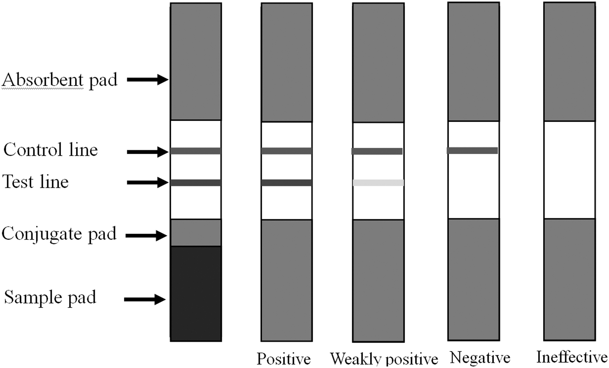
Schematic diagram of one-step strip and principle of result judgment on one-step strip assay for BRV. Compared with the control line, the test line would be visible (weak or dark color), which indicated that the test result was weakly positive or positive. If the test line was invisible, this indicated that the sample did not contain the detected object and the test result was negative. BRV, bovine rotavirus.
Sensitivity and specificity of the strip test
The sensitivity of the test strip was evaluated by detecting different densities of BRV. Samples containing different concentrations (BRV of 1.5 × 10⁴ TCID50/0.1 mL was diluted in a series of 1:5−1, 1:10−1, 1:15−1, 1:20−1, 1:25−1, and 1:30−1, respectively) of BRV were prepared to optimize working solution. The prepared strips were used to test different concentrations of BRV and the results were judged by the naked eyes. The same procedure was repeated three times.
The specificity test was also conducted with standard negative sample, standard positive sample, and samples containing bovine viral diarrhea virus (BVDV), porcine rotavirus (PRV), porcine epidemic diarrhea virus (PEDV), bovine parvovirus (BPV), transmissible gastroenteritis virus (TGEV), or bovine respiratory syncytial virus (BRSV) by the optimized working solution to evaluate the specificity of the test strip. Hundred microliter of sample was added to the sample pad. The result was judged by the naked eyes after 10 min. The same procedure was repeated three times.
Detection of clinical samples
The specific primers were designed based on the conserved regions of the VP6 gene of BRV NCDV strain in GeneBank. A total of 220 fecal samples from different farms where diarrheal disease outbroke (Jilin Province, Liaoning Province and Heilongjiang Province) were collected, which were placed in sterile polyethylene tubes, numbered, and transported by car freezer (4°C) to laboratory. These samples were then detected by reverse transcription-polymerase chain reaction (RT-PCR) and the developed strips, respectively. The results of strips were compared with those of RT-PCR.
Results
Evaluation of purified polyclonal antibody
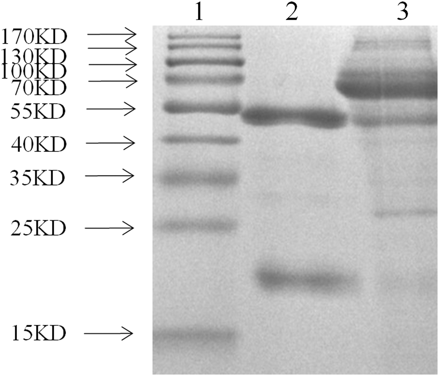
Sera were collected 7 days after the last boost of the rabbits. After purifying by standard ammonium sulfate precipitation, an aliquot was analyzed by SDS-PAGE (Fig. 2). It showed that there was almost no hybrid protein, which indicated the IgG protein was purified effectively. The light chain and heavy chain of IgG were clear and visible, and the sizes of them were 54 and 23 kDa, respectively. BandScan software analysis showed that the purity of pAbs was 93%. The protein concentration was 2.16 mg/mL by measuring with trace protein concentration meter.

SDS-PAGE analysis of purified polyclonal antibody. Lane 1: standard protein marker; lane 2: purified pAbs; lane 3: unpurified pAbs. The IgG protein was purified effectively. The light chain and heavy chain of IgG were clear and visible, and the sizes of them were 54 and 23 kDa, respectively. IgG, immunoglobulin G; pAbs, polyclonal antibodies; SDS-PAGE, sodium dodecyl sulfate-polyacrylamide gel electrophoresis.
Evaluation of purified monoclonal antibody
Purified mAbs were also analyzed by SDS-PAGE. The result showed that the mAbs were purified effectively and there was almost no hybrid protein. Both light chain and heavy chain were clear and visible, and the sizes of them were 50 and 20 kDa, respectively (Fig. 3). BandScan software analysis showed that the purity of mAbs was 95%. The protein concentration was 2.325 mg/mL by measuring with trace protein concentration meter.
SDS-PAGE analysis of purified monoclonal antibody. Lane 1: standard protein marker; lane 2: purified mAbs; lane 3: unpurified mAbs; the mAbs were purified effectively. The light chain and heavy chain were clear and visible, and the sizes of them were 50 and 20 kDa, respectively. mAbs, monoclonal antibodies.
Determination of colloidal gold particles
During the preparation, the colloidal gold solution changed from colorless to wine red. The 20 nm colloidal gold had good stability and no precipitation occurred in 2 months. The results of TEM showed that the colloidal gold particles prepared in this study were well dispersed and uniform in size (Fig. 4). The colloidal gold particles were spherical with an average diameter of 21.05 ± 4.87 nm. No aggregation of colloidal gold particles occurred, which indicated that the colloidal gold particles were stable in solution. Because of surface resonance of the colloidal gold particles, the peak of the colloidal gold curve was at 520 nm according to the UV/Vis spectra. The peak width was narrow (Fig. 5), which met the standard of colloidal gold to use as probes.

Transmission electron microscopy image of gold nanoparticles. Transmission electron microscope observation showed that particles had varying sizes and shapes with an average diameter of 21.05 ± 4.87 nm. The colloidal gold particles prepared were well dispersed and no aggregation occurred, which indicated that the colloidal gold particles were stable in solution.
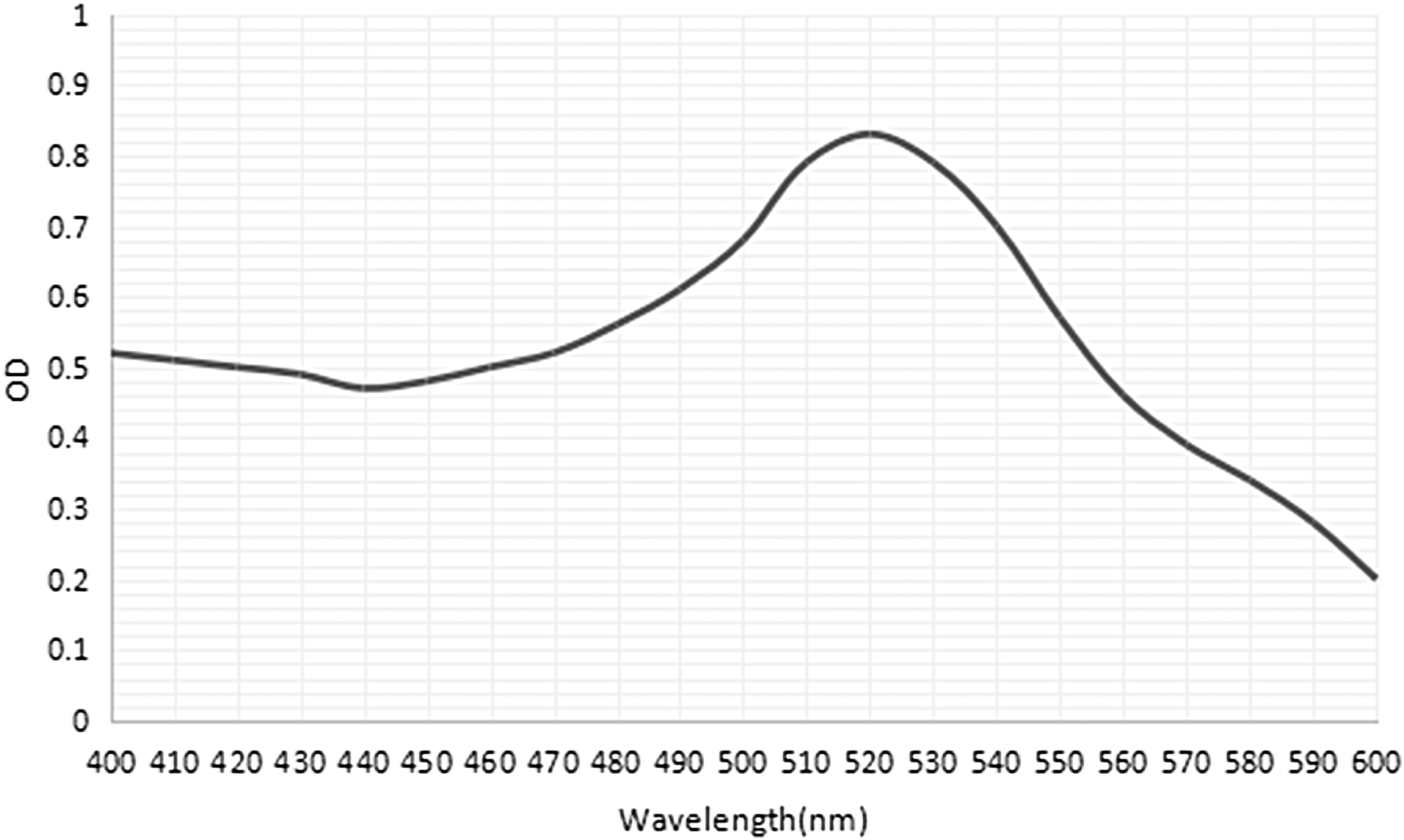
Ultraviolet-visible spectra of colloidal gold. The peak of the colloidal gold curve was at 520 nm due to surface resonance of the colloidal gold particles. The peak width was narrow and met the standard of colloidal gold to use as probes. OD, optical density.
Optimization and characterization of antibody-gold conjugates
The optimum pH value of the colloidal gold solution was the minimum pH value that the solution remained reddish. It was determined that the optimum pH to stabilize colloidal gold particles was 8.5. At the pH of 8.5, the optimum of purified antibody was determined. The result showed that the minimum of purified mAbs to make the solution remain reddish was 64.38 μg/mL. The optimum concentration of mAbs added was 130% of the minimum to stabilize the solution. Therefore, the optimum concentration of purified mAbs for the conjugation was 83.69 μg/mL so that enough mAbs could conjugate with the gold particles and stabilize the colloidal gold.
Optimization of reaction reagents
According to the hybridization mentioned above, the result showed that 0.01 M PB was more effective as the coating solution than the others in treating antibodies. The bands of the quality control line and the test line were more clearly visible when treating the antibodies with 0.01 M PB (Fig. 6A). Three percent BSA, 5% BSA, 3% skim milk, and 5% skim milk solution were chosen as blocking solution, respectively, in the experiment (Fig. 6B). The background was lighter when 3% BSA and 5% BSA solution were used as blocking solution. Furthermore, the sample blocking time was shorter when using 3% BSA compared with 5% BSA, which was more suitable for rapid test.
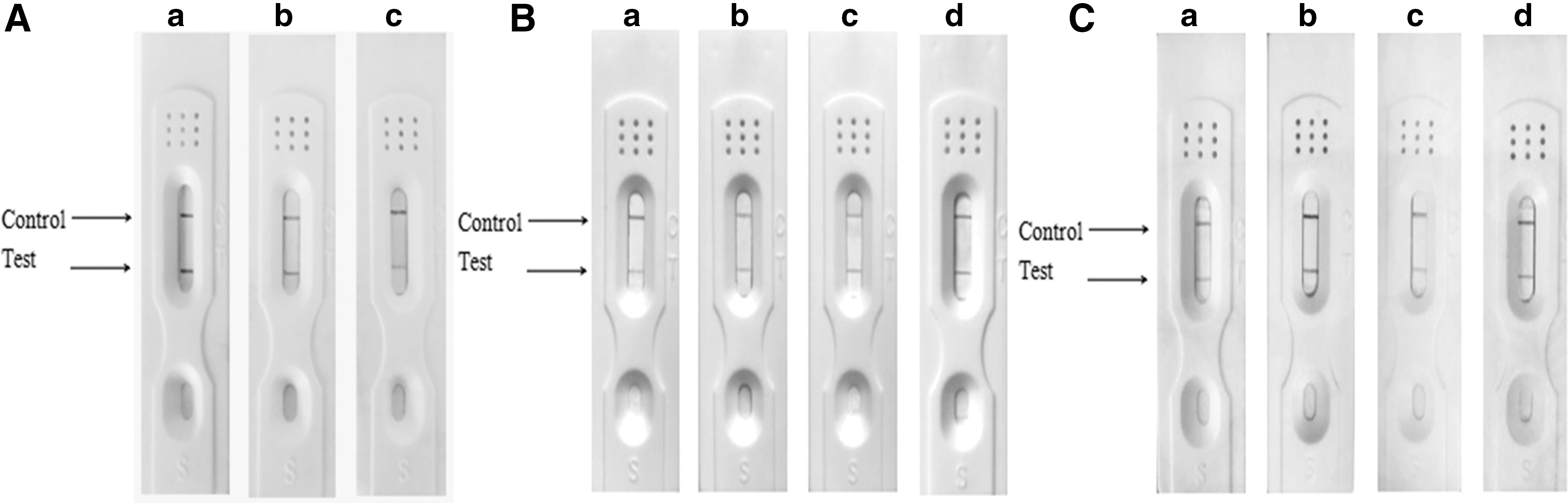
Optimization of reaction reagents.
The sample pads were soaked in different treatment solutions, respectively, and the effects were detected. As shown in Figure 6C, the test line was more clearly visible when the sample pad was soaked in solution B. Based on this result, solution B (containing 0.05% Tween-20, 5% sucrose, 0.5% BSA, and 0.5% Triton X-100) was selected to soak the sample pads.
Sensitivity and specificity
The sensitivity of the test strip was analyzed by testing cell culture of BRV diluted serially (BRV of 1.5 × 10⁴ TCID50/0.1 mL was diluted in a series of 1:5−1, 1:10−1, 1:15−1, 1:20−1, 1:25−1, and 1:30−1, respectively). The prepared strips were used to test different concentrations of BRV. The result showed that the minimum detectable amount of the test trip was about 1 × 10³ TCID50/0.1 mL. Similar results were observed in three repetitions (data not shown).
To identify the specificity, the test strips were used to detect standard negative sample, standard positive sample, and samples containing BVDV, PRV, PEDV, BPV, TGEV, or BRSV. If the sample contained BRV, there would be two red lines in the test region and the control region (Fig. 7). For other samples that did not contain BRV, there would be a single red line in the control region. The results suggested that the test strips showed high reactivity and specificity in the detection of BRV. There was no cross-reaction in the detection of other viruses.
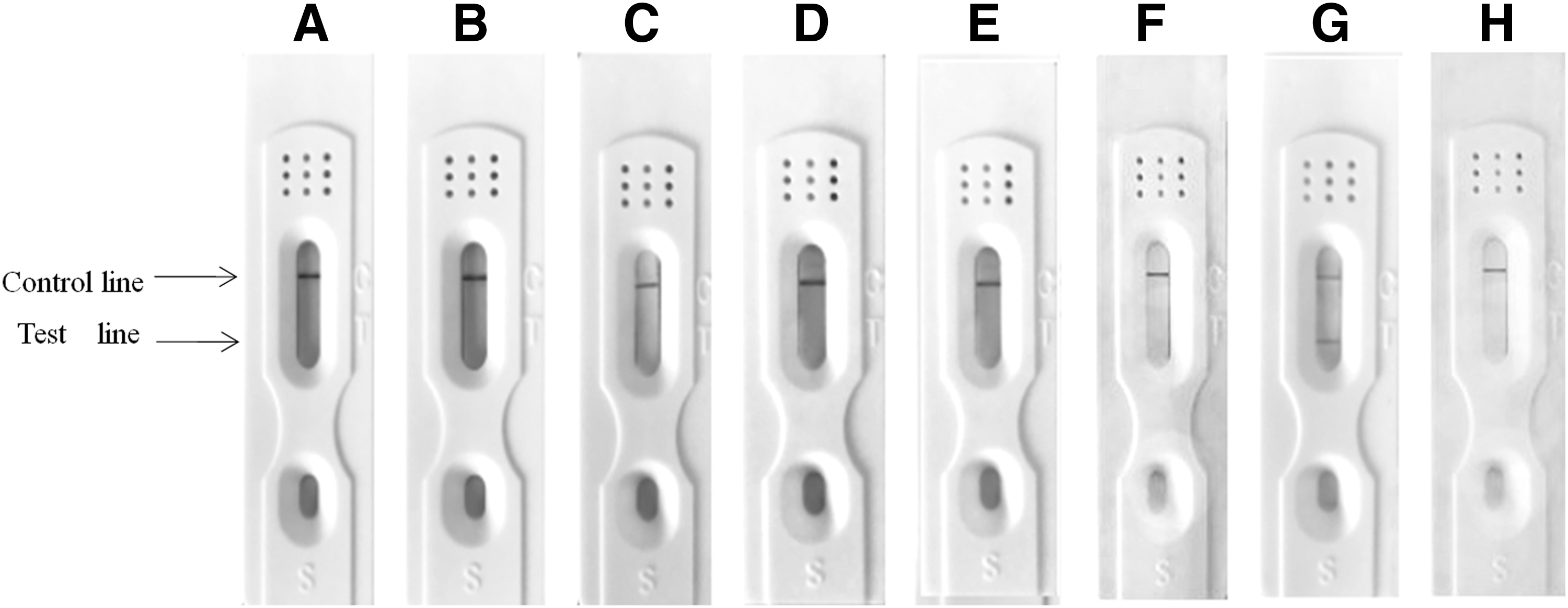
Specificity assay of the test strip. Samples containing BVDV
Clinical sample testing
Two hundred twenty fecal samples were collected from different farms (Jilin Province, Liaoning Province, and Heilongjiang Province). Each sample was detected by the test strip (Fig. 8A) and RT-PCR (Fig. 8B), respectively. The test results of the two methods were compared (Table 1). The results showed that the coincidence rate between the test strip and RT-PCR was 100%.
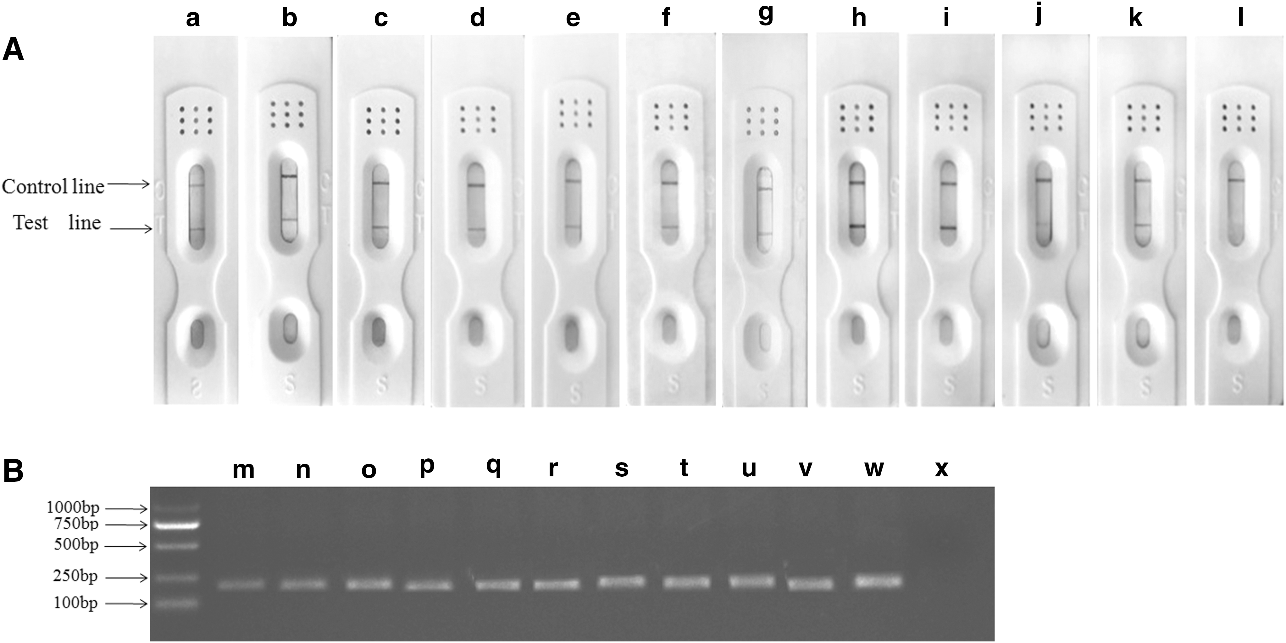
Detection of BRV in clinical samples by the test strip
Comparison of the Test Strip with Polymerase Chain Reaction Method for Detection of Clinical Samples
Discussion
BRV infection usually occurs in young calves, especially newborn calves. The sick calves suffered from severe diarrhea and dehydration leading to death. The conventional methods for detecting BRV are mostly completed in the laboratories with some instruments by skilled technicians. The infection from BRV of group A is the main cause of acute diarrhea in newborn calves (34). The VP6 protein is the group (subgroup)-specific antigen protein of BRV (9,34,38). In this study, we prepared monoclonal antibody against VP6 protein. Basing on this, colloidal GICG was combined to prepare a rapid diagnostic strip for detecting BRV. The test strip can be operated in the field without professional personnel and equipment.
The colloidal GICG assay is a rapid and simple method to detect pathogens in the field. This method has been widely used to detect many pathogens, such as reovirus, avian influenza virus, and mycoplasma (1,23,36). Colloidal GICG is a solid-phase labeled immunoassay technique, which uses staining emulsion and colloidal gold as the makers (27). GICG is characterized by simple operation, time-saving steps, small sample consumption, and easy result interpretation. It is suitable for the qualitative, semiquantitative, and quantitative detection of a large number of objects in nonlaboratory environment.
Colloidal gold particles not only can be deposited tightly on the adsorption line for better identification but also can adsorb protein antibodies initiatively (13). Many studies have shown that colloidal gold particles suitable for labeling mAbs are about 20–30 nm in diameter (14,16,21,25). The smaller the diameter of colloidal gold particles is, the better the mixing degree on the adsorption line, which can improve the detection sensitivity. In this study, we prepared the 20 nm colloidal gold particles, which were dispersed and uniform in size. The dispersed colloidal gold particles were easy to flow on the membrane. The prepared colloidal gold particles had good stability and no precipitation occurs in 2 months.
Colloidal GICG strip has been developed into a method for qualitative analysis and rapid visual evaluation in detecting pathogens. Therefore, the color intensity of the control line and the test line must be strong enough to observe the difference between test sample and negative control. The reaction conditions of the immunochromatographic test strip are important parameters affecting the quality of the strip (23,35). In this study, we optimized different reaction conditions, including the amount of mAbs, the pH of colloidal gold solution, coating solution, blocking solution, sample pad treatment solution, antibody concentration in control line, and antibody concentration in detection line.
To improve the specificity of the strip test, mAbs were used instead of pAbs on the test line. The specificity assay revealed that the established test strip had high specificity and no cross-reaction with other clinical pathogens. The limit of detection for BRV was 1 × 10³ TCID50/0.1 mL by the developed test strip. In detecting clinical samples, we compared the results of test strip and PCR method. The two methods showed good correspondence. Although PCR is a method with higher sensitivity and lower detection limit (1), the test strip is both simple and rapid. More importantly, the test strip also had good specificity. Unlike conventional detection methods, colloidal gold-based immunochromatographic strip can be performed easily without special equipment, which only requires simple visual judgment.
Conclusion
In this study, a novel colloidal GICG strip was developed to rapidly detect BRV. The test strip exhibited high specificity and sensitivity for BRV detection. To our knowledge, this is the first report about detection of BRV by an immunochromatographic strip. It is a low-cost, an on-site, and an easy method to perform, which does not require special equipment or skilled personnel. This work revealed that the established test strip could be used as a rapid detection method for BRV, which had potential application value in the diagnosis and management of BRV infection.
Footnotes
Acknowledgments
We thank Chuang Xu from Heilongjiang Bayi Agricultural University and Jing Bai from Northeast Agricultural University for help with sampling.
Ethics Approval
The research was carried out in accordance with the principles of the Basel Declaration and recommendations of Guidelines on Animal Experimentation, the Ethical Committee for animal sciences of Heilongjiang Province. The protocol was approved by the Ethical Committee for animal sciences of Heilongjiang Province.
Authors' Contributions
Z.L. and F.Z. developed the test strip. T.T. and X.Y. prepared the mAbs and pAbs. M.W., R.W., Y.J., and W.C. optimized the reaction conditions. L.W. and H.Z. collected the clinical samples. X.Q., Y.L., Y.X., and L.T. conceived the project. X.Q. was the grant holder and drafted the article, which was then revised by all authors. All authors have read and approved the final article.
Author Disclosure Statement
No competing financial interests exist.
Funding Information
This work was supported by grants from the National Key Research and Development Plan Project (No. 2016YFD0500904), the Natural Science Foundation of Heilongjiang Province (C2016028) and “Academic Backbone” Project of Northeast Agricultural University (18XG21).