
Abstract
Human cytomegalovirus (HCMV) is a paradigm for pathogen-mediated immune evasion. The immune response to HCMV has been intensively studied for many years and still remains the focus of attention for numerous research groups. UL23 is an early gene of HCMV, belonging to the US22 gene family, encoding protein UL23. However, no monoclonal antibodies against to HCMV UL23 protein have been reported to prepare for the research. In this study, we prepared a highly specific monoclonal antibody against UL23 protein by alternately immunizing BALB/C mice with both UL23 recombinant protein and HCMV Towne. Recombinant protein UL23 was used as a detection antigen to screen 305 strains of hybridoma cells. One of them was identified to secrete IgG1 mAb named as 26C5. Western blotting results showed that not only the overexpressed UL23 protein in 293T cells but also the viral UL23 protein in HCMV-infected human foreskin fibroblast cells specifically were recognized by 26C5 mAb. Notably, we found that UL23 protein were enriched by 26C5 mAb in coimmunoprecipitation experiment with high potency and the native form of UL23 protein localizing primarily in the cytoplasm were recognized by 26C5 mAb in immunofluorescence assay with high specificity. The monoclonal antibody obtained in this study lays the foundation for further study of HCMV UL23 protein.
Introduction
Human cytomegalovirus (HCMV), a member of the human herpesvirus family, is the largest known DNA virus in humans, consisting of linear double-stranded DNA with an average length of 235 kb (7). Studies have shown that HCMV infection rates range from 40% to 90% globally and up 100% in some developing countries (2,3). Although HCMV infection rarely causes clinical symptoms in a healthy host, HCMV can establish a potential infection in hosts (9). However, HCMV infections pose a life-threatening risk in immunocompromised patients, such as transplant recipients and patients with uncontrolled HIV infection (10).
The replication of the HCMV genome is sequential, and the replication cycle is divided into three stages: immediate early, early, and late (4,8). HCMV encoding protein UL23, one of the early proteins, caught our attention. UL23 belongs to the US22 family of the HCMV genome. Richard Adair discovered that UL23 is localized in the cytoplasm around the nucleus in HCMV-infected human fetal foreskin fibroblast (HFFF)-2 cells by thin-section immunogold electron microscopy (1). Our previous studies have shown that the UL23 protein specifically interacts with interferon-γ-inducible protein Nmi through a yeast two-hybrid screen and coimmunoprecipitation (Co-IP) in human cells (5). However, there is no commercial antibody of UL23 so far, leading to many limitations of the further study of UL23.
In this study, we successfully prepared two strains of hybridoma cells secreting IgG1 mAbs, which named as 5C8 and 26C5, respectively. 5C8 as well as 26C5 were able to recognize the UL23 protein overexpressed in 293T cell, while only 26C5 could detect UL23 in HCMV-infected cells. In addition, 26C5 also showed sensitivity for UL23 protein in Co-IP. Consistent with previous observations, UL23 protein was found in the cytoplasm using 26C5 mAb by immunofluorescence.
Materials and Methods
Ethics
The animal protocol for this research was approved by the Institute of Laboratory Animal Science, Jinan University. The authors declare their compliance to publication ethics.
Cells, virus, and antibodies
Human foreskin fibroblasts (HFFs) cells were obtained from Lonza, Inc. (Allendale, NJ), while HEK 293T cells were purchased from American Type Culture Collection (Manassas, VA). Both of them were resuspended in Dulbecco's modified Eagle medium (HyClone) containing 10% fetal bovine serum (FBS; Gibco) and 1% penicillin/streptomycin (Gibco) and incubated at 37°C in a humidified incubator with 5% carbon dioxide (CO2). Mouse myeloma cells SP20 were kept by the laboratory and maintained in RPMI 1640 with L-glutamine (Gibco) supplemented with 10% FBS and 1% penicillin/streptomycin at 37°C in 5% CO2. HCMV Towne was provided by professor Fenyong Liu (University of California, Berkeley). Mouse anti-alpha Tubulin (Proteintech), mouse anti-HA (Proteintech), rabbit anti-Nmi (Abcam), rabbit anti-GFP (Beyotime), goat anti-mouse (Proteintech), and goat anti-rabbit (Proteintech) antibodies were used in the experiments.
Expression and purification of pUL23
To obtain high-purity antigen, UL23, and his-tag were fused and expressed in Escherichia coli BL21 (DE3). According to the high affinity between his-tag and Ni2+, UL23 protein was purified with Ni Sepharose in our laboratory (6).
Generation of mAbs
Three BALB/c mice (female, 8 weeks old) were immunized intraperitoneally with 500 μg of pUL23 emulsified in Freund's complete adjuvant. Mice were boosted without adjuvant 1 month after the first immunization. Spleen cells were removed 7 days postbooster, pooled and homogenized for hybridoma production. The splenocytes were fused with SP2/0 myeloma cells at the ratio of 4:1 using 50% (v/v) polyethylene glycol. The hybridomas were cultured in complete RPMI supplemented with 20% fetal calf serum and hypoxanthine–thymidine (HT) medium and then plated out in 96-well plates. Hybridoma cell culture supernatant was tested for antibody production by an indirect enzyme-linked immunosorbent assay (ELISA). Cells with positive culture supernatant were cloned by limiting dilution.
Enzyme-linked immunosorbent assay
A 96-well plate was coated with 100 μL/well of purified pUL23 at 2.5 μg/mL in carbonate/bicarbonate buffer at 4°C overnight. After three washes with PBS containing 0.05% Tween-20 (PBST), the plates were blocked with 200 μL/well blocking buffer (NaCl 2.92 g, NaHCO3 0.84 g, Tris 12.1 g in 100 mL of ultrapure water, pH 8.0) at 37°C for 2 h. The plates were washed again. One hundred microliters supernatant from each hybridoma was added to each well and incubated for 1 h at 37°C. HT medium was tested in parallel as negative assay controls. The plates were then washed three times with PBST, then incubated with conjugated goat anti-mouse IgG (1:2,500) for 1 h at 37°C. Following incubation, the plates were washed three times with PBST and incubated with 100 μL/well of 3,3′,5,5′-tetramethylbenzidine (TMB) solution for 20 min. The reaction was stopped with 50 μL/well sulfuric acid (2 mol/L). The plates were read using a microplate reader at 450 nm. Wells with optical density (OD) 450 nm values twofold higher than the negative controls OD 450 nm were considered as positive wells.
Identification of monoclonal antibody subclass
ELISA plates were coated with different subclasses of murine monoclonal antibodies. Ascitic fluid was taken for primary antibodies. The types of monoclonal antibodies were detected using ELISA capture according to the instructions of monoclonal antibody subclass identification kit.
sodium dodecyl sulfate-polyacrylamide gel electrophoresis and Western blotting analysis
239T cells were transfected with the pCDNA3.1(+)-UL23 plasmid and pCDNA3.1(+) plasmid according to the procedure of GenStar eukaryotic cell transfection reagent instructions. Twenty-four hours later, cells were lysed and total protein was collected. HFF cells were infected with HCMV-Towne or UL23 deleted mutant (multiplicity of infection [MOI] = 2) and incubated at 37°C and 5% CO2. At 24 h postinfection, cells were harvested. Total protein was extracted from cells using western and IP cell lysate followed by centrifugation at 20,000 g for 15 min. The supernatants were collected and heated at 99°C for 10 min in 5 × SDS buffer to sodium dodecyl sulfate-polyacrylamide gel electrophoresis (SDS-PAGE) analysis. Proteins were transferred into a polyvinylidene fluoride (PVDF) membrane using a Trans-Blots Turbo™ Transfer System (Bio-Rad). Then the membranes were blocked with Tris-buffered saline with Tween-20 (TBS-T) (20 mM Tris-HCl [pH 7.5], 137 mM NaCl, and 0.1% Tween-20) containing 5% skim milk for 1 h at room temperature. The blots were incubated with antibodies against pUL23 (1:200 dilution) overnight at 4°C. After three washes with TBS-T buffer, the blots were incubated with a goat anti-mouse antibody (1:5,000 dilution) for 2 h at 4°C. After extensive washes, the antibody-protein complexes were detected by chemiluminescence using imaging-analyzing system. α-Tubulin was chosen as the endogenous control.
Co-IP assay
293T cells were transfected with pcDNA3.1(+)-Nmi plasmid and pcDNA3.1(+)-UL23-HA plasmid. Total protein was collected after 24 h. Then protein lysates were incubated with 26C5 or anti-HA antibody at 4°C overnight. Washing five times with lysis buffer, protein A/G agarose beads were used to conjunct with antibody at 4°C for 12 h. Then cell lysates were boiled in 5 × SDS-PAGE loading buffer after washing.
Immunofluorescence assay
HFF cells infected with HCMV (MOI = 1) were grown in confocal dishes. After 48 h, the infected cells were fixed in 4% paraformaldehyde at room temperature for 20–30 min, permeabilized with 0.1% Triton X-100 in PBS for 10 min. Then the cells were blocked with 5% bovine serum albumin. pUL23 was detected using the monoclonal antibody generated in this study. A Cy3-labeled Goat Anti-Mouse IgG was used as a secondary antibody at a 1:500 dilution. After each of the above steps, cells were washed by PBS three times. At last, cells were stained by 4′,6-diamidino-2-phenylindole (DAPI) for 5 min, and washed by PBS. Images were attained under fluorescence microscope.
Results
Production of monoclonal antibodies
Blood from the tails of immunized mice was collected and the serum antibody titer was measured by ELISA. As shown in Table 1, the highest antiserum titer of five immunized mice reached 1:16,000, indicating effective immunization. After fusion process, 305 hybridoma cells were plated out in 96-well plates. Hybridoma cell culture supernatant was tested for antibody production by ELISA. In this study, two hybridoma cells secreting mAbs against UL23 were obtained and designated as 5C8 and 26C5.
The A450 nm Value of Different Immunized Mouse Antiserum Titer
Subclass identification of monoclonal antibodies
The Mouse Immunoglobulin Isotyping Kit (Proteintech) was used for identification of the monoclonal antibody. The 5C8 and 26C5 monoclonal antibodies showed positive in wells coated with IgG1 and Kappa antigens (Fig. 1), indicating that the subtypes of both 5C8 and 26C5 are IgG1 and the light chain belong to kappa.
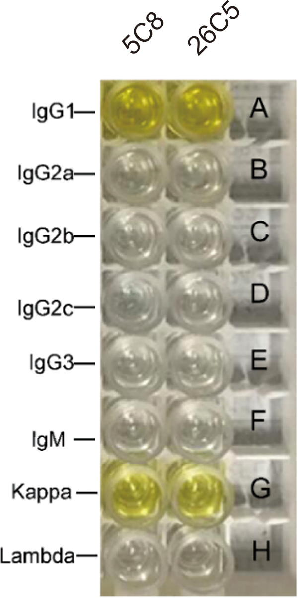
Subclass identification of UL23 monoclonal antibodies. Wells A–F indicates the heavy chain of the sample
Purification of monoclonal antibodies
Since both of 26C5 and 5C8 are IgG1 monoclonal antibodies, the ascites was purified by octanoic acid-ammonium sulfate precipitation method. The purified ascites was subjected to SDS-PAGE. As the results showed, the purified monoclonal antibodies from the ascitic present two bands near 25 and 50 kDa (Fig. 2), which are consistent with the expected molecular size of the IgG light chain and heavy chain.
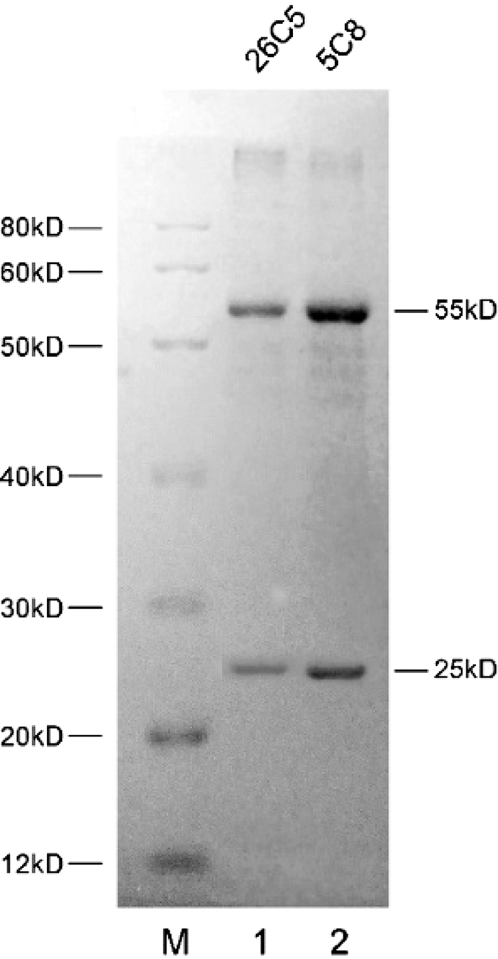
Sodium dodecyl sulfate polyacrylamide gel electrophoresis (SDS-PAGE) of purified ascites. M, protein Maker; 1, purified hybridoma cell 26C5 ascites; 2, purified hybridoma cell 5C8 ascites.
Identification of monoclonal antibodies by western blotting
Monoclonal antibodies 5C8 and 26C5 were used as primary antibodies for western blotting to detect UL23 protein. For monoclonal antibody 5C8, an ∼35 kDa band was detected in 293T cells transfected pcDNA 3.1(+)-UL23, while no signal was detected in 293T cell or 293T cell transfected with pcDNA 3.1(+) (Fig. 3A). Thus, 5C8 can specifically recognize overexpressing UL23 protein. However, UL23 cannot be detected in HFF cells infected with HCMV. On the contrary, a prominent band at 60 kDa was detected in both uninfected and infected cells (Fig. 3B, lanes 1–3). These results indicate that 5C8 antibody could recognize a 60 kDa endogenous protein of HFF cells, failing to recognize the viral protein UL23 in cells infected with HCMV. For monoclonal antibody 26C5, 35 kDa band was both detected in 293T cells overexpressing UL23 protein and HFF cells infected with HCMV, indicating that 26C5 recognized native form of UL23 protein in cells infected with HCMV. Thus, 26C5 was selected to further application to detect UL23.
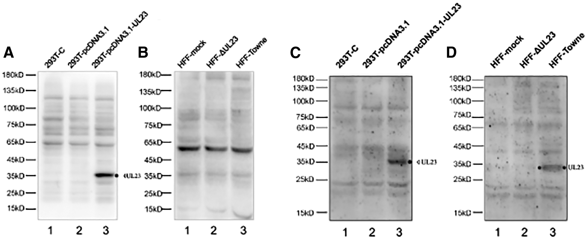
Identification of 5C8 and 26C5 monoclonal antibodies by western blotting.
Identification of the interaction of UL23 and Nmi using monoclonal antibody
Our previous studies have shown that UL23 protein specifically interacts with Nmi (5). In this study, protein lysates from the UL23-HA and Nmi overexpressing cells were immunoprecipitated with 26C5 or anti-HA antibody, and immunoblotted with HA antibody, Nmi antibody, and 26C5, respectively. The Input group shows successful transfection of the Nmi plasmid and the UL23-HA plasmid (Fig. 4 lanes 1–3). A small amount of endogenous Nmi could also be discovered when the Nmi plasmid was not transfected (Fig. 4, lane 2). Consistent with our previous study, Nmi could be detected when 26C5 or HA antibody was used to IP (Fig. 4, lanes 6 and 9), suggesting that 26C5 can enrich UL23 protein with high potency and specificity.
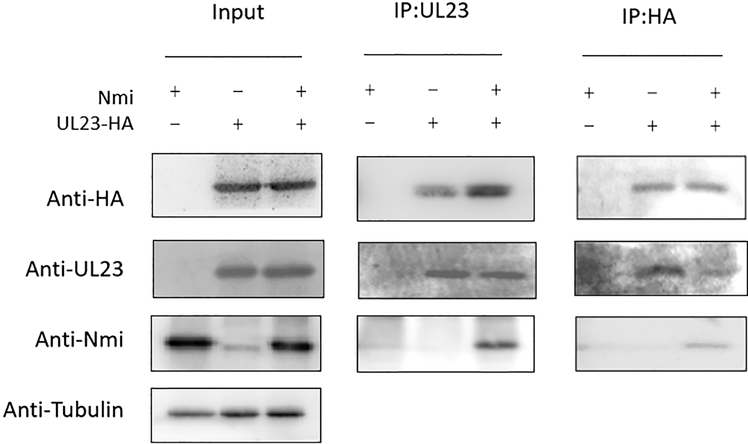
Detection of the interaction of Nmi and UL23 by coimmunoprecipitation assay using UL23 monoclonal antibody 26C5.
Detection of UL23 protein during HCMV infection by indirect immunofluorescence of monoclonal antibody
It is reported that UL23 is localized in the cytoplasm around the nucleus in HCMV-infected HFFF-2 cells (8). To determine whether 26C5 can apply to indirect immunofluorescence, monoclonal antibody 26C5 was used to detect subcellular localization of UL23 protein. HFF cells were infected with HCMV Towne and UL23-deleted mutant, respectively. GFP fluorescence was observed in infected cell, indicating successful infection of HCMV. UL23 protein, which localized surrounding the nucleus, was not detected in uninfected cells and cells infected with UL23-deleted mutant, while red fluorescent of the UL23 protein was detected in HFF cells infected with HCMV Towne (Fig. 5). These results demonstrate that 26C5 can apply to immunofluorescence assay to detect UL23 protein with high specificity.

Indirect immunofluorescence of UL23 monoclonal antibody 26C5. HFF cells were infected with HCMV Towne or Towne-ΔUL23 (UL23-deleted mutant). Immunofluorescence staining was performed using purified 26C5 at a dilution of 1:200 and Cy3-conjugated goat anti-mouse IgG to detect UL23 (red). DAPI was used for nucleus staining. DAPI, 4′,6-diamidino-2-phenylindole; EGFP, enhanced green fluorescent protein. Color images are available online.
Discussion
Due to the large size of HCMV genome and the extremely complex structure, research on HCMV and its protein function has been facing great difficulties. Up to date, only a small number of HCMV viral proteins have been studied intensively. As we know, specific antibodies are essential for the study of proteins. However, it is difficult to prepare a recombinant viral protein having a natural structure and activity as an immunogen for preparing antibodies.
Our previous study shows that HCMV UL23 protein interacts with the host interferon-inducible protein Nmi and inhibits the activity of interferon-γ-dependent JAK/STAT1 signaling pathway (5). To further study its mechanism, UL23 antibody with high potency and high specificity is necessary. However, we have commissioned company to produce UL23 antibody, but results were unsatisfactory. This greatly limits our research on UL23. We later successfully prepared UL23 polyclonal antibody (6). The pAb can recognize HCMV-encoded UL23 protein and UL23 protein overexpressed in cells. However, monoclonal antibody has the advantage of higher specificity, uniformity, and scalable production compared with polyclonal antibody. In this study, a high-specificity IgG1 mAb against UL23 protein was successfully prepared and applied to western blotting, Co-IP analysis, and immunofluorescence experiment.
26C5 successfully detected UL23 protein in cells infected with HCMV (Figs. 3D and 5). In contrast, 5C8 with higher antibody titers could only detect UL23 overexpressed in cells (Fig. 3A, B), indicating that mAb 26C5 can detect native form of UL23, while 5C8 cannot. We hypothesize that the native form of the viral protein UL23 does not expose its epitope recognized by 5C8 mAb.
The titer of UL23 polyclonal antibody was ∼1:521,000 (6), while 26C5 mAb was only 1 × 10⁴. When UL23 polyclonal antibody and monoclonal antibody 26C5 were used to detect viral UL23 protein in western blotting assay, the polyclonal antibody has higher potency after purification. However, even though the titer of monoclonal antibody is lower than polyclonal antibody, mAb has higher specificity in indirect immunofluorescence experiment to locate UL23 effectively. Moreover, 26C5 could also enrich UL23 protein in Co-IP assay with high potency and specificity. Therefore, UL23 polyclonal antibody and monoclonal antibodies were particularly useful in their respective applications.
In summary, we successfully generated an IgG1 mAb against UL23 protein. The mAb 26C5 were specific to the recombinant UL23 and the HCMV expressing UL23 protein. With high specificity, 26C5 could be used to detect UL23 protein location in indirect immunofluorescence experiment and applied in Co-IP assay to enrich UL23 protein.
Footnotes
Author Disclosure Statement
No competing financial interests exist.
Funding Information
This article was funded by the National Natural Science Foundation of China (Grant No. 31770178), Natural Science Foundation of Guangdong province (Grant No. 2016A030311048), and National Major Projects of Major Infectious Disease Control and Prevention, the Ministry of Science and Technology of the People's Republic of China (Grant No. 2017ZX10103011-007), China Postdoctoral Science Foundation (Grant No. 2019M653276).