




Abstract
Abstract
The 9th European Zebrafish Meeting took place recently in Oslo (June 28–July 2, 2015). A total of 650 participants came to hear the latest research news focused on the zebrafish, Danio rerio, and to its distant evolutionary relative medaka, Oryzias latipes. The packed program included keynote and plenary talks, short oral presentations and poster sessions, workshops, and strategic discussions. The meeting was a great success and revealed dramatically how important the zebrafish in particular has become as a model system for topics, such as developmental biology, functional genomics, biomedicine, toxicology, and drug development. A new emphasis was given to its potential as a model for aquaculture, a topic of great economic interest to the host country Norway and for the future global food supply in general. Zebrafish husbandry as well as its use in teaching were also covered in separate workshops. As has become a tradition in these meetings, there was a well-attended Wellcome Trust Sanger Institute and ZFIN workshop focused on Zebrafish Genome Resources on the first day. The full EZM 2015 program with abstracts can be read and downloaded from the EZM 2015 Web site zebrafish2015.org .
Introduction
A
At the closing remarks by Peter Aleström at noon on Thursday, the EZM 2017 venue announcement revealed that Hungary will be the next host country. Máté Varga (Eötvös Loránd University, Budapest) invited the EZM 2015 participants to Budapest in the summer 2017. After the formal conference closure, an EUFishBioMed Society Assembly was held. The Oslo Congress Center turned out to be an ideal venue for the event, right in the center of the city and close walking distance to all the hotels. The organizing company Congress-Conference AS was excellent in all respects. We were also fortunate that the weather was nice throughout the event.
The 9th European Zebrafish Meeting
The 9th EZM program started with two short but insightful opening addresses by the conference hosts and rectors of the University of Oslo, Ole Petter Ottersen, a neurobiologist, and of the Norwegian University of Life Sciences, Mari Sundli Tveit (Fig. 1). The first morning's session on zebrafish genome and phenome was chaired by Andrea Pauli and Christian Mosimann. The first keynote lecture entitled “Big Data in Biology” given by Ewan Birney from the EMBL-associated European Bioinformatics Institute (Hinxton, UK), the main European hub for biomolecular data, discussed the growing problem of how to deal with ever-increasing data sets in zebrafish and medaka research. He pointed out that the two main technology drivers for this glut of data come from DNA sequencing and imaging, one of the main reasons that keep attracting more and more researchers to these transparent fish systems. Birney pointed out that a key focus of current genomics is the elucidation of the functional consequences of sequence variation among individuals and populations. In this regard, medaka scientists have made headway by producing numerous inbred lines. A major task for the zebrafish community will be to concentrate its efforts to generate similar resources for zebrafish genetics. In the first plenary lecture, José Luis Gómez-Skarmeta (Universidad Autónoma de Madrid, Spain) discussed details of how the generation of distinctive cell types that form different tissues and organs requires precise temporal and spatial control of gene expression. One mechanism that his research has revealed to be important is the epigenetic landscape of regulatory elements upstream of the key master genes, with a striking conservation of their 3D architecture in the chromosomes of both zebrafish and mice. In the second plenary lecture, Steve Harvey (Wellcome Trust Sanger Institute, UK) reported promising preliminary results with single-cell transcriptomics to characterize the responses of cells to spatial signal during early embryo patterning. Then, Antonio Giraldez (Yale, USA) discussed control mechanisms of the maternal zygotic transition (MZT) with ribosome footprinting interactome capture and using CLIP-seq for mapping of RNA-binding protein interactions. Didier Stanier (MPI, Bad Nauheim, Germany) reported a novel mechanism for genetic feedback acting upon loss-of-function gene mutations, which may explain some of the discrepancies between the observed phenotypes caused by gene knockdowns (e.g., morphants) versus genetic mutations (e.g., CRISPR gene knockout). To identify the reasons underlying the phenotypic differences between mutants and knockdowns, they made mutations in zebrafish egfl7, an endothelial extracellular matrix gene of therapeutic interest. The egfl7 morpholino-injected animals (morphants) exhibit severe vascular defects, but egfl7 mutants do not show any obvious phenotypes. They identified a set of proteins and genes that were upregulated in mutants but not in morphants. Overall, their data argued for the activation of a compensatory network to buffer against deleterious mutations, which was not observed after translational or transcriptional knockdown. Jeroen Bakkers (Hubrecht Institute, Utrecht, The Netherlands) described an exciting method they have developed, RNA tomography, which is ideal to obtain whole-genome transcriptome information in a spatially resolved manner in embryos or tissues. This new approach combines traditional histological techniques with low-input RNA sequencing and mathematical image reconstruction to generate a high-resolution, genome-wide atlas of gene expression in the embryo or tissue of interest. So far, they have applied this approach to three early stages of zebrafish development, but they have also recently extended the method to identify genes expressed in isolated tissues, such as the regenerating zebrafish heart.
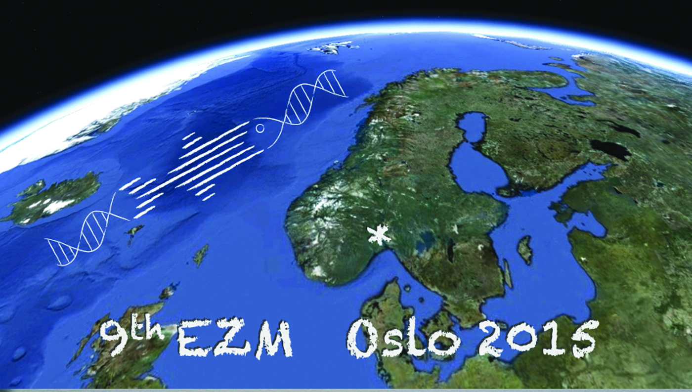
Displays the symbol for the 9th European Zebrafish Meeting 2015 in Oslo Norway. EZM2015 gathered 650 zebrafish scientists, students, exhibitors and others with interest in the subject area coming from 35 countries and 5 continents.
Day 1 afternoon sessions encountered 101 posters and selected short talks in three parallel sessions on Omics (O-I), New Technologies and Imaging (O-II), and Husbandry (O-III). In ORAL I—Omics (moderators Shawn Burgess and Ferenc Müller), Elisabeth Busch-Nentwich presented a novel approach for mutant characterization by multiplexed mRNA sequencing and identification of differential transcript abundance. Cecilia Winata discussed translational regulation of maternal transcripts before and after the midblastula transition and presented evidence for widespread cytoplasmic polyadenylation of transcripts coupled to translational regulation. Andrea Pauli continued in a similar theme and presented mechanisms for translational regulation uncovered on the role of a sequence determinant, which influence translation of open reading frames within protein-coding genes. Bernard Peers presented a comparative transcriptome profiling of pancreatic cell types and demonstrated the strength of the approach in uncovering conserved gene sets acting in regulating endocrine and exocrine pancreatic cell populations among vertebrates. ORAL II—New Technologies and Imaging (moderators Johan Ledin and Jan Huisken). Among highlights in this session, Christian Mosimann reported a workflow for saturating somatic loss-of-function mutagenesis by injection of Cas9–sgRNA ribonucleoprotein complexes with high activity and low toxicity, thereby constituting an alternative to morpholinos for rapid assessment of loss-of-function phenotypes. Flavia De Santis presented a strategy for conditional knockout and permanent labeling of cells by combining Gal4/UAS-mediated expression of Cas9 with the use of the Cre/LoxP toolbox. Herwig Baier reported the adaptation of the Brainbow technique to target expression of an optogenetic actuator and a calcium indicator in subsets of neurons. A developed four-color labeling kit was named Optobow. Finally, Marjo den Broeder introduced a method for label-free stimulated Raman scattering microscopy to visualize lipid content in adipocytes with submicrometer resolution. ORAL III—Husbandry (moderators Stefan Schulte-Merker and Frederic Sohm) dealt with issues related to the development of standardized husbandry, health, and animal welfare guidelines (this will be separately published in Zebrafish.
The second day's morning session was chaired by Zhiyuan Gong and Gareth Griffiths and focused on zebrafish as a model for biomedicine. The strength of using this fish model to study human diseases was well demonstrated by the keynote speaker for this session, Leonard Zon (Harvard Medical School, Boston, USA), who covered an impressive range of diseases, such as those affecting hematopoiesis and skin cancer. He showed, for example, how the ability to visualize the birth of blood stem cells from the aorta early during embryogenesis in the fish provides a screening system facilitating new treatments, such as for derivatives of prostaglandin E2 to stimulate increases in stem cells. The zebrafish is indeed a powerful system for rapidly screening drugs; he described how a number of drugs also appeared to provide promising therapies for human therapies in clinical trials, for example, for melanoma treatment. He appropriately referred to this strategy as “from tank to bedside.” More details of the signaling pathways regulating the process by which hematopoietic stem cells (HSCs) arise transiently from the endothelial cells lining the aorta were described by David Traver (San Diego, USA). One key player in the regulation of temporal and spatial development of HSCs during zebrafish embryogenesis is the cytokine tumor necrosis factor alpha (TNFα) through activation of TNF-receptor 2. TNFα secreted from primitive neutrophils was necessary for both HSC specification and maintenance. The effect was dependent on the expression of the Notch ligand Jag1a on neighboring endothelial cells to promote induction. Coinjection of Tnfr2 MO with either Notch1a or Notch1b MOs identified the former as the main receptor for Jag1a. Further downstream, Notch triggers the development of HSC via NF-kB transcriptional regulation. Ewa Snaar Jagalska (Leiden, The Netherlands) followed up on the use of zebrafish as a cancer model by focusing on the method of injecting human prostate cancer cells into zebrafish embryos. The absence of an adaptive immunity in the zebrafish embryos and larvae allows such foreign cells to grow into tumor-like masses from cancer stem cell precursor cells. An important stimulatory role was identified for the tyrosine kinase Syk in the dissemination of the prostate tumor cells in the fish and subsequently in a mouse model. This opens the door to preclinical testing of Syk and mir25 inhibitors against prostate cancer. The microRNA mir25 was also identified as a suppressor of tumor cell growth. The use of zebrafish for analyzing human and zebrafish tumors was also impressively demonstrated in a number of shorter oral and poster presentations during the meeting. Antonio Pagan, from Lalita Ramakrishnan's group (Cambridge, UK), described the use of infecting zebrafish with Mycobacterium marinum as a model for human tuberculosis, an approach pioneered by this group. He described details of the mechanisms by which the granuloma, initially an aggregate of infected macrophages, can become necrotic, allowing the live bacteria to be released extracellularly, a process that in humans occurs primarily in the lungs. Although zebrafish breathes via the gills rather than the lungs, the main target of human TB, the mechanisms unveiled in the fish are increasingly accepted as being highly relevant for understanding human TB. Whereas the number of bacterial pathogens that have been monitored in the zebrafish larval system is likely around 40 by now, the use of zebrafish for analyzing viral infections has been much less used. An exception is the work of Jean Pierre Levraud (Pasteur Institute, Paris, France), who has pioneered the infection by human alpha viruses, chikungunya virus (CHIKV) and sindbis virus (SINV). He described in detail how the infections are regulated by type 1 interferons; initially, the infection is systemic, but even in embryos a few days old, the interferons restrict viral growth while the viruses enter the central nervous system, where they persist. It is really striking how a vast range of human diseases are now being analyzed in detail in zebrafish models; this was strikingly evident in a myriad of talks and posters in the biomedicine sessions. It was also impressively demonstrated in the last plenary talk in this part given by Pierre Drapeau (University of Montréal, Canada), who described a zebrafish model for amyotrophic lateral sclerosis (ALS), the crippling motor neuron disease (made famous by Stephen Hawking). Genes identified as being mutated in ALS can be identified and mutated in zebrafish, leading to motor neuron and motility defects. Using a screen of more than 3800 clinically approved drugs in this system, the drug pimozide was found to be highly effective in both zebrafish and a mouse model of ALS. Impressively, this drug is now being tested in a phase II clinical trial.
Day 2 counted 132 posters and had selected short talks in three parallel sessions entitled ORAL IV—Cancer and Regeneration, ORAL V—Therapeutics, Angiogenesis, and Cell Development, and ORAL VI—Morphogenesis and Cell Signaling. In O-IV (moderators Ewa Snaar-Jagalska and Hanne C. Winther-Larsen), the power of the zebrafish in regeneration and cancer was highlighted. There were two talks on cancer: Liz Patton started by presenting their work on the identification of a small molecule in zebrafish that targets melanocytes for cell death and how these cells are now repurposing this compound to target ALDH-positive cells in cancer. Felix Oppel presented new genetic models for pediatric high-grade glioma and MPNST and showed exciting evidence that combinations of genetic mutations in p53, nf1, atrx, and h3f3a can contribute to glioma development and progression. Zebrafish provide a unique model to study regeneration, and five talks addressed the cellular and molecular mechanisms that regulate regeneration in zebrafish. Michael Brand presented work on brain regeneration following a traumatic brain injury assay. Using lineage tracing methods, they have previously shown that ventricular radial glial stem cells develop into new neurons. However, they find no evidence of regeneration from the HSCs, suggesting that while the brain can regenerate following injury, this is not due to conversion of the HSC lineage. J.S. Barbosa presented work on regeneration in the telencephalon following injury and used imaging to show that adult neuronal stem cells contribute to repopulation of the neurons. Gene expression studies revealed that AhR signaling is important in a subset of neuronal stem cells and that this pathway is conserved in mammals. Katharina Lust presented their work on the regenerative potential of Müller glia cells in the medaka retina and described new cellular mechanisms of regeneration compared with zebrafish. Christopher Antos presented findings on the development and regeneration of joint cells in the zebrafish fin skeleton and their discovery that the phosphatase calcineurin is required for setting the tissue boundaries between bones and joints. Two presentations on heart regeneration highlighted the importance of zebrafish in unraveling the mechanisms in heart repair and regeneration. Gilbert Weidinger reported that in contrast to mammals where bone morphogenetic protein (BMP) signaling can be detrimental to heart repair, Bmp signaling in zebrafish regulates processes in cardiomyocytes that specifically occur in response to injury and lead to repair. Juliane Münch examined the role of Notch in heart regeneration and discovered that Notch has a role in separating the damaged area from the rest of the heart, cardiomyocyte proliferation, fibrosis, and inhibiting inflammation. O-V (moderators Annemarie Meyer and Tor Gjøen) included talks on subjects ranging from studies of nitrogen metabolism (Cox) to angiogenesis (Lockwood). Using optical projection tomography (OPT), and a transparent transgenic line, liver tumor progression could be traced in live animals. Jon Hildahl reported on the successful use of nanoparticulate antibiotic treatment combined with the drug efflux inhibitor thioridazine to treat mycobacterial infections. In a new model for retinopathies, neovascularization of the retina and the choroid could be followed in great detail after exposure to hypoxia. O-VI (moderators Jochen Wittbrodt and Viola Lobert) focused on three signaling pathways that are integral to development: Wnt, Bmp, and Nodal. Dafne Gays showed that intestinal smooth muscle cells arise from the lateral plate mesoderm, using genetic lineage tracing, and further discussed the role of miR145 and foxo1a in the differentiation process. Anja Hagemann focused on the intracellular trafficking of Wnt, showing that Ap2 is involved in its endocytosis. Anastasia Eskova described the interesting observation that the mutant Mau, where aquaporin 3a is mutated, exhibits defective patterning of pigment. Stephan Heermann showed us some stunning 4D live microscopy films revealing the formation of the optic cup, a process that is defective in the human eye disease coloboma and regulated by Bmp.
The third conference day was dedicated to fish models in aquaculture and environmental studies, including toxicology, with Gert Flik and Petter Arnesen as chair persons. Norway's third export commodity, after oil and gas, is farmed fish, especially salmon. And of course zebrafish, and medaka, can provide excellent model systems for other fish; experiments with, for example, salmon are time consuming and expensive. So it was rationale for the host country to dedicate one of the four morning sessions for the first time in an EZM to cover the broad topic—laboratory fish models for aquaculture. Appropriately, Petter Arnesen is from the Norwegian company, Marine Harvest, one of the largest seafood companies in the world. He summarized the big problems facing aquaculture from a commercial perspective and opened the door to how model fish research could contribute to solving these problems. The keynote lecture was given by Laia Ribas (Barcelona), who gave an excellent broad perspective of the potential, as well as limitations of zebrafish to be used as a model system for different aquaculture fish. Among the important issues for which she believes that zebrafish can provide an important model system are reproduction, stress, pathology, infection, toxicology, nutrition, and growth. The Wednesday session also covered new technologies and tools for zebrafish and medaka research. Jochen Wittbrodt from the Heidelberg University gave an overview of the creation of a population genomic resource by establishing near-isogenic wild lines in medaka. He also introduced the high-throughput morphometric analysis for deciphering complex traits by exploiting X-ray tomography and automated segmentation. Shawn Burgess continued by presenting a high-throughput targeted mutagenesis pipeline for zebrafish using the CRISPR/Cas9 technology in combination with cloning-free single-guide RNA and streamlined mutant identification systems. The CRISPRz Web site is available at http://research.nhgri.nih.gov/CRISPRz and provides a very useful searchable interface for validated CRISPR gene targets.
Back to fish biology, Simon MacKenzie (University of Sterling) entertained the audience by discussing the concept of thermal preferences in fish. Such behavior is triggered by various stress conditions, and MacKenzie showed us convincingly that zebrafish is capable of expressing emotional fever. In addition, coupled with adaptive responses to host–pathogen interactions, the fish that were offered a choice of temperature expressed better survival rates than those without such a choice during an infection. The 9th EZM was technically challenged with a live video broadcast of Christian Lawrence from the Boston Children's Hospital. His talk focused on lessons to learn for the experimental model fish community from aquaculture, based on his long experience in rearing zebrafish embryos, especially important he pointed out is when to start feeding larvae to obtain the highest performance levels and thereby the most reliable scientific output. Unfortunately, the connection across the Atlantic was cut off near the end of his talk, but the full lecture could be viewed online and downloaded at the EZM 2015 Web site. The aqua model session was closed by Hanne C. Winther-Larsen, the only local plenary lecture. She led us through a story from establishing zebrafish as a model organism for aquaculture research, and how the model was used for studies of host–pathogen interaction focused on species of Francisella bacteria, with promising development and trials of vaccine technologies relevant for several aquaculture fish species. The last part particularly caught the attention of the session moderator Petter Arnesen.
Day 3 posters (135) and selected short talks in three parallel sessions entitled ORAL VII Aqua—Reproduction and Toxicology, ORAL VIII—Neurobiology, and ORAL IX—Migration and Development. O-VII (moderators Harald Sveier and Igor Babiak) was started off with Lex Nederbragt from the Norwegian Sequencing Facility. His talked focused on the new high-quality Atlantic cod reference genome. This work has been a challenging task as the cod genome contains an extreme number of repetitive regions. Xinhua Shu enlightened us on the topic of pollution and ecotoxicology of pharmaceuticals in water flushed out via the drain and how this might affect fish in the water column. Another highlight from this session was the talk by Arturas Kavaliauskis demonstrating the benefits of using nanoparticulate vaccines consisting of polyI:C and chitosan against viral infection problematic in aquaculture. In O-VIII (moderators Camila V. Esguerra and Finn-Arne Weltzien), Emre Yaksi showed, using two-photon calcium imaging and quantitative behavior, that different taste categories give dissimilar brain stem responses and can generate different behaviors. Raphaël Candelier gave an interesting presentation on the development of two-photon light-sheet imaging of zebrafish brains, combining the advantages of near-infrared illumination, to avoid activating retina sensory cells, with the high data throughput advantage of light-sheet microscopy. Dario Acuña-Castroviejo demonstrated that zebrafish can be used as a model for Parkinson's disease and showed neuroprotective properties of melatonin. CM von Berg-Maurer showed for the first time the activity of corticotropin-releasing hormone (CRH) cells in vivo using two-photon calcium imaging in larval zebrafish with transgenic label of CRH cells while simultaneously applying stressful input of different intensities. In O-IX (moderators Jarema Malicki and Trude M. Haug), Mary Mullins showed that Balbiani body precursor components are first localized with the formation of the chromosomal bouquet, suggesting that these two oocyte meiotic features are linked on the pathway toward oocyte polarity. JGM Bergboer investigated sensory cilia function in zebrafish olfactory sensory neurons. Using a combination of transgenic Gcamp5 (calcium biosensor) and ift88 (cilia mutant) zebrafish, they showed the crucial role of cilia in sensing of pheromones (bile acids). The day 3 program ended with the DANIO-CODE workshop, a forum for developments in the annotation of the zebrafish genome. Ferenc Müller provided an overview of a consortium that formed in London in 2014, aiming at coordination of the generation and management of zebrafish genome annotation resources. Müller encouraged all to join. ZFIN will host a track hub for genomic data sets, and consortium members work on establishment of a data coordination center. A small number of presentations followed, including computational analysis of nucleosome positioning during zygotic genome activation (Miler Lee) and a report on a new class of small RNAs called site RNAs associated with silencing of genes.
The day 4 morning session was devoted to development and neurobiology, chaired by Mary Mullins and Jean-Stephane Joly. The keynote lecture was given by Rainer Friedrich, who gave an outstanding presentation on his group's progress in elucidating neural circuit formation and brain function in the zebrafish. He reported on the remarkable complete reconstruction of the zebrafish larval and adult olfactory bulb using serial block-face scanning electron microscopy. Using calcium-imaging techniques, his group examined neuronal activity in the olfactory bulb and then probed neural circuit function with inducible optogenetic methods. Altogether, he demonstrated the value of the zebrafish to perform a system-wide neural analysis of olfaction. The first plenary presentation was by Jan Huisken reporting on their recent cutting-edge advances in selective plane illumination microscopy or SPIM, which is amazing. He showed how their latest iteration of SPIM can now simultaneously detect multiple fluorophores, such as GFP, YFP, Hoechst, and BODIPY, through a clever algorithm of spectral unmixing. He also discussed their ability to deconvolve the many images acquired by real-time microscopy to significantly reduce the disk space needed for storage. Claire Wyart focused on very original cell types of the CNS that are in contact with the cerebrospinal fluid (CSF). Behavior and locomotion have long been known to be modulated by the content and flow of the CSF. However, while the mechanisms underlying the modulation of neuronal networks by CSF are unknown, they took advantage of the transparency and genetic accessibility of zebrafish larva to investigate the sensory interface between CSF and spinal circuits. CSF-contacting neurons sense bending of the spinal cord and in turn relay information to locomotor central pattern generators. Altogether, the identification of a novel sensorimotor loop interfacing CSF to motor circuits opens new paths for the investigation of thermo-, mechano-, and chemosensory modulation of locomotion. Elegant imaging technologies were also used by Lucia Poggi and collaborators to understand asymmetric divisions that generate one self-renewing progenitor and one neuron-committed cell. She reported for the first time on the in vivo dynamics of anillin, a key component of the F-actin furrow, and its contribution to progenitor cell divisions in the zebrafish retina. Retinal ganglionic cell (RGC) progenitors position the anillin midbody at the apical domain to perform a correct cell division and asymmetric distribution of daughter cell fates.
Olivier Kah discussed the potential functions of aromatase in glial cells. Aromatase indeed has a strong activity in these cells. This is an exciting research topic due to several unique features that challenge what was concluded in other vertebrates. Radial glial cells persist throughout life and act as progenitors in the brain of developing and adult fish. Expression of GFP in transgenic Tg(aromatase/cyp19a1b:GFP) fish was directed to radial glial cells of adults. Moreover, brain aromatase activity is correlated with peripheral sex steroid levels. Hence, Kah suggested that, in addition to classical functions in brain sexual differentiation and sexual behavior, aromatase expression in radial glial cells could be involved in the modulation of proliferation in the brain.
Florence Marlow discussed her group's latest advances in understanding how the germ plasm is assembled in the early embryo. She showed that the microtubule motor protein Kif5ba plays a key role in bringing the germ plasm and the buckyball protein to the cleavage furrows of the early embryo in specifying the primordial germ cells. They also identified the Dazap2 protein in a yeast two-hybrid screen for proteins that interact with the buckyball and showed its function, through generation of a maternal dazap2 mutant, in maintaining the perinuclear germ granules associated with the germ cells in zebrafish.
The workshop Zebrafish in Teaching (moderator Charles Press) gathered many participants in the day 3 lunch break to hear about the use of zebrafish in teaching and education. The session began with reminders that this journal Zebrafish has issued a call for papers for a special issue on “Zebrafish in Education” (deadline November 2015) and that a new forum for the life science teacher resource community, the Zebrafish Education Network, has been established at www.lifescitrc.org. Daria Filipek (IIMCB, Poland) presented their experience from their outreach program for 5th- to 6th-grade elementary school students. The program uses an educational movie and laboratory exercises to introduce school students to the biology of zebrafish and practical aspects of scientific work. Kiyoshi Naruse (NIBB, Japan) presented how medaka is used in the scientific education of 5th-grade elementary school students in Japan. Phenotypic observations of spontaneous mutant strains of medaka are readily performed in the classroom and readily applicable to PCR genotyping. Finn-Arne Weltzien (NMBU, Norway) introduced the initiative “School of Fish,” aiming at a Norwegian research school in the pleiotropic application of small fish models in biological research. Charles Press (NMBU, Norway) presented the integrated concept of e-ZFbook that is a teaching resource suitable for secondary school, undergraduate, and postgraduate education. The e-ZFbook resource will use Web sites, apps, short movies, and practical teaching materials to facilitate the use of zebrafish in scientific education.
Before the EZM 2015 closing remarks, the three poster awards were announced by the poster award committee (Shawn Burgess, Florence Marlow, Jean Pierre Levraud, Zoltan Varga, Jan L. Lyche, and Hanne C. Winther-Larsen). One poster was selected from each poster session. The winners had a strong influence from the Benelux countries with winner of session P1 Leonie Kamminge (The Netherlands), session P2 Samrah Masud (The Netherlands), and session P3 Anne Houbrechts (Belgium) and covered different topics from cardiac integrity, Salmonella-induced autophagy, and activation during early development, respectively. The EZM social program included a reception on the Monday evening in the Oslo City Hall, made famous as the site of the annual ceremony to celebrate the Nobel Peace prize, with the Mayor of Oslo, Fabian Stang inviting the EZM 2015 delegates to food and drinks. The conference dinner on Wednesday at the Opera House, well located at the Oslo waterfront, included opera songs and ended with everyone singing the EZM 2015-dedicated song, “Zebrafish Blues” (see zebrafish2015.org for pictures and music). All in all, we can safely conclude that the 9th European Zebrafish Meeting was a resounding success.
Footnotes
Disclosure Statement
No competing financial interests exist.