




Abstract
This study aimed to evaluate the viability of gametes in zebrafish (Danio rerio), at different rigor mortis stages. Viability assessments were conducted on oocytes at various developmental stages using LIVE/DEAD and the 3-(4,5-dimethylthiazol-2-yl)-2,5-diphenyltetrazolium bromide (MTT) reduction assay. For sperm evaluation, both kinetic (computer-assisted sperm analysis) and morphological assessments (Rose Bengal staining) were performed. Results demonstrated that rigor mortis progression significantly impacted oocyte viability during post-rigor stages, with the following viability rates: pre-rigor (70.43 ± 12.31%), fresh/control (46.43 ± 12.54%), post-rigor (27.62 ± 22.29%), and rigor mortis (comparable to fresh/control). Conversely, sperm kinetics exhibited nuanced responses to the rigor mortis stages, with specific parameters showing sensitivity, whereas the others remained relatively stable. Sperm motility was higher in the fresh/control (63.23 ± 19.03%) and pre-rigor (58.96 ± 14.38%) compared to the post-rigor group (3.34 ± 4.65%). This study highlights the significance of the pre-rigor for successful gamete collection and preservation. These findings provide valuable insights for conservation efforts and optimization of genetic resource management for endangered fish species. This study aimed to develop effective assistive reproductive techniques by elucidating the interplay between rigor mortis and gamete quality, contributing to the broader goals of species conservation and maintenance of genetic diversity in fish populations.
Introduction
Preserving the genetic material of endangered aquatic species and valuable reproducers in fish farms is a pressing concern. One promising approach to address this challenge is the collection of gonads following animal death, which enables the utilization of reproductive material otherwise lost. Previous investigations have demonstrated the potential to generate offspring using frozen–thawed sperm through techniques such as intracytoplasmic injection and transplantation of testicular tissue and germ cells.1–3
Building on these findings, studies on species such as stinging catfish (Heteropneustes fossilis), 4 striped catfish (Pangasius sutchi), 5 and rosy barb (Puntius conchonius) 6 have highlighted the viability of post-mortem stored at −20°C. In these studies, sperm collection occurred over several days after death, followed by cryopreservation, and viability was assessed through parameters such as motility, eosin–nigrosin staining, scanning electron microscopy, and fertilization attempts. These results emphasize the feasibility of extending the reproductive potential of aquatic species under specific conditions.
Further expanding on post-mortem reproductive viability, analyses conducted with rainbow trout (Oncorhynchus mykiss) subjected to varying post-mortem intervals (0, 6, 12, and 24 h at 10.5°C) explored several critical factors. These included muscular ATP levels, histology, immunohistochemistry, and enzymatic dissociation of testicular tissue, ultimately demonstrating the successful transplantation of spermatogonia into 30 dpf larvae. 7 These findings reinforce the potential of advanced reproductive technologies for conserving genetic material in fish.
Among aquatic species, zebrafish (Danio rerio) stands out as a prominent model organism in developmental biology and biomedicine. Widely used for toxicity assessments, environmental risk evaluations, and pharmacological studies,8,9 zebrafish have significantly advanced our understanding of sexual determination and gonadal development in fish.10–12 However, as in other species, postovulatory aging of zebrafish oocytes impacts egg quality and increases the incidence of ploidy anomalies in embryos. 13 Similarly, sperm quality also declines over time, although this window of optimal fertility can be extended under specific storage conditions, such as low temperatures. 14
Despite these advancements, the relationship between rigor mortis stages (pre-rigor, rigor, and post-rigor) and the quality of gametes collected post-mortem remains poorly understood. This study aims to bridge this gap by investigating how rigor mortis dynamics influence the viability of collected gametes, thereby enhancing the predictability of successful assisted reproductive techniques.
Materials and Methods
Experimental design
The gonads of 200 zebrafish (80 females and 120 males) were collected and were randomly distributed among the following groups: Control/fresh and treatments pre-rigor mortis; rigor mortis; and post-rigor mortis. A total of 40 individuals were analyzed, with the same samples used for both mitochondrial activity and sperm morphology analyses. In all analyses, each individual was considered a replicate.
The study received approval from the Animal Care Committee at the Universidade Federal do Rio Grande do Sul (UFRGS) (Project number: 43232), in accordance with the Brazilian Directive for the Care and Use of Animals for Scientific and Didactic Purposes—DBCA, the CONCEA (National Council for Control and Animal Experimentation) Euthanasia Practice Guidelines, and according to ARRIVE guidelines. 15
Fish care and euthanasia
The fish used in this study were obtained from Delphis Pet Store and maintained in 40 L aquariums at a stocking density of 7 fish/L. The water was filtered and dechlorinated, with constant aeration, temperature maintained at 27°C ± 2°C, pH stabilized between 7.0 and 7.4, and a photoperiod of 14 h of light and 10 h of darkness. All conditions were monitored, including ammonia levels, nitrite levels, and pH, using Labcon® Test Kit. The fish were housed in the AQUAM Laboratory, Faculty of Agronomy, at UFRGS. Fish were fed twice daily with Tetra® TetraMin Flakes, equivalent to 6% of their body weight, and once daily with newly hatched Artemia nauplii. The fish were fasted for 24 h before the experiment and euthanized using a lethal dose of tricaine methane sulfonate (0.6 mg/mL, pH 7.0–7.4). 16
Rigor mortis stages in zebrafish (Danio rerio)
Based on the work of Dunford et al., 17 and the general understanding of rigor mortis in fish, we developed a specific methodology to assess the different stages of this process in zebrafish. Rigor mortis is characterized by the loss of muscle plasticity and extensibility due to alterations in the contraction–relaxation cycle occurring in three stages: pre-rigor, characterized by the beginning of muscle stiffening; full rigor, when stiffness is fully established; and post-rigor, when rigidity resolves and muscles regain flexibility. 18 In this study, we defined these stages based on the post-mortem changes observed in zebrafish. While rigor mortis in larger fish is typically assessed on a horizontal surface, we adapted the methodology by using a plastic protractor to measure changes in body angle, which serves as a reliable indicator of the different stages. This adaptation tailors the analysis to the specific characteristics of this experimental model.
After euthanasia and biometry of the animals (average weight: 0.585 ± 0.181 g; average length: 3.64 ± 0.503 cm), each animal was held by the caudal peduncle at intervals of 30 min to 1 h after death and measured against a plastic protractor (180°) to measure the angle to determine the stage of rigor mortis (control/fresh: 40°; pre-rigor mortis: 35°; rigor mortis: 0°; post-rigor mortis: 40°).
The fish reached the rigor mortis stages according to the room temperature (21.5 ± 2.12°C) and humidity (66 ± 6.3%). According to the animals reaching the angle on the protractor, corresponding to the rigor mortis stages previously established, the gonad collection was performed. In the experimental conditions described, the fish reached pre-rigor mortis within 1 h, rigor mortis within 8 h, and post-rigor mortis at approximately 15 h.
Ovarian and testicular tissue collection
The fish were maintained in Petri dishes with drops of aquarium water, to avoid skin dryness. Furthermore, the gonads were collected according to the rigor mortis stage.
Positioning the fish in dorsal decubitus, and using microdissection scissors, a small incision was made in the cranial direction on the ventral surface of the abdomen of each animal to remove the intestine and expose the gonads. The ovarian tissue was extracted, placed in 90% Leibovitz L-15 medium (pH 9.0), weighed (average: 0.071 ± 0.034 g), fragmented in three slices (dimensions of 10 × 2 mm), and weighed individually (average weight of fragments: 0.023 ± 0.010 g). Each fragment was placed in microcentrifuge tubes containing 90% L-15 (400 µL). This step was performed for 20 min. The testis was collected, weighed (average weight: 0.011 ± 0.017 g), and macerated in microtubes containing Hank’s Solution (HBSS) (50 µL).
Analysis
Mitochondrial activity
To evaluate mitochondrial activity in living cells, thiazolyl blue tetrazolium bromide (MTT) staining and spectrophotometry were used. Based on the reduction of MTT to formazan crystals by mitochondrial succinate dehydrogenase, this method is only effective in living cells.
The ovarian tissue fragments were subsequently placed in 90% L-15 (400 µL), and the liquid was removed. In males, the sperm was homogenized in HBSS solution (50 µL; pH: 7.5; osmolarity: 300 mOsm/KgH2O) and centrifuged at 1000 g for 10 min using a Refrigerate Centrifuge (NT 81 Novatecnica®), and the supernatant was discarded. The samples were incubated with MTT (400 µL) for 120 min at 28°C. The resulting formazan crystals were dissolved in dimethylsulfoxide (Me2SO) (400 µL), and the protocol was modified by adding paraformaldehyde 14% (20 µL), acting as a fixer, and cooling the sample to 4°C up to the day of analysis. This adaptation of the protocol was justified by the time of the last treatment (post-rigor) and the absence of a microplate reader at the time of analysis. The protocol was adapted for all samples and treatments. On the next day, the samples were thawed and pipetted into 96-well microplates (100 µL each well) for absorbance analysis at 570 nm on the SpectraMax® M2 Microplate reader. The color intensity of the samples indicates higher mitochondrial activity. Each sample was analyzed in triplicate.
Live/dead viability stain protocols
The staining protocol was used to determine the number of viable cells present in the ovarian and testicular tissue fragments at different post-mortem stages. Fluorometric methods in diacetate (FDA), SYBR-14, and propidium iodide (PI) test the integrity of the cell membrane by alternating inclusion (FDA/SYBR-14) and exclusion staining (PI). FDA permeates the plasma membrane and is hydrolyzed by intracellular esterases. SYBR-14 (LIVE/DEAD Sperm Viability Kit®) is a green fluorescent stain that has an affinity for DNA; when penetrating living cells, they lose their acyl groups and bind to the DNA of the cell. In both staining protocols, viable cells showed bright green staining. PI cannot penetrate viable cell membranes, resulting in bright red-stained cells (dead cells).
For viability assays, one ovarian tissue fragment from each female was treated with FDA and PI. 19 Each fragment (10 females/treatment: fresh/control; pre-rigor mortis; rigor mortis; post-rigor mortis: 40 samples) was incubated with FDA (0.02 mg/mL; 300 µL) and PI (0.2 mg/mL; 30 µL) in the dark for 4–5 min at RT (25 ± 1°C). Follicle stages were determined according to the characteristics described by Selman et al. 20 For the sperm membrane integrity assay, a sample of milt was diluted and macerated in Hank’s Solution (20 µL) (10 males/treatment: Fresh/control; pre-rigor mortis; rigor mortis; post-rigor mortis: 40 samples) and incubated with SYBR-14 (0.02 mm; 300 µL) for 4 min, and PI (1.19 mm; 0,5 µL) for 1 min in the dark, at RT (25 ± 1°C). All cells present on the glass slide in each group (control and treatments), in both females and males, were evaluated and analyzed under a fluorescence microscope (OPTON; Model TNI-06T-PL; 20× objective lens). The percentage of cell viability in each oocyte phase (primary growth stage [PG], cortical alveoli [CA], and vitellogenic [Vtg]) within the fragments and in the spermatozoa was calculated as follows: (cell viability [%] = [number of damaged membranes (oocytes/spermatozoa)/total number (oocytes/spermatozoa) × 100]).
Sperm kinetic parameters (computer-assisted sperm analysis)
The sperm samples were collected fresh and at the three rigor mortis stages (pre-rigor mortis, rigor mortis, post-rigor mortis); to evaluate the sperm kinetics, the milt was diluted and activated in Hank’s Balanced Salt Solution (50 µL) and activated with distilled water (1:10 µL). In a Neubauer hemocytometer chamber, the solution (5 µL) was added and covered with coverslip. A high-speed camera (Basler AC640–120uc, 120 fps, Ahrensburg, Germany) was fixed under a transmitted light microscope (Nikon Eclipse E200) at 1000× magnification. Five videos were recorded for each fish using the Pylon Viewer 4 software (Version 4.1.0.3660 64-Bit; Basler, Ahrensburg, Germany) at a capture rate of 100 frames per second. Each video was 20 s long and analyzed at 0.5 s (50 frames), starting 10 s after activation. The videos were edited using the VirtualDub software and analyzed using the computer-assisted sperm analysis plugin in ImageJ. The evaluated parameters were motility rate (%), curvilinear velocity (µm·s−1), mean travel velocity (VAP, µm·s−1), straight-line velocity (VSL, µm·s−1), straightness (STR, %), wobble (WOB, %), progression (PROG, µm), and beat cross frequency (BCF, Hz). The settings used for analysis were a (amplitude threshold) = 5, b (brightness threshold) = 60, c (contrast) = 50, d (detection sensitivity) = 8, e (elongation ratio) = 5, f (frame rate) = 10, g (gradient threshold) = 15, h (Head Size) = 5, i (integration time) = 1, j (jump distance) = 25, k (kinematic parameters) = 5, l (Linearity Filter) = 10, m (motility threshold) = 80, n (noise reduction) = 80, o (object size) = 50, p (Progression Threshold) = 60, q (quality control) = 100, r (region of interest) = 431, s (speed threshold) = 0, and t (track duration) = 0 (adapted from Wilson-Leedy; Ingermann, 2007). 21
Spermatozoa morphology
Sperm were diluted in HBSS Solution and fixed in a 4% buffered formaline. 22 An aliquot from each sample (30 µL) was homogenized in Bengal Rose staining solution (3 µL). 23 Two drops (15 µL) were placed on a glass slide, using the drained drop method (adapted from Sanches et al.). 24 The slides were analyzed under an optical microscope at 1000× magnification (Nikon® Eclipse Si, Tokyo, Japan), and sperm samples (n = 200 spermatozoa/slide) were evaluated for sperm morphology for the following classifications: normal spermatozoa, loose head, degenerated head, macrocephaly, microcephaly, short tail, distally curled tail, strongly curled tail, broken tail, bent tail, short tail, and proximal and distal gout (adapted from Milliorin et al., 2011). 25
Statistical analysis
Normality (Shapiro–Wilk and/or Kolmogorov–Smirnov) and homogeneity (Bartlett and/or Levene) tests were verified. Variables that met the assumptions were analyzed using a one-way analysis of variance (ANOVA). When a significant difference was observed, the means were compared using Tukey’s test. Nonparametric data were analyzed using Kruskal–Wallis analysis, followed by Dunn’s test. To evaluate the types of sperm pathologies observed at different stages of rigor mortis, a two-way ANOVA was used, considering the effects of the type of pathology (head, cytoplasmic gout, and tail), the stage of rigor mortis, and the interaction between factors. When a significant effect was observed between any of the factors, a mean test was performed. All analyses were performed considering a significance of 5% (p < 0.05). Parametric variables are presented with bar graphs (mean and standard deviation), and nonparametric variables are presented with Box and Whiskers style graphs (minimum–maximum, median). Graphs were created and analyzed using statistical software.
Results
In the analysis of cell viability in oocytes at the PG stage (Fig. 1A), the highest rate of viability (p < 0.05) was observed in the pre-rigor mortis group (70.43 ± 12.31%), compared to the fresh/control (46.43 ± 12.54%) and post-rigor mortis (27.62 ± 22.29%) groups, with no significant difference (p > 0.05) compared to the rigor mortis group. For oocytes in the CA (Fig. 1B), primary Vtg1 (Fig. 1C), and tertiary Vtg3 (Fig. 1E) stages, the highest viability rates (p < 0.05) were observed in the fresh/control (CA: 43.44 ± 17.05%; Vtg1: 43.75 ± 11.54%; Vtg3: 49.64 ± 27.26%), pre-rigor mortis (CA: 59.08 ± 17.10%; Vtg1: 31.24 ± 13.57%; Vtg3: 40.24 ± 16.60%), and rigor mortis (CA: 50.99 ± 29.57%; Vtg1: 37.37 ± 20.13%; Vtg3: 54.98 ± 21.84%) groups. In contrast, the post-rigor mortis group exhibited averages of CA: 15.22 ± 13.44%; Vtg1: 13.37 ± 8.784%, and Vtg3: 5.145 ± 10.28%.

Oocytes cell viability (%) at different rigor mortis stages using the fluorometric method in diacetate (FDA) and propidium iodide (PI). Oocyte phase:
In the analysis of mitochondrial activity, there was no difference between females (p = 0.9121) (Fig. 2) and males (p = 0.9417) (Fig. 3A) in the experimental groups. The viability of zebrafish sperm collected at different stages of rigor mortis was not significantly different between the experimental groups (p = 0.0822) (Fig. 3B). A higher percentage of spermatozoa with normal morphology was observed in the fresh/control group (55.25 ± 6.084%) (Fig. 3C) compared to the rigor mortis group (28.90 ± 8.491%) and the post-rigor mortis group (18.80 ± 3.599%).

Oocytes mitochondrial activity by thiazolyl blue tetrazolium bromide (MTT) in different rigor mortis stages. Kruskal–Wallis test: p = 0.9121; Kruskal–Wallis statistic = 0.5308.
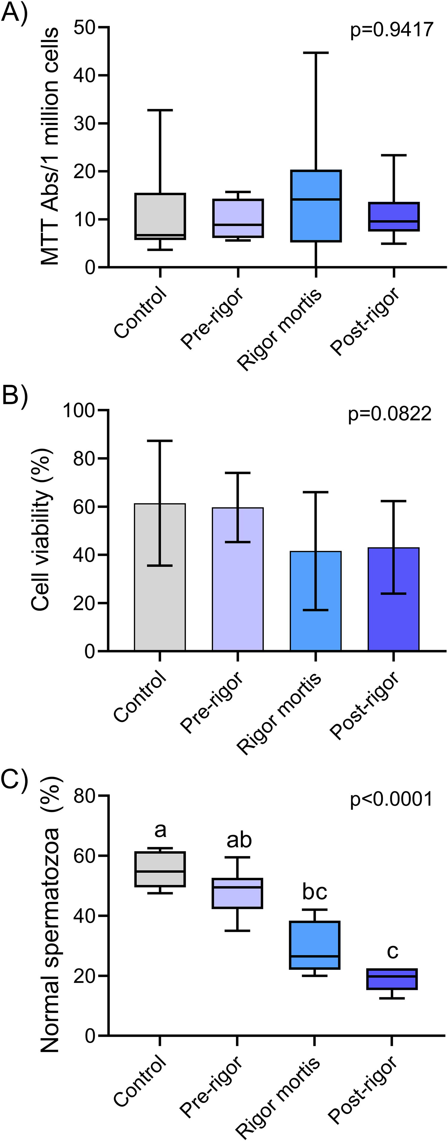
Sperm quality analysis in different rigor mortis stages.
The rigor mortis phase significantly impacted various variables related to sperm motility, including VCL, VSL, VAP, WOB, PROG, and BCF. However, there was no significant difference in the STR between the groups. The motility rate was higher (p < 0.05) in the fresh/control (63.23 ± 19.03%) and pre-rigor mortis (58.96 ± 14.38%) groups compared to the post-rigor mortis (3.34 ± 4.65%) group (Fig. 4A). The motility variables VCL (127.7 ± 14.42 µm/s) (Fig. 4B) and WOB (85.61 ± 3.045%) (Fig. 4F) exhibited higher means (p < 0.05) in the fresh/control group compared to the rigor mortis (75.45 ± 13.22 µm/s; 46.49 ± 12.85%) and post-rigor mortis (42.16 ± 57.76 µm/s; 28.51 ± 39.04%) groups.

Sperm motility and kinetics analysis (CASA—computer-assisted sperm analysis).
VSL, VAP, and PROG showed similar values. Animals in the fresh/control group showed the highest values (p < 0.05), with VSL at 109.5 ± 15.37 µm/s (Fig. 4C), VAP at 99.86 ± 11.96 µm/s (Fig. 4D), and PROG at 3263 ± 445.5 µm/s (Fig. 4G). The pre-rigor mortis group also exhibited elevated values, with VSL at 95.49 ± 13.62 µm/s, VAP at 85.88 ± 12.95 µm/s, and PROG at 3011 ± 408.8, differing from the other groups. BCF was significantly higher (p < 0.05) in samples from the rigor mortis group (49.60 ± 3.953 Hz) compared to the fresh/control (31.42 ± 4.027 Hz) and post-rigor mortis (18.24 ± 25.03 Hz) groups (Fig. 4H).
The percentages of spermatozoa with degenerated heads, microcephaly, and loose heads differed among the experimental groups. The highest values (p < 0.05) of spermatozoa with degenerated heads were observed in the rigor mortis (10.75 ± 3.352%) and post-rigor mortis (8.700 ± 2.288%) groups (Fig. 5A). The post-rigor mortis group exhibited a higher incidence of microcephaly (8.400 ± 2.331%) (Fig. 5C), while the fresh/control group had a lower percentage (1.903 ± 1.172%), which differed from that of the rigor mortis group. The percentage of spermatozoa with loose heads was significantly higher (p < 0.05) in the post-rigor mortis group (Fig. 5D) than in the pre-rigor mortis group. Macrocephaly (Fig. 5B) and cytoplasmic gout (Fig. 5E and F) did not differ among the experimental groups.
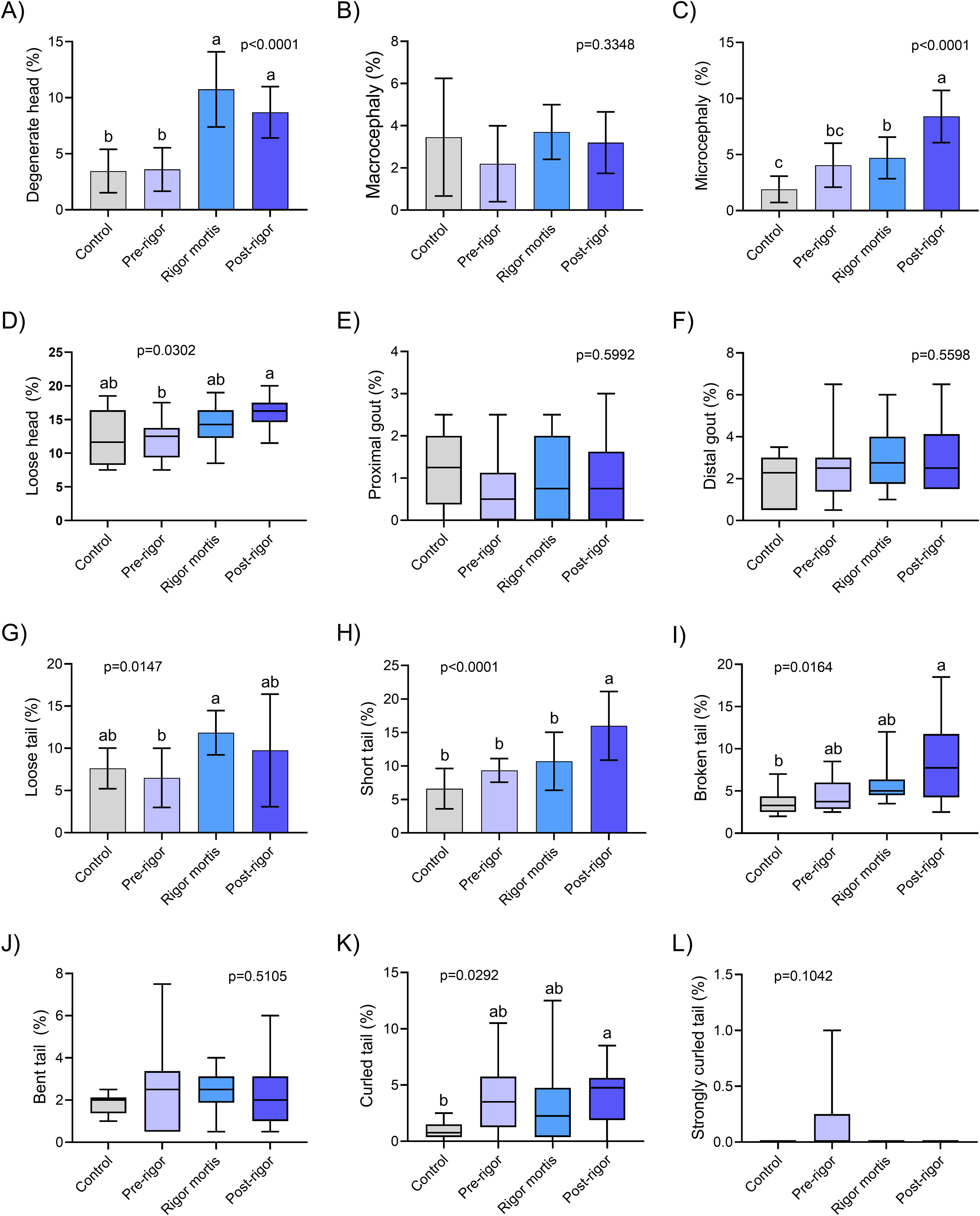
Sperm morphology and pathology.
Differences were observed in the percentage of spermatozoa with loose, short, broken, or bent tails among the experimental groups. The highest percentage of loose tails (11.85 ± 2.625%) (Fig. 5G) was found in the rigor mortis group, which was significantly different from the pre-rigor mortis group (6.500 ± 3.512%). The post-rigor mortis group exhibited a higher percentage of short tails (Fig. 5H) (16.00 ± 5.132%). Broken tails were predominant in sperm samples collected from animals in the post-rigor mortis group (8.350 ± 5.028%) (Fig. 5I), whereas spermatozoa with bent tails (Fig. 5J) were more frequent in the post-rigor mortis group (4.150 ± 2.667%), which significantly differed from the fresh/control group (0.9013 ± 0.7739%). No significant differences were found in other morphological alterations of the tail among the experimental groups (Fig. 5K, L).
There was a significant effect of the experimental group, the type of alteration, and the interaction between the factors. The highest values (p < 0.05) of head alterations were observed in the rigor mortis and post-rigor mortis groups. Regarding tail anomalies, significant differences were observed among all experimental groups, with the post-rigor mortis group showing the highest value (p < 0.05), followed by the rigor mortis, pre-rigor mortis, and fresh/control groups. No significant differences were observed between the experimental groups in terms of morphological alterations related to cytoplasmic gout. Morphological alterations in the head and tail were observed in the fresh/control, rigor mortis, and post-rigor mortis groups, with the highest rates (p < 0.05), which differed significantly from the cytoplasmic gout-type alterations. In the pre-rigor mortis group, a higher percentage of tail changes was observed (p < 0.05), followed by head changes, with the lowest rate for cytoplasmic gouts (Fig. 6).

Sperm area pathology. Two-way analysis of variance (ANOVA) = effect area pathology (p < 0.0001; F(3, 108) = 45.04), effect treatment (p < 0.0001; F(2, 108) = 429.2), and effect interaction (p < 0.0001; F(6, 108) = 10.34). Different uppercase letters indicate the difference between treatments within the same area by Tukey’s test. Different lowercase letters indicate difference between areas within the same treatment by Tukey’s test.
Discussion
Assessment of cell viability in oocytes using LIVE/DEAD and MTT assays offers complementary insights, albeit with provisional conclusions. Structural analysis using LIVE/DEAD staining revealed diminished viability during the peak degradation stage (post-rigor) across all cellular developmental stages compared to the alternative assessment criteria (fresh/control, pre-rigor mortis, and rigor mortis). However, the MTT assay failed to corroborate this observation as it yielded no statistically significant differences among the evaluated stages.
The incongruity between the two assays underscores the intricacies associated with the assessment of cell viability amid rigor mortis progression. While the LIVE/DEAD assay focuses on cell membrane integrity, the MTT assay evaluates metabolic activity. This divergence suggests that, notwithstanding the conspicuous morphological changes evident during the heightened degradation phase, metabolic activity may not be uniformly impacted, thus contributing to discordant outcomes. The absence of significant differences in the MTT results across rigor mortis stages elicits thought-provoking inquiries. It is plausible that oocytes in advanced developmental stages sustain consistent metabolic activity even in the aftermath of rigor mortis. This implies heightened metabolic resilience compared to the less mature growth stages.
In summation, the findings arising from the LIVE/DEAD and MTT assays, delving into cell viability, unveil the intricate cellular dynamics inherent in rigor mortis. These disparities underscore the need to embrace multifaceted evaluation criteria to comprehensively grasp cellular viability within dynamic contexts such as rigor mortis. According to Gryshkov et al., 26 exclusive assessment of cell membrane viability provides a less comprehensive perspective on the number of viable or nonviable cells. Thus, the concomitant utilization of these analyses with metabolic evaluation assays is imperative, given their demonstrated heightened sensitivity compared to isolated approaches such as the trypan blue staining method. 27 Remarkably, in oocytes, Marques et al. 28 observed that the MTT assay exhibited enhanced sensitivity for distinguishing between treatments with higher viability than those with lower viability. This phenomenon may be attributed to the elevated mitochondrial concentration inherent to oocytes during specific developmental stages.
It is reasonable to postulate that cellular degradation may induce an increase in metabolic activity, which could potentially be linked to the presence of active bacteria in these environments (e.g., lactic acid–producing bacteria). 29 These bacterial strains possess oxidoreductase enzymes, which may introduce confounding variables into the MTT assay. However, a comprehensive investigation is necessary to determine whether the bacterial consortia involved in rigor mortis can directly affect the observed outcomes of this assay. Moreover, rigorous exploration is essential to establish whether bacteria participating in rigor mortis can stimulate measurable mitochondrial activity within the confines of this specific analytical framework.
In males, analyses of cell membrane integrity (LIVE/DEAD) and cellular metabolic activity (MTT) revealed no qualitative differences between the various rigor mortis stages and fresh/control milt. However, upon evaluating the outcomes related to sperm kinetics and morphology, discernible qualitative deterioration was observed in these reproductive cells from animals with rigor mortis and post-rigor mortis. In kinetic assessments, across all analyzed variables, spermatozoa obtained from pre-rigor animals did not deviate from samples collected from fresh/control animals. These findings underscore the fact that spermatozoa exhibit kinetic attributes conducive to viability similar to those sourced from live animals. The significance of these findings lies in the prospect of establishing germplasm banks from deceased animals up to the pre-rigor mortis stage, without compromising sperm quality. For the cryopreservation of zebrafish sperm, ensuring that the motility of a fresh sample falls within the range of 80%–95% is minimal essential.30,31 In this regard, sperm motility analyses yield results similar to those in the control sample demonstrating the potential utility of this objective.
The assessment of sperm kinetics provided valuable insights into the dynamic aspects of sperm motility in relation to the different stages of rigor mortis. Our findings revealed the noteworthy influence of the rigor mortis phase on various parameters that contribute to sperm motility. Significant variations were observed in key metrics, including VCL, VSL, VAP, WOB, PROG, and BCF. These alterations reflect the complex interplay between the physiological and biomechanical factors that regulate sperm movement.
It is essential to highlight that the effect of rigor mortis on sperm motility was not uniform across all parameters. While certain variables demonstrated substantial declines in post-rigor mortis samples, others exhibited intermediate responses, and a few parameters remained relatively unchanged. The overall motility values, along with VSL, VAP, and PROG, showed a more subtle pattern, with the fresh/control and pre-rigor mortis groups displaying higher averages compared to the rigor mortis and post-rigor mortis groups. This difference is probably linked to the physiological and biomechanical changes that occur during rigor mortis. In the fresh or pre-rigor mortis stage, sperm cells retain a more intact cellular and structural environment, which supports more efficient movement and directional control. In contrast, parameters such as VCL and WOB showed significant declines in the rigor mortis and post-rigor mortis samples, indicating reduced sperm propulsion and disrupted trajectory. These variations may reflect alterations in the mechanical properties of sperm cells and their microenvironment during the different stages of rigor mortis.
Furthermore, the elevation of BCF in the rigor mortis group suggests an intriguing connection between beat frequency and the state of rigor mortis. The higher BCF observed in rigor mortis samples could indicate a compensatory mechanism aimed at sustaining motility under challenging conditions. However, the physiological basis underlying this phenomenon requires further investigation.
The observed disparities in sperm kinetics between the rigor mortis stages and fresh/control and pre-rigor mortis groups underscore the multifaceted nature of sperm motility and susceptibility to physiological changes. It is worth noting that the absence of significant differences in STR emphasizes that sperm motility alterations primarily affect curvilinear aspects rather than the overall directional stability of sperm movement.
These findings offer valuable insights into both basic reproductive biology and practical applications of assisted reproductive technologies. Distinct alterations in sperm kinetics during rigor mortis stages may have implications for sperm quality assessment, cryopreservation protocols, and breeding programs. Further research is needed to unravel the precise mechanisms underlying these kinetic changes and explore potential strategies to mitigate their impact on sperm function and fertility outcomes.
The increased percentages of spermatozoa with degenerated heads observed in both the rigor mortis (10.75 ± 3.352%) and post-rigor mortis (8.700 ± 2.288%) groups suggest potential consequences of cellular degeneration due to autolysis.32,33 This association between degenerated heads and rigor mortis progression implies a connection between tissue architecture breakdown and membrane integrity disruption, aligning with autolytic processes. Additionally, the higher prevalence of microcephalic spermatozoa in the post-rigor mortis group (8.400 ± 2.331%) compared to the fresh/control group (1.903 ± 1.172%) indicates significant morphological changes, potentially linked to autolytic mechanisms. The decrease in structural integrity and a higher proportion (p < 0.05) of spermatozoa with detached heads in the post-rigor mortis group relative to the pre-rigor mortis group suggested a possible impact of rigor mortis progression on head detachment and structural integrity. These findings highlight the susceptibility of spermatozoa to structural disruptions during the post-mortem period, which affect their functional viability. Tail morphology also showed variations; the rigor mortis group had a higher proportion of spermatozoa with loose tails (11.85 ± 2.625%) compared to the pre-rigor mortis group (6.500 ± 3.512%). Moreover, the pronounced presence of short tails in the post-rigor mortis group (16.00 ± 5.132%) underscores a potential correlation between rigor mortis progression and tail morphology alterations, further implicating autolytic processes. The sperm membrane, which is composed of a lipid layer containing phospholipids and cholesterol, is vital for membrane organization.34–36 During autolysis, this membrane becomes susceptible to lipid peroxidation by reactive oxygen species, resulting in loss of membrane fluidity and integrity. 37
These morphological deviations have implications for sperm motility and hydrodynamics because morphological abnormalities can negatively affect the mechanical dynamics of sperm propulsion.38,39 The interplay between structural disruption and altered motility highlights the complex relationship between form and function, underscoring the need for a comprehensive understanding of morphological intricacies in the context of sperm performance.
Conclusion
We conducted a comprehensive analysis of cell viability and sperm kinetics at different stages of cellular degradation during rigor mortis. The findings of this study reveal an intricate interplay between morphological changes and sperm functionality across these distinct stages. Remarkably, the pre-rigor mortis stage emerged as a pivotal period for the collection and preservation of gonads, exhibiting robust qualitative indices that can potentially enable the application of cryopreservation strategies. In contrast, more advanced stages of degradation, such as rigor mortis and post-rigor mortis, demonstrate marked qualitative deterioration in reproductive cells. Even under challenging conditions, the potential to harness these cells for conservation purposes persists, offering a means of mitigating the adverse effects of population loss. Understanding these cellular dynamics, coupled with insights into the potential impacts of microbiological activities and their underlying mechanisms, underscores the complexity and significance of assessing reproductive viability in rigor mortis. Furthermore, our findings expand our knowledge of reproductive biology in threatened species, bolstering the foundation for effective conservation strategies and genetic management. The practical implications of this study may resonate with targeted population revitalization and the long-term preservation of vulnerable species, underscoring the relevance of these findings within the realm of conservation biology and biological sustainability.
Footnotes
Acknowledgment

Authors’ Contributions
R.V.D.: Conducted practical activities in all experimental stages, prepared figures and tables, and wrote the first draft of the manuscript, and adjusted the drafts until the final text. L.S.M.: Planned and supervised the execution of the research activity and reviewed the drafts of the manuscript. T.R.F.: Helped in the execution of the practical activities and data collection. N.d.S.T.: Helped in the execution of the practical activities and data collection. R.B.R.: Conducted the statistics, data analysis, prepared graphs, discussed the results, and reviewed the drafts of the manuscript. J.L.B.: Helped in the interpretation and discussion of the results and reviewed the drafts of the manuscript. R.S.d.S.: Helped in the execution of the practical activities and data collection. D.H.S.S.: Contributed to the interpretation of the results as well as in the final revision of the manuscript. D.P.S. Jr.: Provided the animals and laboratory structure for conducting the experiments, as well as supervised the activities and monitored all experimental stages, and was responsible for the final draft of the manuscript. All authors reviewed and approved the final version of the manuscript.
Disclosure Statement
No competing financial interests exist.
Funding Information
This study was supported by Coordenação de Aperfeiçoamento de Pessoal de Nível Superior (CAPES).