
Abstract
In view of the scarcity of literature data on the use of radiological imaging techniques in forensic veterinary medicine, while at the same time the number of reported crimes against animals involving the use of firearms is rising, this paper attempts to assess the usefulness of radiography and computed tomography (CT) in the post-mortem diagnosis of gunshot wounds (GSW) in comparison to classic necropsy. The design of the experiment was as follows: preparation of the research material (13 dog carcasses), shooting of the material from different distances (1.5 and 12 m, plus one contact shot to the head) and using different types of ammunition, followed by X-rays and CT scans in each case to examine the injuries resulting from the shot. The final steps of the experiment were photographic documentation and autopsy by the Virchow method. In the examined material, post-traumatic bone lesions and the presence of metallic foreign bodies were successfully imaged by both radiography and CT. GSW analysis using CT provided much better data quality and some additional information. Two general conclusions can be drawn from the results of the experiment. First, damage caused by gunshots is correlated with the calibre, initial velocity and kinetic energy of the projectile, as well as the distance from the muzzle of the gun to the object shot. Second, radiological examination is useful in preparing forensic veterinary opinions. Used as a complement to classic necropsy, they increase the possibility of an accurate post-mortem diagnosis of shooting victims.
Introduction
Firearms have accompanied human beings for about seven centuries. As one of the more important offensive and defensive attributes, they have been a subject of interest for a wide range of people, and consequently a vast number of diverse models have appeared over the centuries. Despite significant changes in the mechanisms of operation or appearance of individual types of firearms, their applications have remained largely unchanged. From the start, they have been used for offensive, defensive and hunting purposes or, increasingly, for sport and as collectibles.
The legal status of firearm possession in Poland is strictly regulated by the Arms and Ammunition Act 2003, which precisely classifies types of firearms and ammunition, and also clearly defines the rights of Polish citizens to possess them. An important problem here, however, is the criminal environment, in which weapons and ammunition are traded illegally, increasing the number of firearms in Poland. This unfortunate upward trend has resulted in an increase in accidental and deliberately inflicted injuries. These injuries affect not only people but increasingly animals as well.
The aim of veterinary forensics is to provide the judicial authority with comprehensive information based on a thorough analysis and supported by scientific documentation. Such information is necessary to establish the facts, present the objective truth and issue a just sentence. 1 As an applied science, forensic veterinary medicine deals in part with explaining the mechanisms and effects of various types of injuries to the body of an animal, including mechanical injuries, among which we classify gunshot wounds (GSW), which often result in death. 2
The fundamental activity of forensic veterinary medicine, making it possible to determine the cause of death of an animal, is the necropsy. The pathological changes revealed during the necropsy are often an important aid for judicial bodies in answering the questions that arise during the proceedings. In the case of the death of an animal due to a gunshot, the examiner’s goal is not only to determine the causes of death, but also to locate and extract objects such as bullets or cartridge elements, which then have the status of material evidence, and to determine the direction of their penetration, which is the basis for reconstruction of the event. In a conventional necropsy, identification of foreign bodies in the carcass is difficult and time-consuming, as it requires extensive dissection of soft tissues, and the risk of fragments being overlooked is quite high. Therefore, a classic X-ray has for many years been a recommended stage of a post-mortem examination in the case of GSW. Performed before the body cavities have been opened, it enables prior planning of the necropsy procedures and faster extraction of foreign bodies, reducing the chance that they will be overlooked. Advances in medicine, however, have led to the replacement of conventional radiography by more modern post-mortem imaging (PMI) techniques, including computed tomography (CT), due to its broad possibilities. In the case of deaths due to gunshots, CT can not only detect bullets and cartridge elements, but also precisely determine their spatial location and their track in the victim’s body, which can help to establish from where the shot was fired.
The literature on the use of CT or other PMI techniques (radiography, magnetic resonance imaging (MRI) or ultrasound) for the purposes of forensic veterinary is currently very scant. For this reason, and due to increasing access to firearms, resulting in more and more frequent cases of fatal shooting of animals, we attempted to compare images of post-traumatic lesions and the location of foreign bodies in the case of gunshots to the head and thorax of dogs examined by classic forensic necropsy and by post-mortem radiography and CT examination. The study assessed the relationship between the extent of GSW and the calibre of the projectile and the distance from which the shot was fired. In the case of a gunshot to the chest, the construction of the projectile was taken into account as well. The results were used to assess the suitability of these post-mortem diagnostics methods for the purposes of forensic veterinary medicine.
Methods
The research material consisted of 13 mixed-breed domestic dogs bred by amateur breeders, which due to advanced age and progressive atrophy (marasmus senilis) had a poor prognosis which was an indication for euthanasia. The dogs were euthanized with the consent of their owners. The dogs were euthanized by intravenous injection with pentobarbital (Biowet, Puławy, Poland).
The initial stage of the experiment involved shooting the experimental surface, that is, the head and thorax of the dogs, from varying distances in order to obtain a GSW image characteristic of the type of ammunition used and the distance from the muzzle of the gun to the research material.
The research material, in the form of seven heads previously separated from the torso at the height of the cervical vertebrae, as well as six carcasses, was placed in numbered plastic bags and, following completion of the necessary formalities, transported to the shooting range. At stands specially prepared for this purpose, 13 shots were fired: seven at the heads and six at the left side of the thorax. The material was positioned perpendicular to the line of fire. The shots were fired from two distances – 1.5 and 12 m – measured using a laser range finder (PRO, Bielsko-Biała, Poland), plus one contact shot to the head. A brief description of the ammunition used during each shot is presented in Tables 1 and 2. The ammunition most often identified during necropsies of dogs shot in the chest was classified into three types, depending on the construction of the projectile: LRN (lead round nose, a bullet with no a jacket and a conical tip), FMJ (full metal jacket) and JHP (jacketed hollow point, a jacketed bullet with a small spherical hole in the tip). Hence, the assessment of GSW in the thorax, apart from the distance of the shot and the calibre of the bullet, also took into account its construction.
Characteristics of ammunition used for shots to the head.
Characteristics of ammunition used for shots to the thorax.
LRN: lead round nose, a bullet with no a jacket and a conical tip; FMJ: full metal jacket; JHP: jacketed hollow point, a jacketed bullet with a small, spherical hole in the tip.
After the shots were fired, the research material was placed in sealed plastic bags and taken for image analysis of the GSW. Radiographic examinations were carried out using a fully automated DX-D 600 radiography system (Agfa, Mortsel, Belgium) with a fixed digital caesium iodide detector (43 cm×43 cm) and an NX acquisition station with the Musica 3 image processing algorithm. Dorsoventral and lateral digital X-rays were taken. Tomographic imaging was performed using an Astelion Advance Edition 16-row CT system (Toshiba, Tokyo, Japan). The acquisition parameters were as follows: spiral sequence, 0.5 mm layer thickness, 120 kV voltage, automatic current modulation in the range 50–320 mA, adaptive iterative dose reduction iterative reconstruction algorithm and 0.75 seconds rotation time. The elements being scanned were placed as close as possible to the longitudinal axis of the gantry, and the narrowest possible field of view (FOV) was used to achieve the best possible resolution and the smallest possible voxel size. Diagnostic images were reconstructed on the acquisition console using standard filters for evaluating soft tissues and bone structures, as well as additional protocols for high-resolution reconstruction of skull base and dentition structures with compensation algorithms for the presence of foreign bodies.
Next, a post-mortem necropsy was performed. At each stage of the necropsy, observations were compared with the previously obtained X-rays and CT acquisition results. The necropsy was performed in a necropsy room in accordance with the general post-mortem procedure, which includes two stages: external examination and internal examination. The first step was macroscopic assessment of the GSW, which include the entry wound and the abrasion collar that may accompany it and, in the case of a perforating wound, the exit wound as well. The necropsy involved locating mechanical damage caused by the projectile in the tissues of the animal, which made it possible to determine its path of flight in the body. An essential stage of the necropsy was the extraction of foreign bodies from the tissues, including projectiles as well as projectile fragments. Radiological examinations (X-rays and CT scans) were extremely helpful at this stage, as they accurately revealed foreign bodies with high X-ray attenuation (metal elements).
Results
Gunshots wounds to the head
Forensic necropsy
In six of the seven cases analysed, ball cartridges fired from different distances had sufficient kinetic energy after passing through the tissues to exit the test material. Superficial damage without penetration of the cranium was found in one case – a shot with a shotgun shell from a distance of 12 m. A shot with a ball cartridge, irrespective of the calibre, in each case was found to cause numerous comminuted fractures in the skull, as well as destruction of the brain tissue. The contact shot with a .38 Wadcutter calibre bullet caused mechanical damage to the brain tissue in numerous locations, but due to the rapid loss of energy when the bullet encountered barriers of bone, it did not exit the test material. So, there was no exit wound. The shot from a distance of 1.5 m with a .22 calibre Long Rifle bullet completely destroyed the brain tissue due to the tendency of the projectile to ricochet in the tissues, and also caused the appearance of bullet fragments, each of which moved through the material with a specific kinetic energy until the energy was exhausted. This also caused bullet fragments to remain in the tissues, as they lack sufficient energy to pierce the barrier formed by bones. The shot with a shotgun shell from a distance of 1.5 m created the entry wound with the largest diameter, and also caused the most damage to the skeleton, brain tissue and other soft tissues. In the case of the shot with the same shell from a distance of 12 m, no damage to the skeleton or brain tissue was found. Numerous shot pellets were identified in the subcutaneous and muscle tissue in the vicinity of the wound.
Radiography and CT
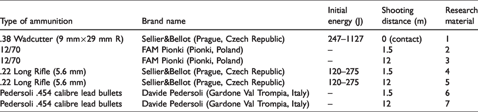
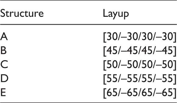
In each head examined, post-traumatic bone lesions and the presence of metallic foreign bodies were successfully imaged by both radiography and CT. It should be noted, however, that GSW analysis using CT provided much better data quality and some additional information. Three-dimensional models created based on data from CT acquisition made it possible to determine the spatial location of the foreign bodies precisely, and in material no. 1 (Figure 1), also to determine the track of the bullet, which penetrated the head from the frontal bone through the left cerebral hemisphere to the external occipital protuberance of the cranium, where it was identified. The 3D reconstructions played a particularly important role in the analysis of GSW in the form of numerous comminuted fractures in the skull, as in the case of the shot from 1.5 m using a Long Rifle .22 calibre bullet (5.6 mm). The spatial model of research material no. 4 illustrated the bone damage clearly and precisely. In this way, CT eliminated the problem of verbal description, which in the case of such complex post-traumatic lesions is usually difficult and, even with attached photographic documentation, is not always possible to relate and re-create observations from the necropsy for other people, such as a judge or prosecutor, during legal proceedings (Figure 2). This aspect of CT spatial reconstruction was also underscored in the case of GSW from a 12/70 shotgun shell, where it would be impossible to describe the exact location of such small and numerous metallic bodies – pellets – in a necropsy protocol (Figure 3).
Three-dimensional volume-rendered computed tomography (CT) image illustrating material no. 1 – the contact shot to the head with a .38 calibre bullet 9 mm×29 mm R. The bullet track is marked with an arrow.
Three-dimensional volume-rendered CT image illustrating material no. 4 – the shot to the head with a .22 Long Rifle 5.6 mm from a distance of 1.5 m.
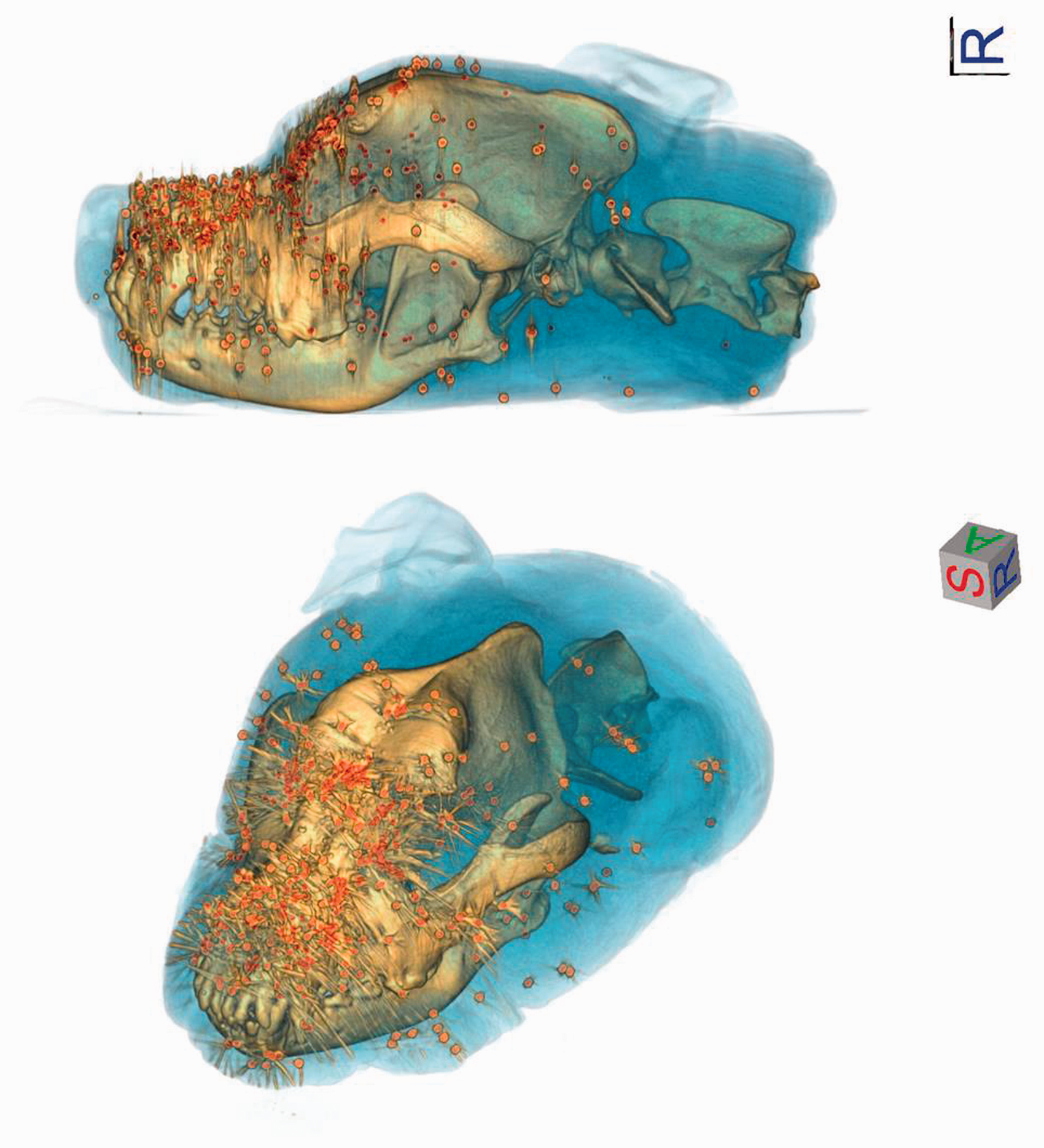
Three-dimensional volume-rendered CT image illustrating material no. 3 – the shot to the head with a 12/70 shotgun shell from a distance of 12 m. The shot pieces are marked in red.
Gunshots wounds to the thorax
Forensic necropsy
The .22 Long Rifle cartridge (5.6 mm) of the LRN jacketless type did not cause such serious injuries as the other jacketed bullets (FMJ and JHP). In the case of this bullet, there were no differences in the extent of the GSW depending on the distance of the shot. The kinetic energy of the .22 Long Rifle fired from distances of 1.5 and 12 m caused it to fragment on contact with the hard obstacle of bone, which may also have been due to the lack of a jacket. This energy was not enough to cause serious damage, such as bone fractures, but in the case of a gunshot in a living organism, fragments of the lead bullet that are not removed may cause serious health consequences in the future. Parabellum 9 mm×19 mm cartridges of the FMJ type, irrespective of the distance of the shot, caused perforating bullet wounds. The shot from a distance of 1.5 m caused a fracture in one rib and perforation of the myocardium. For the shot from 12 m, two fractured ribs were observed, as well as damage to the heart as before. The shot fired from the greater distance caused more serious damage to the bone tissue, probably due to differences in the animals’ individual constitution. Parabellum 9 mm×19 mm JHP cartridges, like FMJ bullets, caused perforating wounds, irrespective of the distance of the shot. The extent of these wounds was much greater than in the four previous cases. This is due to the greater kinetic energy of JHP bullets compared to the other types used in the experiment and to their ability to fragment after hitting the target. In the case of the bullet fired from a distance of 12 m, the bullet track was smaller than the one formed by the shot fired from 1.5 m. The larger bullet track created by the shot fired from 1.5 m is due to more energy being transferred by the projectile to the penetrated tissues.
Radiography and CT
In every case of material (nos. 3–6) in which there was damage within the skeletal system, images of post-traumatic lesions were successfully obtained by both radiography and CT. The CT images, as in the case of GSWs to the head, proved more useful than the X-rays. The spatial models based on them provided valuable information used in planning the conventional necropsy, making it considerably easier and faster to perform. CT proved particularly useful in the case of material no. 6, shot from a distance of 12 m with a Parabellum 9 mm×19 mm JHP cartridge, where a comminuted fracture of the shoulder was identified, together with a fracture of the spinous processes of the thoracic vertebrae (Figure 4). The radiological methods also enabled successful imaging of the presence of metallic foreign bodies in the form of projectiles or their fragments (Figures 5 and 6). This made it easier to find ‘material evidence’. In the case of material nos. 1 and 2, they identified small fragments of the .22 Long Rifle bullet that the examiner was unable to extract during the necropsy. The results of the experiment on an animal model clearly indicate the possibility of extensive damage to the tissues and skeleton as a result of a gunshot to the head or chest, from a distance of both 1.5 and 12 m, and even more so in the case of a contact shot. The type, nature and extent of injuries found in the cranial cavity and thoracic cavity are the basis for inferring the cause of death resulting from the injuries incurred.
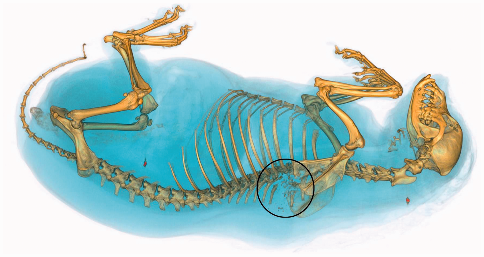
Three-dimensional volume-rendered CT image illustrating material no. 6 – the shot to the thorax with a Parabellum 9 mm×19 mm (jacketed hollow point (JHP)) from a distance of 12 m. Post-traumatic lesions within the skeletal system are marked with a circle.
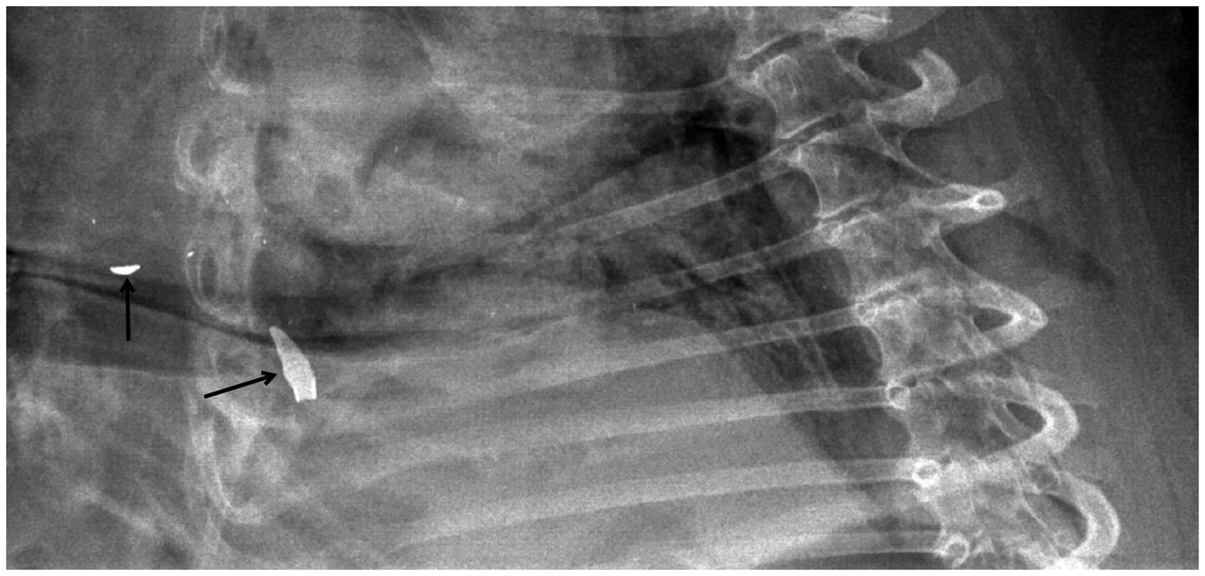
Lateral thoracic radiograph of material no. 5 – the shot to the thorax with a Parabellum 9 mm×19 mm (JHP) from a distance of 1.5 m. Metallic foreign bodies are marked with arrows.
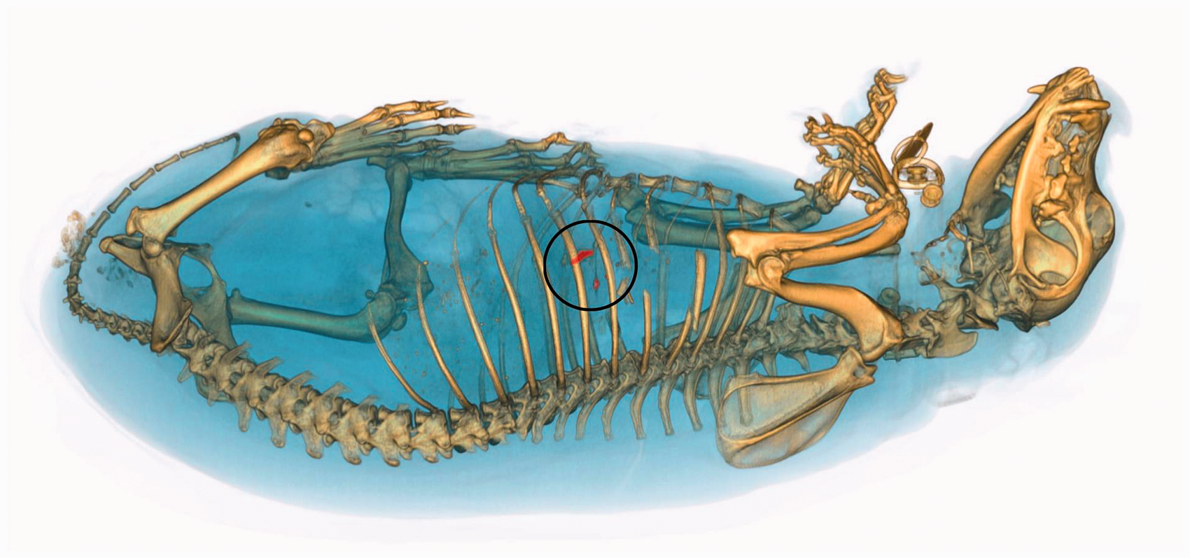
Three-dimensional volume-rendered CT image illustrating material no. 5 – the shot to the thorax with a Parabellum 9 mm×19 mm (JHP) from a distance of 1.5 m. Metallic foreign bodies are marked with a circle.
Discussion
According to Ball et al. 3 and Felsmann et al., 4 possession of modern firearms, especially short firearms, is becoming increasingly common. Their use causes injuries that pose a serious threat to both the health and life of the gunshot victim. The authors cited above also claim that easier access to firearms leads to an increase in the number of shootings of animals. The use of firearms in hunting is intended to kill the animal as quickly as possible. Unfortunately, the number of cases in which an animal is shot in circumstances other than hunting has been increasing from year to year, which we have observed in our own practice. 2 The assessment of GSW is very complicated, and each case requires a thorough and individual approach. For this reason, veterinarians should broaden their knowledge of terminal ballistics, as any act in which a firearm is used against an animal, except for situations such as hunting or necessary defence, is in violation of the law.
Okoń et al. 5 analysed mechanical injuries in animals (including GSW) on the basis of data collected from necropsies conducted over five years. Of 73 deaths, 31 were found to be a consequence of deliberate human activity, and 21 of the victims were dogs. GSW accounted for 35% (11 cases), and the victim was most often a young dog or cat. Risselada et al. 6 analysed penetrating injuries in dogs and cats. Four (25%) of the cases were GSW: three in the chest and one in the abdominal cavity. Listos et al. 7 presented 171 cases of gunshots in animals from a period of nine years, of which 134 victims were dogs.
In our own experiment, we studied GSW in animals, which may be caused either by premeditated action or by accident. A major limitation of the experiment was the use of carcasses as research material (no live material was used). The lack of circulation in the subjects resulted in the absence of extravasations, ecchymosis or blood in the body cavities. Therefore, the pattern of the mechanical damage to the tissues and the level of degradation of the material were incomplete. However, in spite of these limitations, analysis of the results of the experiment suggest that the damage caused by gunshots is associated with the calibre, initial velocity and kinetic energy of the projectile, as well as the distance from the muzzle of the gun to the object shot. Matoso et al. 8 conducted an experiment comparing the specific character of GSW in human skull models caused by three different calibres of projectiles, including the 9 mm×19 mm Luger cartridges used in the present study. Their results revealed morphological differences in GSW, which were correlated with the kinetic energy at the moment of impact. The study showed that the highest projectile velocity caused a round wound, while the smallest caused a wound with an irregular triangular shape. Similar research, with similar results, was carried out by Thali et al., 9 who performed experiments on eight perforated skulls, in some cases with 9 mm×19 mm Luger and .38 calibre bullets.
In the part of our research on GSW to the head, a shot from a ball cartridge, irrespective of the calibre, in each case caused numerous comminuted fractures in the skull, as well as destruction of brain tissue. These results thus confirm those of an experiment carried out by Oehmichen et al. 10 on perforated human skulls. The shots with a .454 calibre bullet fired from distances of 1.5 and 12 m caused damage identical to that caused by the other ammunition used in the experiment fired from the same distance. This suggests the danger of using legal black-powder guns, and calls into question the soundness of legal regulations permitting the possession of this weapon by any adult Polish citizen without the need to register individual firearms. On the other hand, it draws attention to the fact that despite the possibility of purchasing black-powder guns, there has been no significant increase in their use for criminal purposes, which may indicate that restrictive regulations on firearms are unwarranted. The shot with a shotgun shell from a distance of 1.5 m caused the entry wound with the largest diameter, resulting in the greatest damage to the skeleton, brain tissue and soft tissues. This is due to the large diameter of the projectile. When it pierces the skin, it behaves like a fixed cartridge and does not split into individual pellets until it reaches the brain tissue, as evidenced by the high concentration of pellet agglomerates in the occipital area and cervical segment. In the case of the shot to the head with a shotgun shell from a distance of 12 m, numerous shot pellets were found in the subcutaneous and muscle tissue in the vicinity of the gunshot, but there was no damage to the skeleton or brain tissue. It can therefore be concluded that the shooting of an animal (dog) in the circumstances of the experiment need not directly result in death. It is highly probable that it can cause numerous injuries in the surviving animal. Given the location of the gunshot injuries, they must cause the animal pain and suffering, which under the provisions of the Animal Protection Act of 21 August 1997 constitutes a punishable act.
In the second part of the experiment, analysing GSW in the thorax, the extent of the GSW was found to depend not only on the distance from which the shot was fired, the initial velocity of the projectile and its kinetic energy, but also on the type of ammunition – its construction. In the case of an FMJ or LRN bullet, the tissue damage was not as extensive as after a shot with a JHP bullet that fragments on impact with the target. Thoracic GSW were also studied by Thali et al. 11 Autopsy was used to analyse the damage done by a hollow-point bullet fired from about a metre, which caused the sudden death of the victim. The entry wound had a diameter of 3.2 cm, and bullet fragments were found in the bullet track. The autopsy revealed perforation of the victim’s lungs and the right atrium and ventricle. The cause of death was internal bleeding due to myocardial rupture. In our research, the shots fired at distances of 1.5 and 12 m using a JHP bullet had a very similar effect to that described by Thali et al. 11 In the case of the object shot from a distance of 1.5 m, a bullet fragment was found in the bullet track as well. A study by Tanrisever et al. 12 showed differences in the size of the entry and exit wounds. Our research shows that the exit wound is usually larger and has a more irregular shape than the entry wound. Furthermore, according to the observations of the authors cited, the damage caused by a bullet inside the body is much more extensive than the size of the entry wound would suggest.
The necropsy is the basic activity of veterinary forensics, providing, as shown above, extensive information to enable determination of the causes of death and sometimes also the cause-and-effect sequence that led to the death of the animal. In the case of gunshot victims, post-mortem diagnostics also include radiological tests carried out prior to the classic necropsy using radiography, CT and MRI. The usefulness of these methods is the basis for numerous scientific studies in the field of human forensic thanatology, but there have been few such studies pertaining to veterinary forensics. In this study, a comparative analysis of GSW in dogs was performed using necropsy preceded by radiography and CT examination.
Radiological imaging techniques have long been used in forensic medicine to examine GSW. 13 Wüllenweber et al. 14 conducted an analysis of GSW to the head using radiography and CT. The researchers found both techniques to be useful in studying this type of injury. Schumacher et al. 15 also attempted to assess the usefulness of CT in examining GSWs to the head and in explaining the mechanism by which they arise. The results obtained using a CT scanner were consistent with the autopsy results, suggesting that CT is a useful tool for forensic medicine in the diagnosis of gunshot victims. Donchin et al. 16 concluded that post-mortem CT may help to reduce the number of erroneous expert opinions based on autopsy alone.
Currently, radiological imaging methods (including X-rays and CT) play an important role in the forensic examination of GSW. They are particularly important in cases where macroscopic examination is not feasible due to the suspicion of infectious disease or because of advanced decomposition. Both X-rays and CT scans can reveal foreign objects embedded in tissues, such as bullets or bullet fragments, in addition to identifying post-traumatic lesions in the skeletal system. However, it should be emphasised that CT has greater diagnostic value because of the possibility of creating spatial reconstructions. In a comparative study of 10 GSW to the head conducted by Woźniak et al., 17 in 30% of cases, the authors found high correspondence between the autopsy image of the fractures and the reconstruction of the fractures by CT, but only partial agreement in the remaining cases. The reason for the differences in this case, however, may have been insufficient density of the cross-sections made during CT. In our study, images from CT acquisition of bone injuries, in both the head and the thorax, were confirmed during the necropsy. The basic task of post-mortem diagnosis of shooting victims, which is the identification and localisation of projectiles, was also successfully performed by CT. This technique proved more accurate in this regard than the classic necropsy, which failed to identify small projectile fragments. It was also possible in certain cases to trace the track of the metallic foreign body, that is, to determine the track of the wound, which is important for the reconstruction of the event. The GSW analyses carried out using CT were also characterised by repeatability of results and objectivity, which is in agreement with the opinion of other researchers. 18 , 19
In addition to the advantages described above, radiological imaging methods also have certain limitations, confirmed in our own research. First, these techniques do not provide information on the colour, consistency or smell of internal organs, which are important for the pathologist in determining the time and circumstances of the death. 20 Second, imaging methods are less capable than autopsy of detecting hypoxia in tissues or changes in nervous tissue. 17 Hence, it seems indisputable that the basic activity of forensic veterinary medicine should be the conventional necropsy. Radiological imaging techniques should be treated as a supplementary and, at the same time, obligatory stage of the classic necropsy, as their combined use facilitates post-mortem diagnosis of shooting victims and increases its effectiveness.
Most of the sources cited deal with research in the field of forensic thanatology, and therefore pertaining to human bodies. Hence, this work, focusing on analysis of GSW in dogs for forensic veterinary opinion, raises a relatively new topic. 7 ,21–23 Given the number of cases of animals being shot and the growing interest of law-enforcement agencies in increasingly comprehensive investigation of the circumstances of crimes, broader aspects of this topic should be thoroughly investigated.
The extent of GSW is associated with the physical properties of the projectile, as well as the distance from the muzzle of the gun to the object being shot. The present study also demonstrated that a classic necropsy combined with a prior radiological examination, especially a CT scan, enables comprehensive post-mortem diagnostics of shooting victims and is faster and more accurate than the conventional approach. This makes it possible to prepare a comprehensive and objective protocol of the post-mortem examination, which is an important part of the forensic veterinary opinion.
Footnotes
Acknowledgements
The authors would like to thank Mr Michał Tracz for assistance with data collection.
Declaration of conflicting interests
The authors declared no potential conflicts of interest with respect to the research, authorship, and/or publication of this article.
Funding
The authors received no financial support for the research, authorship and/or publication of this article.