
Abstract
Objectives
On the 125th anniversary of the first recognised publication on polymyalgia rheumatica, a review of the literature was undertaken to assess what progress has been made from the point of view of the clinical care of affected patients.
Methods
The authors searched Medline and PubMed using the search terms ‘polymyalgia rheumatica’, ‘giant cell arteritis’ and ‘temporal arteritis’. As much as possible, efforts were made to focus on studies where polymyalgia rheumatica and giant cell arteritis were treated as separate entities. The selection of articles was influenced by the authors' bias that polymyalgia rheumatica is a separate clinical condition from giant cell arteritis and that, as yet, the diagnosis is a clinical one. Apart from the elevation of circulating acute phase proteins, which has been recognised as a feature of polymyalgia rheumatica for over 60 years, the diagnosis receives no significant help from the laboratory or from diagnostic imaging.
Results
This review has shown that, following the recognition of polymyalgia as a distinct clinical problem of the elderly, the results of a considerable amount of research efforts including those using the advances in clinical imaging technology over the past 60 years, have done little to change the ability of clinicians to define the disease more accurately. Since the introduction of corticosteroids in the 1950s, there has been also very little change in the clinical management of the condition.
Conclusions
Polymyalgia rheumatica remains a clinical enigma, and its relationship to giant cell arteritis is no clearer now than it has been for the past 125 years. Diagnosing this disease is still almost exclusively dependent on the clinical acumen of a patient’s medical attendant. Until an objective method of identifying it clearly in the clinical setting is available, uncovering the aetiology is still unlikely, and until then, preventing the pain and stiffness of the disease while avoiding the problems of prolonged exoposure to corticosteroids is likely to remain elusive or serendipitous.
Introduction
The early history of polymyalgia rheumatica
The year 2013 marked the 125th anniversary of what is generally acknowledged as the first description in the medical literature of the condition now called polymyalgia rheumatica (PMR) by the Scottish physician, William Bruce (Figure 1). Bruce described the rheumatic complaints of, and their eventual resolution in, five elderly men attending the spa at Strathpeffer in the Scottish Highlands.
1
More recently, two other Scottish physicians, Watson Buchanan and Walter Kean (Figures 2 and 3), have provided a good history of the Strathpeffer spa and of William Bruce’s role in its development, as well as a critique of his original paper.
2
William Bruce (1835–1920). W. Watson Buchanan. Walter Kean.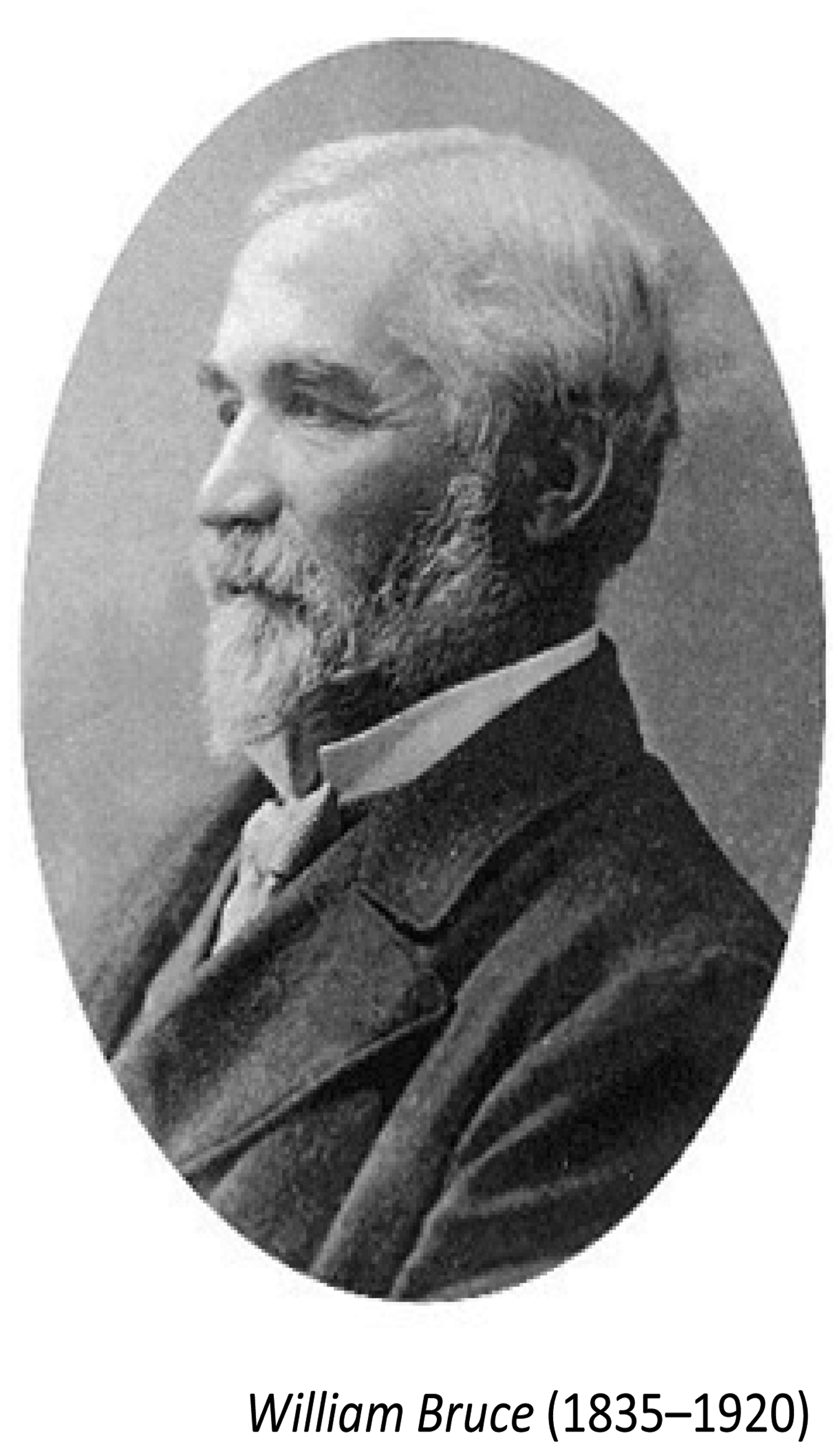

Although it would now be acknowledged generally that PMR is more common in women than in men, it is uncertain whether a better clinical definition of this disease than that of Bruce could be offered today and whether the outcomes achieved are better than those achieved by the cold mineral spa waters of those now rarely used Scottish highland springs.
The historical link between polymyalgia rheumatica and giant cell arteritis
The close association between PMR and giant cell arteritis (GCA) recognised today also has its historical counterpart in the literature reporting the conditions. The first description of GCA in the literature is attributed to Hutchison in 1890, only two years after Bruce’s publication. 3 There is then a long hiatus before either appears again in published work. For PMR, this gap is almost 60 years until 1945 when Holst and Johansen reported a series of patients with what they called ‘Persistent rheumatism’ which from the clinical description given is almost certainly PMR. 4 As reported by Hunder, Porsman reported a further series in 1951 and called the problem ‘A specific type of arthritis’.5,6 Also, Hunder’s early history of PMR indicates that in 1951 Kersley reported a series of patients with probable PMR and his title for the syndrome was ‘A myalgic syndrome of the elderly’. 7 Hunder also indicates that Meulengracht has reported some patients with PMR but his patients are a heterogeneous group and it is difficult to believe he was recognising PMR as a separate pathological entity.5,8,9 It was a few more years before the confusion of the nomenclature was clarified. Several other different names are suggested in papers published over the next few years in addition to the one used by Bruce: ‘Senile Rheumatic gout’. 1 These included ‘Anarthritic rheumatoid syndrome’, ‘Polymyalgia rheumatica’ and ‘Pseudopolyarthrite rhizomelique’.10,11,12 In an effort to emphasise the link between PMR and GCA, an editorial in the British Medical Journal in 1966 suggested yet one more term, ‘Polymyalgia arteritica’, but this did not receive much usage. 13 It was only after the papers by Gordon in 1960 and Bird and colleagues in 1979 that the term Barber had suggested in 1957, ‘Polymyalgia rheumatica’, came into general use.11,14,15
After Hutchison’s description of GCA in 1890, there is again, a gap of more than 40 years in the literature. No further report of GCA appears until Horton and his colleagues in 1932 reported a series of patients with ‘an undescribed form of arteritis of the temporal vessels'. 16 There has been much less confusion about the nomenclature of GCA which has been called generally either ‘Temporal arteritis' based on the most common clinical anatomical site of involvement, or ‘Giant cell arteritis' based on its very characteristic histopathology.17,18
Polymyalgia rheumatica and GCA – one aetiology or two?
What is the nature of this painful disorder of the musculoskeletal system, PMR, and what is the nature of the relationship between PMR and GCA? After 125 years, neither of these questions can be answered in any satisfactory way, nor is it known whether these two disorders belong to one single disease entity, or if they do, whether they represent its whole clinical spectrum of disease.
A review of research in polymyalgia rheumatica
Clinical features
The original paper by Bruce emphasised the advanced age of his patients, the severe pain and stiffness of the muscles of the shoulders, neck and often the pelvic area, the lack of objective clinical signs of joint disease and the recovery from the illness usually within a two-year period. 1 In describing polymyalgia rheumatica today, there are only very few things that could be added to his list.
Advanced age
There is little doubt that PMR is a disease of the elderly. It is rare under the age of 60 years and is almost never seen under the age of 45.19,20
Female preponderance
Unlike Bruce’s original series, there is a very marked preponderance of females in the patients with PMR, the female to male ratio being estimated to be between 2:1 and 3:1 in different reported series.19–23
Morning stiffness
Patients with PMR present primarily with severe symptoms of stiffness and pain affecting the shoulder and pelvic girdle areas.1,4,5,7,24,25 The onset of this is usually rapid – days rather than weeks. The stiffness is often severe enough to prevent patients from rising from a chair or getting out of bed.7,10,25 This stiffness, as with most musculoskeletal inflammation, is most marked in the morning and with PMR usually persists for longer than 1 h.11,24,25 The symptomatic muscles and joints affected in PMR are largely symmetrically affected and the range of motion of the shoulders is often significantly impaired by pain.1,4,7,24,25 The major differential diagnosis for PMR, now and throughout the whole history of this disease, is elderly onset rheumatoid arthritis and, even now, often only time allows distinction to be made between these entities. 26 A number of authors in the reference list of this paper use the term ‘elderly onset rheumatoid arthritis’ and this seems to suggest that this disease is different from rheumatoid arthritis (RA) at other ages. As shown in a recent study, this is not likely to be true, but for convenience, this paper uses the term meaning RA with onset at the age of 50 years or more. 27
Glucocorticoid sensitivity
During the period when PMR was being defined as a distinct clinical entity, the new non-steroidal-anti-inflammatory drugs (NSAIDs), phenylbutazone, indomethacin and oxyphenbutazone, were added to the pharmacopoeia. 28 Also, at this time glucocorticoid steroid drugs were introduced into clinical practice. 29 Although the new NSAIDs provided some relief for the pain and stiffness of PMR, this was overshadowed by the dramatic relief provided by steroids, and by the adverse side-effects of NSAIDs in an elderly population.11,28,30–36 When it then became clear that steroids were needed to prevent the serious vascular risks of GCA whether or not there were symptoms of this disease in association with PMR, steroids became, and remain, the central pillar of pharmacological management of PMR.11,26,32,37–39 PMR is believed to be the most common reason for the long-term use of corticosteroids in the elderly in western societies. The clinical response to corticosteroids is so striking in PMR that it is felt by many physicians that, if this response is not seen, the diagnosis of PMR is put in serious doubt and should be reviewed.20,32–35 Indeed, this steroid responsiveness has been suggested as a diagnostic criterion for PMR and it has been felt that it should be included in any list of such criteria, although the European League Against Rheumatism and American College of Rheumatology (EULAR/ARC) collaborative initiative has not agreed with this.15,38–40 The most typical response to prednisone in PMR is for the patient to experience complete relief of symptoms within two to three days using a small daily dose of the drug, usually less than 20 mg per day.31,41,42
Evidence of certainty that steroids shorten the overall course of the disease is almost completely lacking. Because of the prolonged course of the illness, concern has been raised regarding the long-term side-effects of corticosteroids in the elderly patient populations that suffer most often from this disease.25,28 It is important that all patients with PMR be screened for GCA as the prognosis and management is significantly altered if this is also present. GCA is a significantly greater threat to the patients’ life and quality of life and this diagnosis demands immediate exhibition of considerably higher doses of steroids.25,28,31,33
Absence of musculoskeletal signs
Although the musculoskeletal symptoms may be severe and incapacitating, there is a dearth of significant physical signs10,11,12 Peripheral joint swellings have been described in many patients but these have been, in the majority of patients, more typical of degenerative arthritis. Degenerative arthritis is common in the age group likely to have PMR. Certainly some clinicians have observed inflammatory joint swellings at the time of presentation in some PMR patients but these have proved to be rare, most often monoarticular affecting an MCP, knee or wrist, and transient, especially after the exhibition of steroids.37,43–49 Clinically, the muscles appear to be the site of the symptoms in most patients but, while there may be some loss of muscle strength because of the discomfort, no consistent physical abnormalities of the shoulder or pelvic muscles have been found, and biochemical evidence of muscle damage or necrosis is consistently absent.7,10,47,49 Efforts have been made to locate markers of inflammation in muscle tissue at biopsy, but again these reports are isolated and inconsistent.14,50,51 A small subset of patients in many series presenting as PMR have pitting oedema of the hands and feet. The clinical course of these patients is identical to those with PMR without such fluid accumulation. Whether these represent a different pathological entity is very doubtful, but some authors classify these patients separately as the RS3PE syndrome (Remitting Seronegative Symmetrical Synovitis with Pitting Edema).24,52
Systemic features
As noted by Bruce, significant systemic symptoms are common such as loss of appetite, weight loss and low grade fever, and these symptoms generally parallel the overall course of the disease, but they are neither specific enough nor consistent enough to act as criteria for the diagnosis.1,4,5,10,15,34
Benign outcome
PMR has a benign long-term outlook as was reported by Bruce. 1 The great majority of patients achieve remission within two years of the onset, although a more prolonged and relapsing course has often been reported.1,31,36,53–56 To date, there is no evidence that the use of any pharmacological agent shortens the course significantly. Chuang and his colleagues noted that in their large series the patients who received corticosteroids relapsed more frequently than those treated with NSAIDs, but they pointed out that only the patients with the most mild symptoms were managed without steroid exhibition.39,57
Clinical laboratory studies
Anaemia
No consistent clinical laboratory abnormality that distinguishes PMR has been found to date. It was clear to Bruce as well as to subsequent authors that many of these patients were anaemic and the anaemia is usually normochromic, normocytic in type.1, 7,11,15 There is little evidence that the severity of the anaemia, which is generally considered to be the anaemia of chronic disease, has any prognostic significance for PMR although some exists in relation to GCA.57,58 However, the anaemia may well define a third group of patients who may also be a part of the same clinical constellation as PMR and GCA. Some elderly patients present with a significant normochromic, normocytic anaemia associated with elevated acute phase reactants.58,59 These patients have no symptoms of PMR and no evidence of GCA but may represent a third manifestation of the same disease. 60 In parallel with the other two presentations, treatment with prednisone generally affords significant amelioration of this anaemia. 61
Acute phase response
It was noted in the earliest series of patients with PMR that the erythrocyte sedimentation rate (ESR) was markedly elevated in the great majority of sufferers and, later, this also proved true of other acute phase reactants, C-reactive protein (CRP), plasma viscosity and interleukin 6 (IL6).7,10–12,31,44,47,62–64 Plasma viscosity and ESR show a very close linear correlation, so it is not surprising that they are both elevated in the same clinical conditions.65,66 No evidence has been found of the elevation of these acute phase markers being proportional to the severity of the disease, whether one considers only PMR or also looks at GCA.66–70 A study of patients with PMR symptoms in association with biopsy proven GCA had more systemic abnormalities – higher platelet counts and lower haemoglobin levels as well as higher ESR values than in patients with PMR and no evidence of GCA. 71 While there have been efforts to differentiate among these three parameters as to which is the most discriminating in determining diagnosis and/or remission in PMR, it is not clear from the literature on this that there is advantage in choosing any one.39,63,69–72 An elevated acute response marker is included in all of the proposed criteria for the diagnosis/classification of PMR.69,70,73 Many physicians have used these as markers to monitor patients’ progress and treatment, and to assess whether the disease is in remission.66–70 However, a minority of PMR patients never show any abnormality in these parameters and elderly patients often have co-morbidities that can cause elevations of these acute phase markers without the presence of PMR.25,66,72,74–77 A recent paper argues for fibrinogen as the acute phase marker most responsive to changes in disease activity but the correlation statistics provided do not suggest this will have any major role in guiding the clinical treatment of patients. 78 It is also felt that remission is better estimated clinically rather than by the use of any acute phase markers.76,77 Attempts to tease out the specific parts of the acute phase response that are abnormal in PMR have not yet yielded any more specific useful marker for the disease or its course.64,77
Imaging studies
Electromyography
As noted, there is a lack of objective clinical signs in PMR. There is also no diagnostic imaging method of identifying the disease. Despite the proximal limb girdle being the most consistent site of the symptoms, severe pain and stiffness, there are no consistent changes in the electromyogram of the proximal muscles in PMR. 49
Plain radiography
It was evident in the earliest series of patients with PMR that plain radiographs revealed very little evidence of erosion of the proximal joints.10,11,14,24,43,45 When joint erosion was found on standard radiographs it was likely that the patient had elderly onset RA rather than PMR. Although tomography in conventional radiology may show some erosion of the sternoclavicular joints and of the symphysis pubis and these findings were confirmed in some studies by biopsy, such changes are not specific for PMR.47,79,80 Complications of undetected GCA has been recorded as causing significant changes in conventional radiographs, but these do not add to the specific diagnosis of PMR and plain radiographs are not generally helpful in differentiating PMR from other disorders.59,81
Radioisotope scanning
Some studies have indicated that scanning for musculoskeletal uptake with radioactive isotopes demonstrates inflammation of the proximal joints in patients with PMR. However, this technology does not reliably distinguish PMR from other causes of joint inflammation.47,80–85
Positron emission tomography
Positron emission tomography (PET scanning) has been used as an imaging technique in PMR by a few investigators. 86 It has confirmed the vascular abnormalities of associated GCA, and has indicated additional vascular abnormalities in the peripheral limb vessels in PMR, but it has not offered any lesions specific to PMR. The expense and inconvenience of PET scanning at present is unlikely to allow it to be used in routine clinical management of PMR.86–88
Magnetic resonance imaging
More recently, changes in magnetic resonance images (MRI) of the affected areas have been suggested to be helpful in the diagnosis. Cantini and his co-workers found periarticular inflammation of the shoulder joints and subdeltoid bursitis in all of a small series of PMR patients studied.89,90 A different study using fat suppression MRI found bilateral subacromial/subdeltoid bursitis in all patients with PMR in a further small series of patients.46,91 The neck pain and back pain which are a very frequent feature of PMR are associated with similar findings in the interspinous bursae of the cervical and lumbar spine and of the major bursae around the hip joint.92–94 Cantini and his colleagues noted that careful clinical examination was almost equally efficient at detecting these inflamed bursae close to the hip. 94 However, similar bursitis was present in a significant number of controls and the presence of similar changes in the asymptomatic peripheral joints and tendons of patients with PMR casts doubt on the value of these findings in offering either a specific diagnostic technique or a way of elucidating the pathogenesis of this disease.93–95 The expense and inconvenience of applying MRI studies in patients with PMR is also likely to limit the value of the technology in assessing treatment efficacy and long-term outcomes in PMR. If it can be shown that any of these technologies can elucidate a consistent pattern of images and improve our ability to offer a clear diagnosis of PMR, they may be useful as research tools in future. However, the widespread use of such expensive technology is unlikely in a disease that in most patients proves to be a straightforward clinical diagnosis, amenable to simple inexpensive treatment.
Ultrasonography
Ultrasonography (US) has offered a different, less-invasive imaging modality in the investigation of PMR. A variety of US techniques have been utilised, including B-mode and Doppler. Most Doppler studies employed Duplex US which allows pulsed Doppler, whereby blood flow can be visualised, to be compared on the same screen as colour Doppler which images vascular structures.81,96 B-mode or grey scale scans of the limb girdles in PMR have shown inflammatory lesions of the synovium of the shoulder, acromioclavicular and hip joints as well as the bursae and tendons surrounding them.94–100 The sensitivity of these findings for PMR is very high, but the specificity is less satisfactory as patients with elderly onset RA and controls frequently demonstrate the same type of lesions.98–107 One finding which does seem to be specific for PMR is the two tram line sign. In some PMR patients, the two layers of the deltoid fascia and the two leaflets of the subdeltoid bursa, when viewed anteriorly with the shoulder extended, abducted and internally rotated, are seen as parallel hypoechogenic layers. This sign has been seen to resolve when patients improve clinically after treatment with corticosteroids. 104 Just how prevalent this sign will prove to be in PMR is as yet unknown. Subdeltoid bursitis is very common in PMR and Cantini and his colleagues have stated that, when present bilaterally, this may prove to be an useful addition to the diagnostic criteria for the disease. 103 The US, however, does not readily distinguish PMR from elderly onset RA.105,106 Salvarani’s group has indicated that ultrasonography is less reliable than careful clinical assessment in distinguishing PMR from GCA, but US examination is an important additional method of screening PMR patients to ensure that arteritis is not missed before prescribing the lower dose of glucocorticoids preferred in patients with PMR.94,107,108 The presence of proximal synovial inflammatory changes has proven to be consistent when US findings are compared directly with MRI findings in PMR.94,99
Arthroscopy and tissue biopsy
Douglas and his colleagues reported in 1983 that they found a high percentage of PMR patients had synovitis in the shoulder joints, when they were evaluated by arthroscopy and had biopsy samples of the joint synovium examined, and this was consistent with prior studies.14,24,46,47,82,84 As mentioned earlier, other authors have investigated PMR by examining biopsy specimens of the proximal muscles most closely associated with the symptoms of the disease without finding any changes specific to PMR.14,50,51 However, although pathological evidence of arthritis of the large limb girdle joints, and some biochemical and immunological abnormalities of muscle have been clearly demonstrated in the tissues obtained, the use of such invasive techniques is not justified in the routine diagnosis and management of a disorder that has a benign outcome in the great majority of patients.
Conclusion
Polymyalgia rheumatica remains an enigma, one and one quarter centuries after its first recognisable description in a medical publication. Over the last half century, the amount of research effort in elucidating the aetiology of this disorder and of its relationship to GCA has been prodigious. It is now known that it is more common in women than in men; there is a dramatic response to corticosteroids and there is clear evidence of synovitis, bursitis and tendinitis in the proximal limb girdles. Apart from these few additional facts, almost nothing has been added to the astute clinical observations about the disease by Bruce in 1888. The treatment has changed little in more than half a century and the newer methods of altering inflammatory responses and pathways have not yet proven to influence the outcome of PMR. Whether patients with PMR would consider exposure to the cold water of the Strathpeffer spring preferable to the exposure to oral prednisone for a prolonged period of time has not been established.
Footnotes
Declaration of conflicting interests
None declared.
Funding
Dr P Rooney was supported by a sabbatical stipend awarded by St George’s University School of Medicine.