



Abstract
Objective
To describe the anatomy of the lymph node venous networks of the groin and their assessment by ultrasonography.
Material and methods
Anatomical dissection of 400 limbs in 200 fresh cadavers following latex injection as well as analysis of 100 CT venograms. Routine ultrasound examinations were done in patients with chronic venous disease.
Results
Lymph node venous networks were found in either normal subjects or chronic venous disease patients with no history of operation. These networks have three main characteristics: they cross the nodes, are connected to the femoral vein by direct perforators, and join the great saphenous vein and/or anterior accessory great saphenous vein. After groin surgery, lymph node venous networks are commonly seen as a dilated and refluxing network with a dystrophic aspect. We found dilated lymph node venous networks in about 15% of the dissected cadavers.
Conclusion
It is likely that lymph node venous networks represent remodeling and dystrophic changes of a normal pre-existing network rather than neovessels related to angiogenic factors that occur as a result of an inflammatory response to surgery. The so-called neovascularization after surgery could, in a number of cases, actually be the onset of dystrophic lymph node venous networks.
Lymph node venous networks are an ever-present anatomical finding in the groin area. Their dilatation as well as the presence of reflux should be ruled out by US examination of the venous system as they represent a contraindication to a groin approach, particularly in recurrent varicose veins after surgery patients. A refluxing lymph node venous network should be treated by echo-guided foam injection.
Keywords
Introduction
Neovascularization of the groin after high ligation of the saphenofemoral junction was first described successfully by Glass and Nyamekye, and considered an important cause of recurrence after varicose vein surgery. In fact, neovascularization at the groin level has been found in 40 to 75% of the patients (5 to 34 years) after varicose veins surgery.1–3
Stücker et al. 4 proposed a histomorphologic classification based on the presence of mono or multichannels, three-layered venous wall and nervous fibers to differentiate residual stumps from true neovascularization. True neovascularity was observed in only 26% of the groins. Van Rij et al. 5 also demonstrated the presence of multiple channels in the neovascularization process, which was well shown by cast injection. In addition, El Wajeh et al. 6 demonstrated, by immune-marking, that the so-called “neo channels” were most often pre-existing networks.
Finally, due to the lack of a clear definition and identification criteria, LNVN have not been taken into account in the recurrent varicose veins after surgery (REVAS) consensus. 7 They are neither included in the new terminology of the veins of the lower extremity, 8 nor mentioned in the VEIN-TERM consensus document. 9 In support of these publications, we believe that the presence of LNVN is often confused with true inguinal neovascularization.
Our objective is to describe the anatomy and sonographic aspects of the groin LNVN, and to draw the attention of phlebologists to these under-diagnosed networks.
Material and methods
Dissection and imaging
For this study, we evaluated 400 limbs of 200 non-embalmed cadaveric subjects (mean age: 82). All cadavers were received from the division of the “Don des Corps” in the department of Anatomy, University Paris Descartes, and were bequeathed by informed consent.
The technique was previously described. 10 After exposing the medial marginal vein, a #19 butterfly venous catheter was inserted and directed towards the toes (countercurrent to blood flow). A tube was inserted into the CFV to perform lavage irrigation with soapy water and was repeated several times. Massage of the muscles was performed until a clear liquid was obtained. Then, after ligation of the femoral vein, green neoprene latex was injected (about 120–150 ml per limb), over 30 min.
Dissection was started the next day. A colored segmentation was achieved by painting the veins for a more comprehensive identification of the anatomical elements. 11 This is the original technique of Professor Claude Gillot (who has been working since 40 years on this topic, and dedicated his life for a better knowledge of the venous anatomy, the most complex in the human body).
In addition, we used a series of 100 CT venograms (CTV) to study the 3D morphology of the groin venous complex. The technique of direct CTV used to investigate the venous system has been previously described.12–14 With this technique we can achieve a realistic and accurate 3D reconstruction of the whole venous network. For this purpose, we used Osirix® software 15 (http://www.osirix-viewer.com).The acquisition protocol of multi-slice CT (MSCT) with 64 detectors was: slice width 1 mm, slice increment 0.7 mm, filter B20, matrix 512 × 512. The 3D reconstruction by VRT with Osirix used the following protocol: CT abdomen. color look-up table (CLUT): VR muscles/bones. Opacity: Linear table.
Finally, we assessed groin anatomy during routine ultrasound scanning of chronic venous disease (CVD) patients in upright position with a 7.5 MHz transducer, in both basal conditions and during the Valsalva maneuver. 16
Anatomical description of LNVN
According to Rouvière
17
and Poirier and Charpy,
18
diverse superficial subgroups of lymph nodes are present at the inguinal and sub-inguinal regions. They can be divided into five groups (Figure 1) according to their location around the arch of the great saphenous vein (GSV): lateral-superior (1) medial superior (2) medial inferior (3) lateral inferior (4) and central (5). Some of them are commonly crossed by the tributaries of the GSV termination as well as the anterior accessory saphenous vein (AAGSV).
Location of superficial inguinal nodes surrounding the arch of the GSV, from a dissection of Poirier and Charpy.
18
They can be divided into five groups: lateral-superior (1) medial superior (2) medial inferior (3) lateral inferior (4) and central (5). 6 = great saphenous vein (GSV), 7 = anterior accessory of the GSV (AAGSV), 8 = external pudendal veins, 9 = superficial epigastric vein, 10 = superficial circumflex iliac vein, 11 = posterior accessory of the GSV.
Anatomical characterization of LNVN
LNVN, in healthy subjects as well as in CVD patients, are characterized by three anatomical features
19
:
It is a complex venous network connecting veins and lymph nodes in the groin. One particularly constant lymph node is located between the GSV and the AAGSV (Figure 2). These veins course both around and within the inguinal lymph nodes, crossing them, as shown on the latex casts (Figure 3). They are connected to the CFV by direct perforators,11,16 as shown in Figures 4 and 5. Drawing from an anatomical dissection showing an inguinal node (5) located between the GSV (2) and the AAGSV (2). The vein crossing the node is connected by a direct perforator (6) to the femoral vein. 3 = circumflex iliac tributary 4 = ilio-abdominal tributaries 7 = inter saphenous network connected to the node. Latex casts of LNVN crossing inguinal nodes (N). The plexus shaped venous networks through andinside the nodes are well demonstrated, with their superficial connection (1: great saphenous vein – 2: AAGSV). The perforator (5) is connected to the femoral vein. Anatomical dissection of a right groin after green latex injection showing the sinuous network of the LNVN between the GSV (1) and the AAGSV (2) crossing a node (N) and connected to direct perforators (4) of the femoral vein. Another anatomical dissections of primary LNVN with dystrophic networks connected to the GSV. N = nodes 1 = great saphenous vein 2 = femoral vein 3 = AAGSV 4 = Perforators.


LNVN could have different anatomical and sonographic aspects: In healthy subjects, they are thin and competent and they become dilated, dystrophic and incompetent in patients with primary CVD or more commonly in REVAS.
Results
Anatomical dissections
During our anatomical dissections, LNVN were ever present, but often reduced to a tiny network not greater than 1 mm. This explains why they are not always visible during ultrasonographic examination.
We found dilated and or sinuous LNVN in about 15% of the cadavers who are supposed to have a significant CVD (mean age: 84 years).
CT venograms findings
The findings of our anatomical dissections are confirmed by CTV with 3D reconstruction (Figure 6), showing the AAGSV piercing a lymph node, which drains into the CFV through a direct perforator. However, LNVN are not well investigated with the classical technique by direct CTV.
12
With puncture of a vein of the dorsal foot: a larger volume of contrast with a later acquisition and/or the puncture of a more proximal vein are required. In our 100 direct venograms, it was possible to achieve a 3D modeling of the LNVN in only 20% of cases due to the small size of the networks.
Tridimensionnal modeling of the groin by CT venogram showing LNVN. 1 = femoral vein, 2 = sapheno-femoral junction, 3 = femoral artery, 4 = pudendal vein, 5 = superficial epigastric vein, 6 = subcutaneous abdominal vein, 7 = superficial circumflex iliac vein, 8 = anterior accessory of the great saphenous vein, 9 = posterior accessory of the great saphenous vein, 10 = great saphenous vein, 11 = direct femoral perforator vein, G = lymph node, S = sartorius muscle, M = middle adductor muscle.
Duplex US Investigation13,14
The gold standard for LNVN visualization is color duplex ultrasound (CDU). The typical morphological appearance of lymph nodes is easily recognized, often associated with the presence of a central vein. Figure 7(a) shows this dilated central vein, producing a peculiar image that has been referred to as the “ganglionic eye” sign.
16
LNVN should be assessed in both basal conditions and during the Valsalva maneuver: in Figure 7(b) (green arrow), power Doppler demonstrates primary GSV reflux that fills the central vein of the lymph node at the groin.
DUS identification of LNVN. (a) The ultrasound features of a lymph node are easy to recognize (arrows). When its central vein is dilated, a peculiar image appears (a) which has been referred to as the “ganglionic eye” sign (11). (b) Primary GSV reflux (arrow) fills the central vein of this lymph node at the groin during a Valsalva maneuver, using power Doppler mode. This figure has been reproduced with permission from M Lo Vuolo.
16

Lymph nodes of the inguinal and sub-inguinal groups can have central veins that are large enough to be dissected 20 and can, on occasion, be as large as the superficial epigastric or circumflex iliac veins.
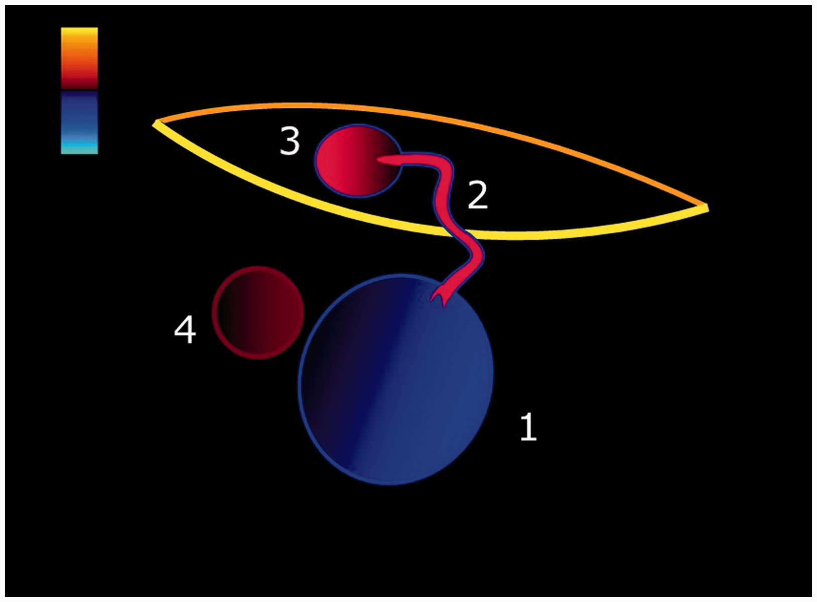
LNVN are complex, with multiple veins that follow either trans-ganglionic or peri-ganglionic paths, and may link diverse structures as shown in Figure 8(a): primary LNVN connections are established caudally with the GSV and even more often to the AAGSV. Connections which are more cephalad may be established with the superficial inguinal veins. Through an insufficient LNVN, primary reflux can supply incompetent GSV or AAGSV trunks.
The complex connections of LNVN at the groin. (a) Primary LNVN caudad connections are established with the GSV (1) and/or the AAGSV (2). More cephalad connections (white arrows up) are established with the superficial inguinal veins (3,4). Connections to the CFV (5) through direct perforators (*) have been anatomically proven (12). A node (6) is consistently located in the groin (22) between the GSV (1) and its AAGSV (2). The saphenous compartment is pictured, demonstrating the muscular fascia (m) and saphenous fascia (s). (b) Through an insufficient LNVN, primary reflux can supply incompetent GSV (7) or AAGSV (8) trunks. This figure has been adapted with permission from M Lo Vuolo.
16

Ultrasound examination can be done in two different groups of varicose vein patients: primary CVD subjects and those referred for assessment of REVAS.
Primary CVD through LNVN (Figures 8 and 9)
This is often under-diagnosed, and has been reported in 6% of patients prior to any operation. 19 This study included 100 non-selected patients investigated prior to surgery for primary CVD. The origin of reflux of the GSV trunk was found to be in the terminal or pre-terminal valve (78%), in a perineal network (10%), in an LNVN (6%) and in a parietal non-systematized network (6%).
Due to the fact that sonographers might not be aware of its presence and significance, the diagnosis of primary refluxing LNVN is often missed. Therefore, its real incidence as an associated point for saphenous truncal reflux is almost certainly underestimated.
Secondary LNVN in recurrent varicose veins after surgery
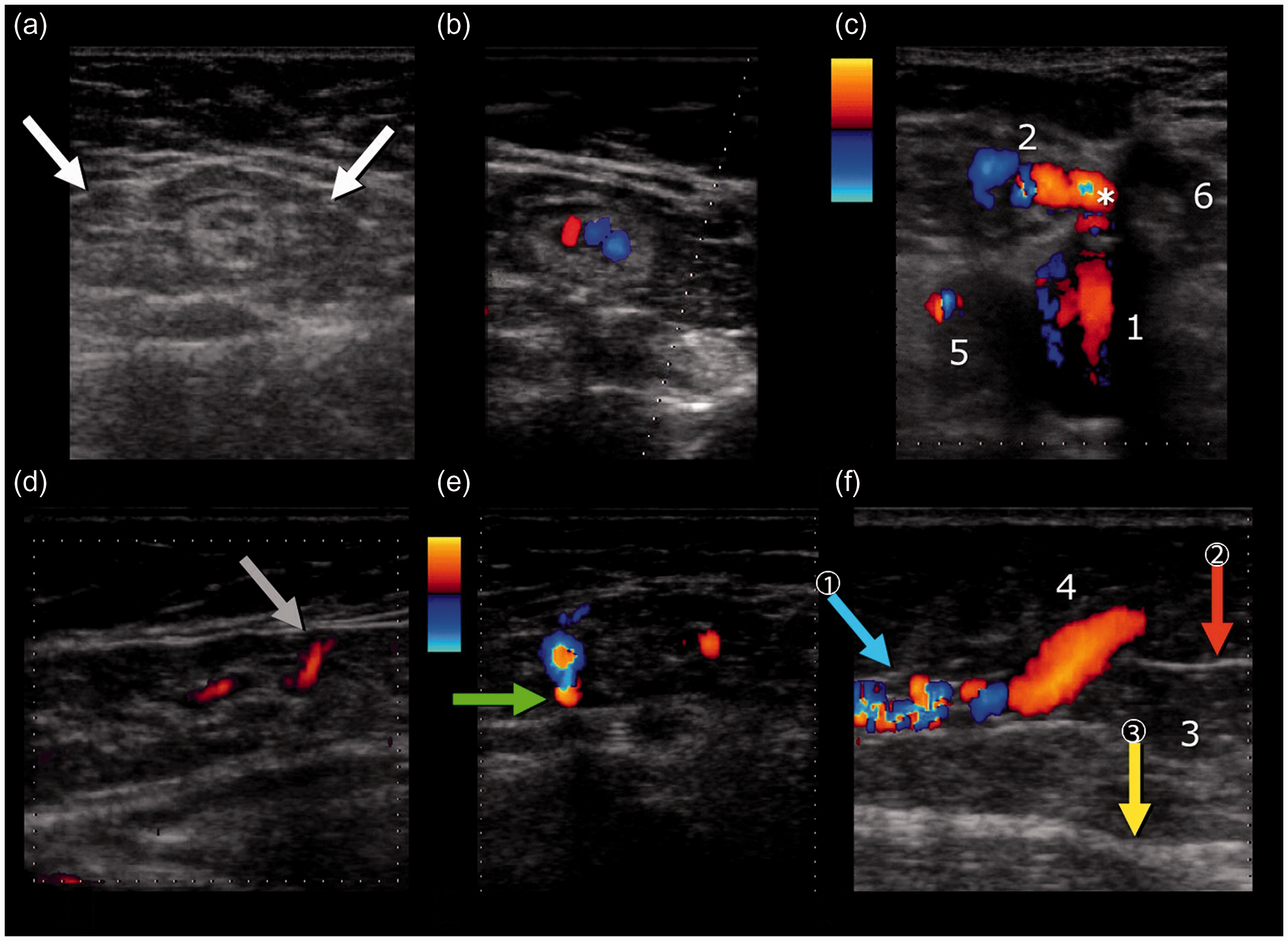
In REVAS patients, a transverse scan at the level of the former SFJ should evaluate the CFV, the muscular fascia and the superficial compartment both in basal conditions and during Valsalva maneuver (Figures 10 to 12). The goal is to demonstrate reflux coming from the femoral vein and filling veins of the LNVN. Once such a finding has been proven, reflux should be traced into the insufficient residual venous trunk connecting with the varicose veins.
Investigation of LNVN by color duplex ultrasound (reproduced with permission from M. Lo Vuolo
16
). During extreme Valsalva, reflux coming from the CFV (F) through a direct perforator (1) fills the primary insufficient LNVN veins around and within a lymph node (N) at the groin. Through superficial communicating veins, reflux from the LNVN (2) fills an insufficient GSV (GS) trunk in the presence of a normal terminal GSV valve. The ultrasound pictures are transverse scans at the groin with both conventional color and power Doppler modes. This figure has been reproduced with permission from M Lo Vuolo.
16
In REVAS patients, the most frequent pattern found at the groin is a single venous channel (2) connecting the CFV (1) with a residual incompetent superficial vein (3). Coincidentally, this is usually the pattern seen when recurrence occurs through LNVN. Femoral artery (4). Saphenous compartment with muscular fascia (bottom curve) and saphenous fascia (top curve). This figure has been adapted with permission from M Lo Vuolo.
16
A transverse scan at the groin (a) which shows a lymph node (white arrows). With color Doppler, a dilated, tortuous central vein is shown (b). With extreme Valsalva (c), transfascial reflux (*) coming from the CFV (1) towards the node’s central vein (2) is demonstrated. The transganglionic course of this LNVN vein (arrow) is clearly shown in this longitudinal view of the node (d). Within the saphenous compartment (e) reflux from the insufficient LNVN is transferred to an insufficient AAGSV (arrow). In the mid-third of the thigh (f), the AAGSV (arrow 1) pierces the saphenous fascia (arrow 2) and leaves the SC (3). The epifascial segment of its incompetent course (4) corresponds with clinically visible REVAS under the skin. Below the exit point of the AAGSV, the SC is found to be empty (3) in this patient who previously underwent saphenous stripping. Muscular fascia (arrow 3). Common femoral artery (5). (c) Another lymph node (6) is seen near the recurrent channel (*). This figure has been reproduced with permission from M Lo Vuolo.
16
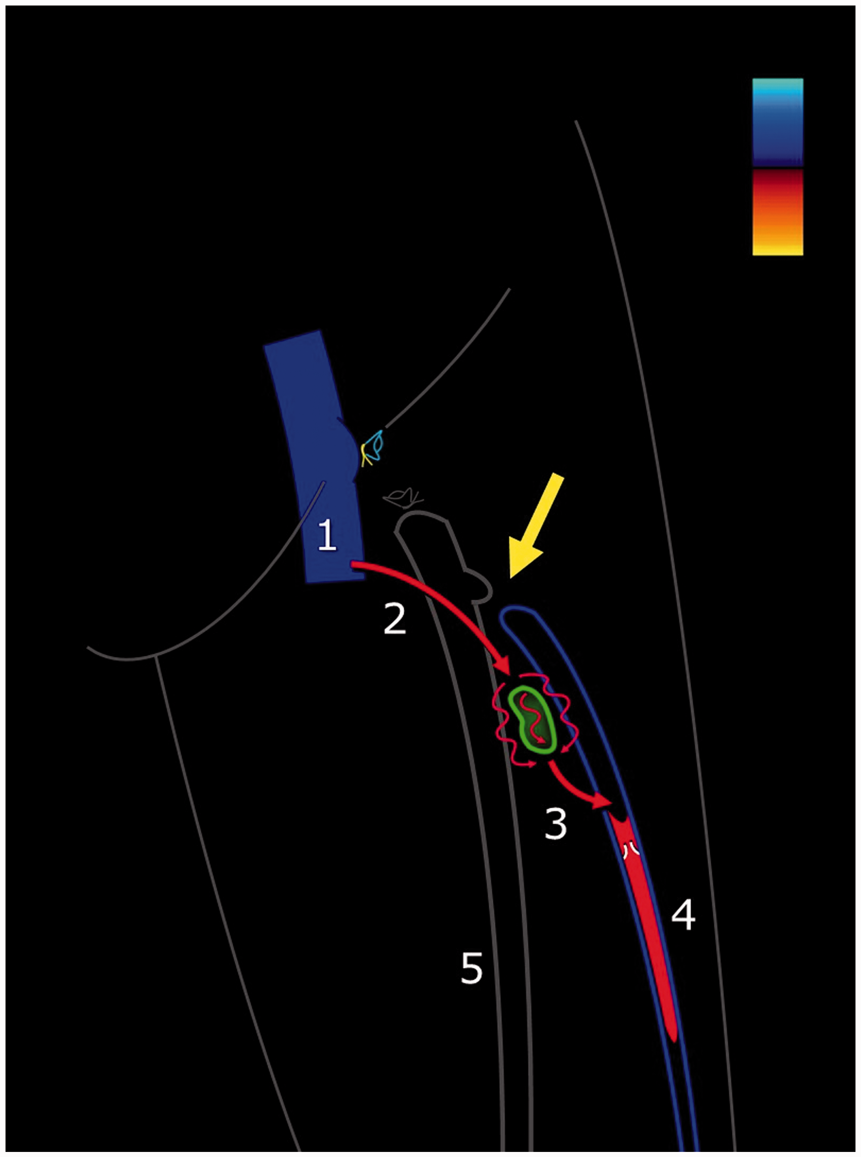
In this scheme by Dr. Lo Vuolo,
16
we can see reflux through a direct perforator (2) coming from the CFV (1) towards the LNVN, to finally drain (3) into an incompetent AAGSV residual trunk (4) which was disconnected (arrow) from the GSV trunk (5) during previous surgery. This figure has been reproduced with permission from M Lo Vuolo.
16

In our experience, LNVN are more often associated with AAGSV truncal reflux than with GSV reflux.
Two case reports illustrating the role of LNVN in primary CVD
The first case report of LNVN responsible for a primary CVD is shown in Figure 13: A 35-year-oldfemale, with two pregnancies and no family history of CVD was classified as Case report of a 36-year-old female with painful varicose veins due to primary LNVN. The skin mapping on the right shows that the reflux of the GSV at the thigh (1) is fed by a refluxing perineal network (2), as well as by LNVN (3) connected to an incompetent AAGSV (4). The echo image shows a large central vein crossing through the node. The 3D veno-CT reconstruction on the left better shows the venous anatomy and the inguinal nodes.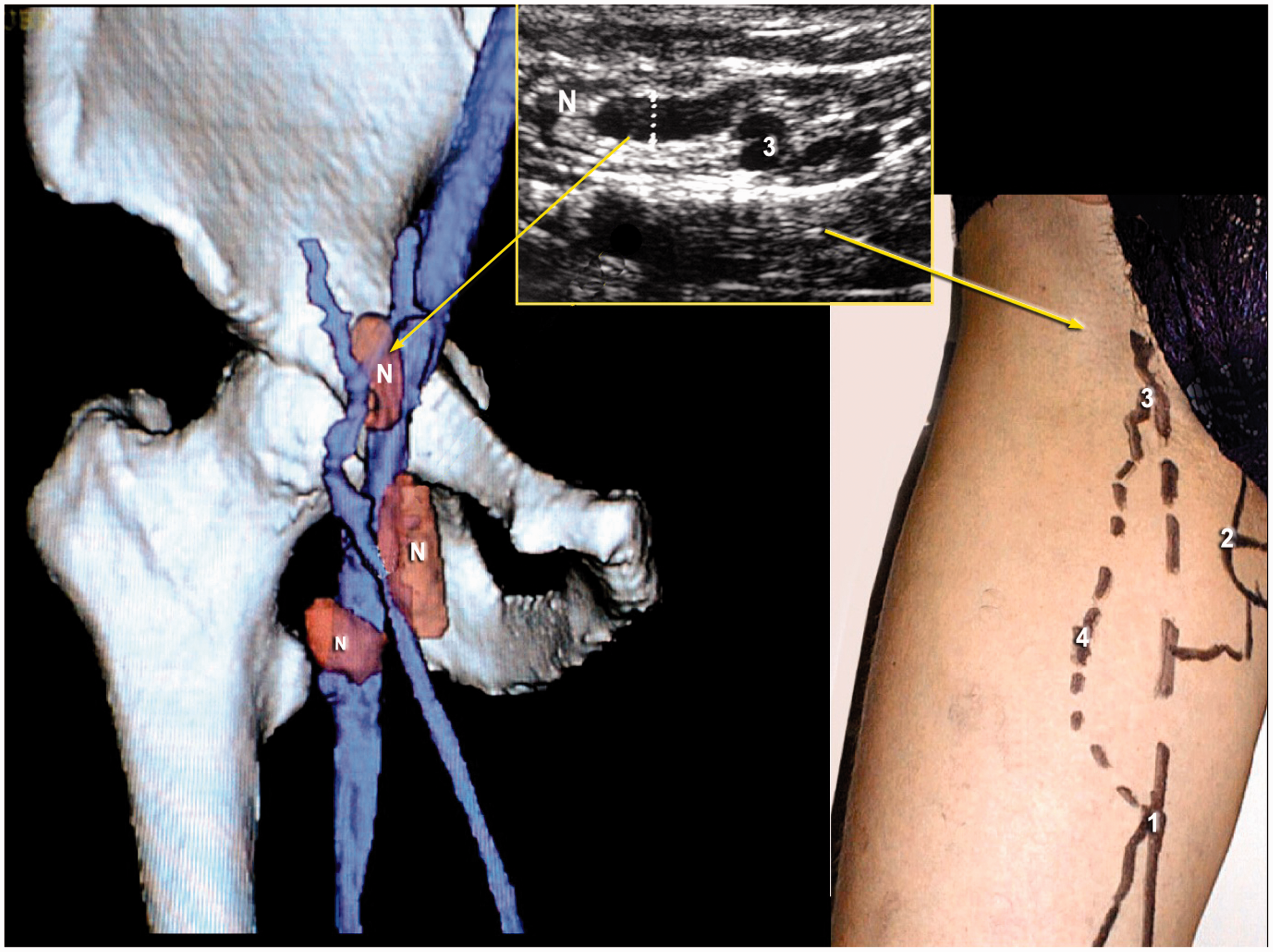
The second case report is shown in Figure 14: A 79-year-old female presented with left lower extremity ulcer, pain, itching and heaviness. The duration of CVD (varicose veins) was more than 25 years. She had a 3 × 4 cm ulcer of the upper medial malleolus for 3 years, and had no previous treatment or proper compression of the limb. Classification was C1-6 s Ep + s As + p Pr.
Case report of a 79-year-old female with an open ulcer in the left lower extremity. The mapping on the right shows a refluxing LNVN (3) feeding the AAGSV (2). The echo images demonstrate, in both transverse and longitudinal scans, a large central vein crossing through the node (1). The nodes in such cases are more echogenic demonstrating the effect of chronic inflammation and fibrosis. Reflux is seen in the LNVN during a Valsalva maneuver.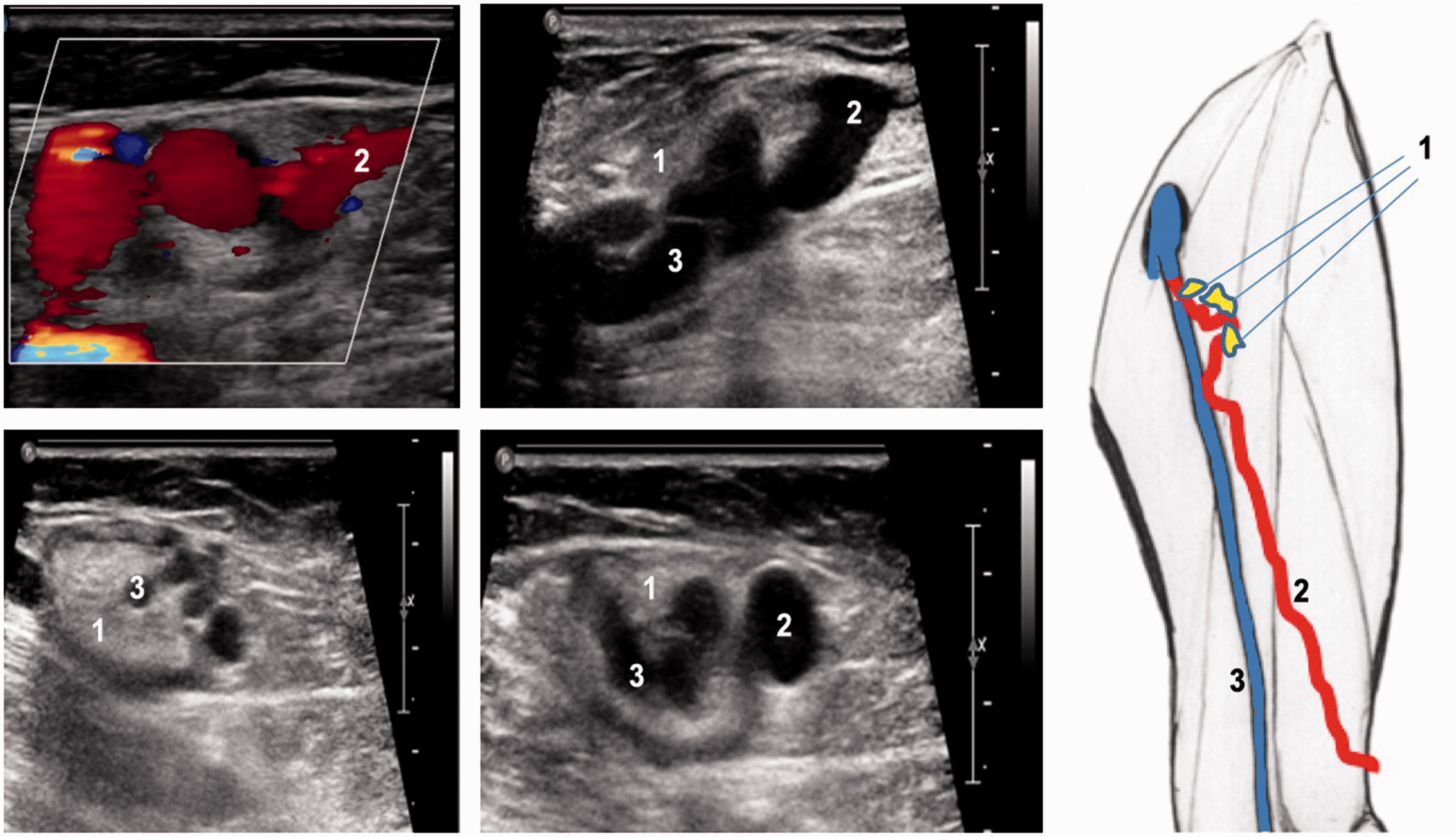
Mapping demonstrated a refluxing AAGSV connecting with LNVN of the groin. In addition, an old thrombus was present along the left small saphenous veins (SSV), and two refluxing perforators (thigh and leg) were connected with non-saphenous varicosities around the ankle close to the ulcer.
Discussion
In REVAS patients, LNVN is considered to be present at a much higher rate. Jones et al. 21 reported a recurrence rate of 52% after 2 years on 133 limbs. De Maeseneer et al. 22 found a rate of 68% after 56 months in a group of patients with recurrent varicosities and Fischer et al. 3 showed a junctional or circumjunctional reflux of the SFJ in 60% of the patients, in a 34-year clinical follow-up study after ligation of the true sapheno-femoral junction and its related tributaries.
LNVN and neovascularization
In the vein-term consensus document, 23 neovascularization was defined as: “presence of multiple new, small tortuous veins in anatomic proximity to a previous venous intervention.”
More recently, the UIP consensus 24 retained the term “neovascularization” to describe the presence of new veins situated at the site of the previous saphenous junction. These veins may be newly formed or can arise from dilation of previously existing groin veins that were invisible (or missed) on duplex ultrasound (DUS) before the operation.
An alternative term for neovascularization at the SFJ could be “groin varicose network”. This includes the possibility that LNVN may or may not be part of the neovascularization pattern.24,25
But it is important to highlight the fact that the diagnosis of neovascularization should rely upon histochemical evidence.
Certainly, our study provides no histological evidence, but it demonstrates that LNVN is an ever-present normal anatomical finding. So it is likely that after surgical disconnection of the SFJ, these pre-existing vessels can dilate as an adaptative mechanism (remodeling) to the newly created hemodynamic scenario.
In this respect, recurrent varicose veins at the groin could be an association of:
Dystrophic changes of normal pre-existing LNVN due to the healing process and inflammatory reaction and local angiogenic factors.
25
Connection of LNVN to the residual or recurrent varicose network below by multiple neovascular channels.
This is confirmed by the immune-histological study of El Wajeh et al. 6 showing that the so-called “neovascularization” is more likely dilatation of pre-existing venous channels: in fact, the presence of nervous fibrils identified by an immunomarker (protein S100) was found in the majority of both redo and control groups.
The explanation for recurrent varicose veins at the groin after high ligation of the sapheno-femoral junction is likely to be multifactorial 26 ; in addition to local factors, the pressure gradient could play a role as suggested by Recek. 27 It appears that neovascularization is a reconnection between peripheral veins (mainly residual varicose networks) and veins at the junction which are ever present anatomically and can be modified by any angiogenic factor (LNVN).
Finally, LNVN and neovascularization could be not be a cause but a consequence of recurrent varicose veins, an “innocent bystander” as stated by Egan et al. 28
Consequences in clinical practice
Sonographers should be aware of the presence and significance of groin LNVN because they can be easily missed, underestimated or misunderstood, particularly following groin varicose veins surgery.
Recognition of a primary LNVN is logically a contraindication to a groin approach; in these cases as well as in REVAS, foam sclerotherapy is recommended. This attitude of avoiding a redo surgery of the groin led to a minimally invasive procedure and a reduction in postoperative complications. 29
Possible physiopathologic hypothesis
Neovascularization of the groin in REVAS patients is likely to be related to the activation of normal or pathological pre-existing anatomical structures of the groin as described previously. 30 This does not exclude the fact that neovascularization can occur in addition to this process. 4
In this hypothesis, the LNVN and neovascularization would be closely related.
This may explain why an invagination without crossectomy or endovenous ablation techniques without a groin approach produces much fewer cases of inguinal neovascularization.31,32
This was shown by a retrospective study of Pittaluga where ligation of the GSV 2 cm below the SFJ, which preserved the proximal non-refluxing tributaries of the GSV, resulted in a very low rate of postoperative neovascularization (1.8%). 33
Additional support for this was recently published in the RCT of Casoni et al., 34 which had a follow-up of eight years.
Limitations of this anatomical paper
As there is no follow-up or clinical studies, no statistical analysis, and due to the lack of histological analysis, this paper cannot depict the different processes involved in neovascularization and LNVN. However, this anatomical study clearly demonstrates that the veins found in the lymph node networks are present in any subject and in particular exist in patients without previous venous procedures. In addition, they are much more common after high SFJ ligation. However, the relationship between LNVN and neovascularization and their respective characterization is unclear, and future clinical studies are needed.
Conclusions
This paper is intended to draw the attention of phlebologists and investigators to under-diagnosed lymph node venous networks. They can be the main leak point of a varicose network and should be ruled out during any venous hemodynamical mapping prior to treatment.
Anatomically, LNVN are characterized by trans-nodal networks connected to the GSV and/or AAGSV, joining the CFV by direct perforators. They exist prior to any treatment and can be found, thin and competent, in any healthy patient.
LNVN can be associated with primary insufficiency in a few patients. On the contrary, they are very commonly found in REVAS following groin surgery, and could be confused with neovascular channels.
LNVN seem to be venous “remodeling”, with dilation and dystrophic changes of normal pre-existing networks, as an adaptive response to surgical approach due to angiogenic factors, and thus could be a consequence rather than a cause of recurrence.
When they are found to be dilated and refluxing, surgery of the groin is contraindicated, and echo-guided foam sclerotherapy is the method of choice.
Footnotes
Declaration of Conflicting Interests
The author(s) declared no potential conflicts of interest with respect to the research, authorship, and/or publication of this article.
Funding
The author(s) received no financial support for the research, authorship, and/or publication of this article.