



Abstract
Background
Computed tomography (CT) is the gold standard for evaluation of pulmonary nodules and is at the same time responsible for the majority of the collective effective dose.
Purpose
To evaluate radiation dose and efficacy of computer-assisted detection (CAD) for solid pulmonary nodules in low dose chest CT performed at 70 kV.
Material and Methods
CAD was performed upon chest CT with 70 kV and 100 kV (gold standard) at manufacture’s recommended tube current of 87 mAs (collimation, 64 × 0.6 mm). Detection rate for pulmonary nodules and size measurements of both techniques were compared to each other. Radiation dosage in terms of effective dose (E) was measured using an Alderson-Rando Phantom.
Results
Seventy-four patients with 301 solid nodules were included in the study. CAD detection rate was similar for 70 kV (94.7%) and 100 kV (92.4%). Mean transversal nodule diameter was 5.5 mm for 70 kV and 5.7 mm for 100 kV with an average volume of 0.12 mL (both techniques). Derived from the phantom measurements patient examinations resulted in an E of 0.51 mSv (70 kV) versus 2.02 mSv (100 kV).
Conclusion
70 kV low-dose chest CT is suitable for CAD based lung nodule analysis at a fraction of the radiation burden of the standard technique. Since the measurements are highly accurate, 70 kV CT could be used for detection of pulmonal lesions as well as follow-up studies.
Introduction
Helical computed tomography (CT) of the thorax represents the current gold standard for the evaluation of pulmonary nodules due to quick acquisition of a three-dimensional image set in a high spatial resolution. While most small nodules are of benign origin, nodule characteristics such as solid appearance, a diameter of >5 mm, and a traceable growth rate raise suspicion for malignancy (1–3). Considering average tumor doubling times and patient risk profiles, follow-up examinations are therefore usually performed within 3–12 months (4). For about a decade, radiologists have been able to make use of dedicated software applications (computer-assisted detection [CAD]) which search for nodules and perform accurate size measurements (5). However, increased concern is raised among patients and physicians since CT is known to be responsible for the majority of ionizing radiation exposure in today’s medicine (6,7). Apart from using automated tube current modulation techniques and iterative reconstruction algorithms, scientific efforts focus on dose savings in chest CT by reducing tube voltage and current (8,9).
Recently a new X-ray tube for CT devices (Straton MX, Siemens Healthcare, Erlangen, Germany) that can perform CT examinations at voltages of 70 kV, has become commercially available, promising a new perspective in low-dose imaging. The purpose of this study was to evaluate 70 kV chest CT regarding radiation dosage and effectiveness of CAD-based solid nodule detection.
Material and Methods
Patients, examination technique
Data from patients who underwent chest CT for cancer staging between May and June 2012 in a 64-slice scanner (Somatom Definition AS, Siemens Healthcare, Erlangen, Germany) were retrospectively evaluated for this study. The image sets used for this analysis were taken from 74 consecutive patients (46 men, 28 women) with an average age of 59 years (range, 19–83 years). Instead of the standard protocol of an 80 kV non-enhanced CT followed by a contrast-enhanced CT scan (100 kV), patients were examined with 70 kV for the non-enhanced scan and then with the usual 100 kV contrast-enhanced scan. The local institutional review board waived the need for informed consent.
By using the manufacturer’s default, the tube current of both scans was set to 87 mAs with the following parameters: activated tube current modulation; rotation time, 0.33 s; pitch value, 0.6; and collimation, 64 × 0.6 mm. Prior to the CT scan, a scout view (100 kV, 35 mA, frontal view) was prepared to plan the examination range. To minimize the risk of motion artifacts image acquisition was performed in end-inspirational breath-hold in caudo-cranial direction. Both data-sets were reconstructed from raw data in the transverse direction (slice thickness, 1.5 mm; increment, 0.7 mm) with a hard, edge enhancing kernel (i70) in an image matrix of 512 × 512. Iterative image reconstruction “SAFIRE” (Siemens Healthcare, Erlangen, Germany) was activated by default (strength: 3).
Computer-assisted nodule detection, background noise
Lung CAD was performed on a dedicated multimodality workstation (Leonardo-Syngo MMWP V.36 A, Siemens Healthcare, Erlangen, Germany) running the software application “Oncology”. Apart from CAD, this software features real-time multiplanar image reconstruction for manual evaluation of the data-set. By default the software was set to evaluate solid nodules only, since the authors are aware of many false-positive results of CAD tools when evaluating ground glass apparent consolidations. First, the 100 kV CT studies were manually analyzed by three readers with 3–8 years of experience in chest radiology in consensus (BS, BB, and MK) regarding the number and location of the pulmonary nodules (lung window, center, –500 HU; width, 2000 HU). Then, both data-sets (70 kV and 100 kV) were analyzed again, this time using the CAD function “automatic lesion detection”. The software automatically displayed the location of the nodules, measured the maximum transverse diameter, and performed volumetric analysis. All CAD results were reassessed by the three readers regarding quantitative false-positive and false-negative findings and the results of both kV-techniques were compared to each other. Lesions that were not detected by CAD were manually tagged and added to the CAD list for size evaluation. According to the recommendations of the Fleischner Society, the nodules were divided into four groups: <4 mm, 4–6 mm, 6–8 mm, and >8 mm (4). If a nodule measured 4 mm in the 100 kV data-set and 3.9 mm in the 70 kV data-set, the lesion was classified in the nodule group “4–6 mm”, since CAD of the 100 kV technique was seen as the gold standard.
To measure background noise, a 10 mm region of interest (ROI) in circular shape was placed in air in front of the right chest. The lung window was used to exclude accidental measurements of the patient's sheet. Standard deviation of HU of three measurements was noted and the average was defined as the background noise. The mean result of both examination techniques was compared to assess the difference in average background noise.
Phantom measurements
The anthropomorphic Rando Phantom (The Phantom Laboratory, Salem, NY, USA) was utilized to determine radiation exposure. One hundred and twenty-six pre-calibrated lithiumfluoride thermoluminescent dosimeters (TLD, The Harshaw Chemical Company, Cleveland, OH, USA) were placed in boreholes of predefined anatomical regions of the head, neck, and trunk (3 TLDs per boreholder). Both the 70 kV and 100 kV examinations were performed upon the phantom which was placed on the examination table in the isocentral supine position. To achieve an appropriate radiation dosage, a similar to the previously performed patient examinations CTDivol value was taken (for the phantom measurements to keep the previously chosen CTDivol). The TLDs were irradiated five times in order to achieve a significant dosage. After pre-annealing the rods at 1008℃ for 10 min, they were read using a dedicated analytical aperture made by the same manufacturer as the TLDs. Each measured result was divided by 5 to achieve the TLD dose. With knowledge of the different organ doses, the effective doses (E) were calculated by following the recommendations of the International Commission On Radiation Protection (ICRP) (10).
Patient effective dose estimation
The dose length products (DLP) of the CT studies were automatically documented after each CT examination in a dose protocol sheet. DLP is a reasonable parameter to estimate patient effective dose (E) by multiplication with a standardized factor k (E = DLP × k). For this study, the effective patient doses were ascertained using the conversion factors that were determined from the phantom measurements.
Statistical analysis
Mean values and ranges were calculated and box and whisker diagrams were calculated for descriptive statistics. Sensitivity and positive predictive value of CAD performance was calculated and paired samples t-test was used to analyze differences in means. Differences in patient effective doses of both techniques were analyzed by using the Wilcoxon Signed Ranks test, since normal distribution was excluded via Kolmogorov-Smirnov test. A P value of α < 0.05 was considered a significant difference.
Results
Performance of automated nodule detection and measurement of both examination techniques in the study population (n = 74 patients).

The measurement of background noise showed a significantly higher (P < 0.01) standard deviation of HU in the 70 kV protocol (110.5 HU; range, 47.8–177.0 HU) versus the 51.9 HU (range, 40.1–111.8 HU) compared to the 100 kV protocol (see Fig. 1 for illustration of the images’ graininess).
Computer-assisted measurement of pulmonary nodules at 70 kV (a) and at 100 kV (b) (1.5 mm slice thickness; image center, –500 HU; image width, 2000 HU). The measured transversal diameter and volumetry differed slightly.
Phantom measurements
Examination parameters and effective dose measurements for thoracic CT at 70 kV and 100 kV (phantom study).

DLP, dose length product; E, effective dose.
Patient radiation dose
The 100 kV protocol showed significantly higher values of the DLP in comparison to the 70 kV technique (P < 0.001). The average DLP for the 70 kV technique was 38.4 mGy × cm (range, 16–96 mGy ×cm), while the DLP in the 100 kV group was 134.7 mGy ×cm (range, 62–313 mGy ×cm). The mean E value of all examinations was 0.51 mSv (70 kV) compared to 2.02 mSv (100 kV, P < 0.001). The minimum E was 0.02 mSv (resp. 0.93 mSv), and the maximum effective dose was 1.29 mSv (resp. 4.70 m Sv) (Fig. 2).
This figure shows the effective dose (E) values of chest CT of both examination techniques. The difference was statistically significant (P < 0.01).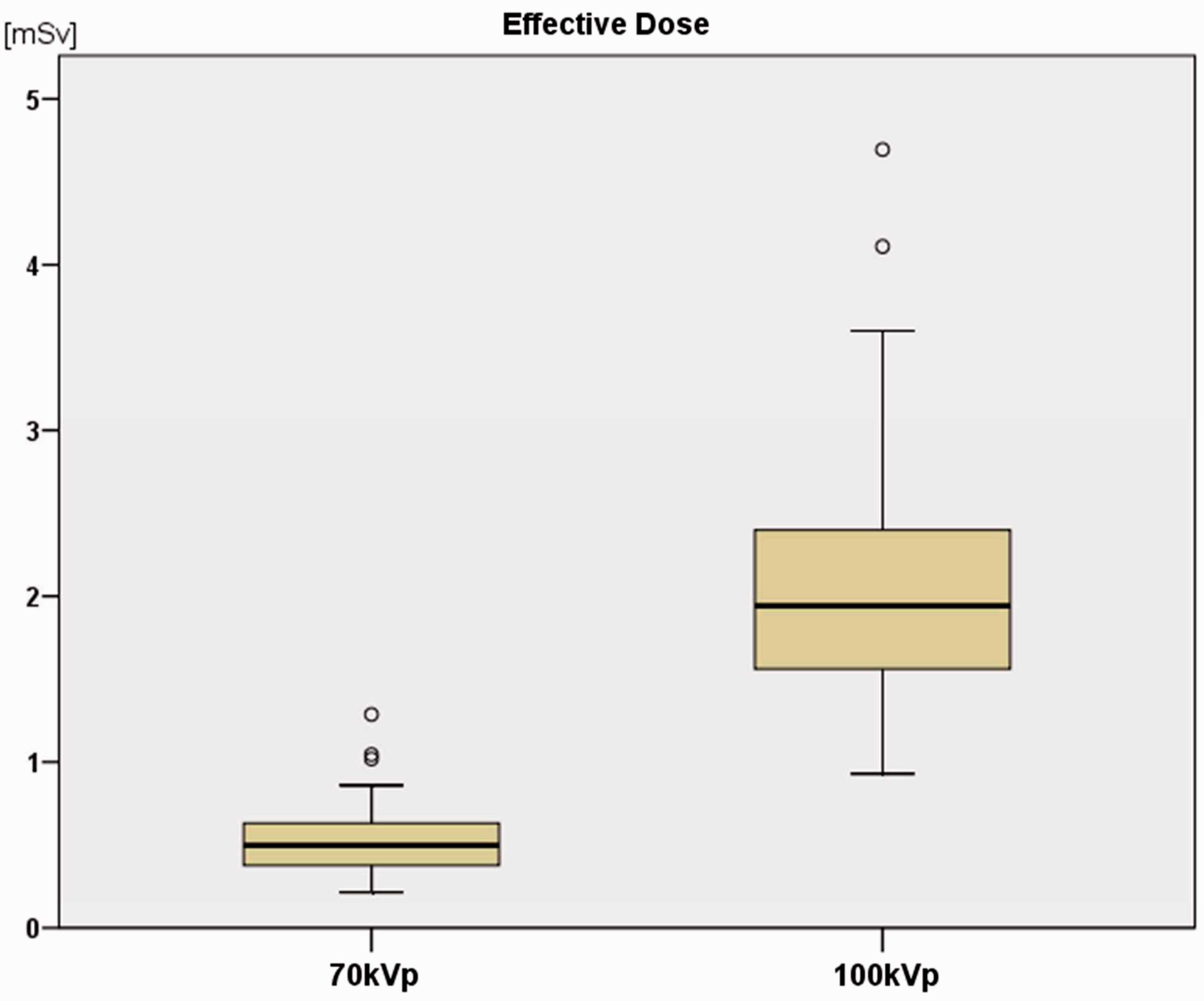
Discussion
Responding to the ever increasing collective dose of patients who undergo X-ray-based imaging, a major challenge for radiologists currently is to keep the radiation at the lowest level necessary. It is a known fact that the reduction of photon energy (tube voltage) is a major contribution in decreasing the radiation burden in chest CT imaging (8,9). However, the loss of diagnostic validity must be avoided by all means, since the accurate and reproducible evaluation of pulmonal lesions is crucial for patient health. The purpose of this study was to analyze chest CT with the recently introduced 70 kV technique regarding CAD performance and radiation exposure.
As expected, the images acquired with the 70 kV technique suffered from increased graininess due to the reduced X-ray power. However, CAD-based nodule detection performed well with the proposed low-dose technique at a comparable, yet slightly increased sensitivity and specificity. The reason for the marginal better result of the 70 kVp series might be from physical nature: due to the decreased tube voltage, attenuation of tissues increase and therefore the increasing difference to lung tissue might turn the balance towards the low kVp protocol. Yet this is hypothetical since a measurement of varying attenuations of these nodules was not part of the study. Also, size evaluation of the lesions regarding the maximum transverse diameter and measured volume was almost identical for the four different nodule groups. Although the size measurements between both techniques reached statistical significant differences, we believe that these results may not be of clinical disadvantage: the CAD measured differences were so small (range, 0.1–0.5 mm) that the spatial resolution of 1.5 mm might be responsible for the limited accuracy. In comparison to the available data in the literature, CAD performance in this study was within standard precision (11,12). However, the results presented here showed a certain amount of false-positive performance and missed a number of nodules: about 5% of the lesions were not detected automatically, which underlines the strong need for manual re-evaluation of the lung parenchyma by the radiologist. Furthermore, the limited positive predictive value found in our study certifies this statement. However, in general CAD-based size measurements are likely to be superior to manual analysis, since even the same readers tend to vary size analysis by up to 30%, especially for lesions <5 mm (11–13). In particular, for follow-up examinations of suspicious lesions, CAD is a powerful tool to objectively identify slowly-growing small pulmonary lesions. Interestingly the computer-assisted nodule performance was marginally worse in the 100 kV protocol, the result was constant when several examination studies were repeated. This difference could be caused by the increased difference in density between (air-filled) lung parenchyma and solid nodules, which is a consequence of a decreased tube voltage in CT imaging. The comparable diagnostic validity found in both techniques of this study suggests that the 70 kV technique is suitable for CAD-based follow-up examinations that investigate the growth behavior of lung nodules.
Different values of established conversion factors (k) that are used to estimate normalized effective dose values (E) in thoracic CT studies.

DLP, dose length product.
The average E of the 70 kV technique in this study was 0.51 mSv, which is in contrast to the standard protocol result of 2.02 mSv. Compared to the data in the literature, the radiation burden is therefore lower by 50–90% (8,16,20). Especially in combination with improved iterative raw data reconstruction techniques (e.g. ASIR, SAFIRE, AIDR, iDose) that suppress image noise effectively, 70 kV imaging could be utilized for various fields of anatomic areas (8,21–23).
Limitations of the current study include the uncertainty of performance of the 70 kV technique for soft tissue discrimination in low-contrast areas, such as mediastinal lymph nodes or muscle tissue. Moreover, the influence of artifact-causing metal implants (e.g. spine screws, metal plates, heart pacer) on the diagnostic validity is still unclear, but it is expected to be worse for the low kV technique. Furthermore, even if the examination-technique-adapted conversion factors of this study are completely accurate for estimating the effective patient dose, the method is still merely an approximation, since the values were derived from a standardized phantom body. The fact that non-enhanced (70 kV) and iodine contrast-enhanced (100 kV) data-sets were compared may have led to an overestimation of the results of the 100 kV scan since increasing the attenuation of certain nodules might have improved the overall CAD performance. Finally, the influence of patient’s body mass index was not part of this study, yet it is unclear whether the low dose protocol may be adequate for obese patients.
Comparing the 70 kV technique to chest X-ray imaging, the effective patient dose is only about three times higher, yet with the benefit of an isovolumetric data-set in submillimeter spatial resolution.
In conclusion, the study shows that 70 kV chest CT is suitable for CAD-based lung nodule detection and evaluation while causing significantly lower radiation burden to the patient.
Future analysis might now be necessary to re-evaluate the benefit of chest CT screening in contrast to the concern associated with the currently diminished radiation-based stochastic risks.
Footnotes
Conflicts of interest
JM Kerl JM is consultant of Siemens Healthcare. M Sedlmair is a research scientist of Siemens Healthcare.
Funding
This research received no specific grant from any funding agency in the public, commercial, or not-for-profit sectors.