



Abstract
Background
Surgery in the lesser pelvis is associated with a high complication rate as surgeons are spatially limited by solid anatomic structures and soft tissue borders. So far, only two-dimensional (2D) parameters have been used for risk stratification.
Purpose
To precisely measure the inner pelvic volume a computed tomography (CT)-based three-dimensional (3D) approach was established and compared to approximations by 2D parameter combinations.
Material and Methods
Thin-layered multi-slice CT datasets were used retrospectively for slice by slice depiction of the inner pelvic surface. The inner pelvic volume was then automatically compounded. Combinations of two to four 2D dimensions determined in 3D volume rendered reconstructions were correlated with the inner pelvic volume. Pearson’s correlation coefficient and Chi square test were used for statistical calculations. Significance level was set at P < 0.05.
Results
In total 142 patients (91 men, 51 women) aged 64.8 ± 10.6 years at surgery were included in the study. Mean calculated pelvic volume was 1031.13 ± 180.06 cm3 (men, 996.57 ± 172.43 cm3; women, 1093.34 ± 178.39 cm3). Best approximations were obtained by combination of the 2D measurements transverse inlet and pelvic height for men (r = 0.799, P < 0.05) as well as transverse inlet, obstetric conjugate, interspinous distance and pelvic depth for women (r = 0.855, P < 0.05).
Conclusion
We describe a precise and reproducible CT-based method for pelvic volumetry. A less time consuming but still reliable approximation can be achieved by combination of two to four 2D dimensions.
Introduction
In clinical routine, surgeons are challenged by operating in spatially limited areas like the lower pelvis with its rigid boundaries. For years, a narrow pelvis has been discussed to be a crucial risk factor for the surgical outcome in urologic, gynecologic, or visceral surgical interventions (1–6). However, presurgical pelvimetry is not yet part of clinical routine. By now there are numerous computed tomography (CT) or magnetic resonance imaging (MRI) based studies evaluating the predictive value of two-dimensional (2D) parameters like the obstetric conjugate, transverse inlet diameter, interspinous distance, sagittal midpelvic diameter, intertuberous distance, and sagittal outlet for intra- or postoperative issues (3,5,7–12). These studies revealed partly controversial results. Targarona et al., Akiyoshi et al., and Wang et al. for instance found a significant correlation of pelvic outlet diameters and operation time, whereas Ogiso et al. and Neill et al. could not confirm this association (3,5,7,11,12). According to Baik et al. and Wang et al., the interspinous distance can serve as prediction factor for cancer involvement of the circumferential resection margin after lower anterior rectum resection, especially in women (8,12). Hong et al., however, did not find any significant correlation for single 2D diameters of the bony pelvis and operation time, intraoperative blood loss, and surgical margin status after open radical retropubic prostatectomy. Only a combination of parameters, i.e. pelvic index, revealed a trend towards significant correlation with the outcome parameters (10). The latter is supported by a recent publication of Bertani et al., showing a significant correlation of anastomotic leakage rate after lower anterior rectum resection and the pelvic index, i.e. a combination of the anteroposterior diameter in the inlet and outlet plane as well as the transverse diameter in the outlet plane (13). However, pelvic volumetry, i.e. a three-dimensional (3D) approach for measurement of the inner pelvic volume considering also soft tissue boundaries like muscle bundles or aponeuroses, has not been performed, yet.
Thus, the aim of the study was to establish a radiological method to determine the exact surgically experienced inner pelvic volume and to test its suitability for daily use. We present a slice by slice technique based on multiplanar reconstructions of thin-layered multi-slice CT scans. As an alternative, if 3D pelvimetry might not be possible because of technical or time reasons, we performed a correlation analysis with different 2D dimension combinations to identify the best gender-specific approximation.
Material and Methods
This study received ethical board approval (EA4/044/14). All patients gave written informed consent for scientific use of their medical data.
Participants
A data search in the local picture archive and communication system (PACS) of the radiological department was performed for all patients who underwent conventional anterior resection of rectal cancer since 2004. Only patients who experienced a thin-layered CT scan either prior or post surgery were included into the study. Scans were performed due to oncologic staging or due to postoperative issues. Exclusion criteria were changes of the bony pelvis or the soft tissue margins caused by prior surgery or radiotherapy to ensure proper measurements.
CT based pelvimetry
CT data were obtained on a 64-slice or a 16-slice scanner (both Siemens Healthcare, Erlangen, Germany). Scans were acquired with a 0.6 mm collimation at 120 kVp tube potential and 250 mAs reference tube current time product with dose modulation, noise level 12. Reconstructed slice thickness was set as 1.0 mm. The CT datasets were processed with help of the software solution Visage 7 (Version 7.1.3.911, Visage Imaging, Inc., San Diego, CA, USA). For pelvic volumetry, 2D multiplanar reconstructions (2D MPR) were realigned – according to the surgeon’s view onto the operating field – parallel to the plane of the pelvic inlet extending from the promontory to the upper rim of the pubic symphysis (Fig. 1a). In that plane, the inner pelvic surface was determined by segmenting it manually. Thereby, inner pelvic boundaries were defined according to the innermost anatomic barriers for surgeons, e.g. bone, muscle, or ligaments/aponeurosis (Figs. 1b and 2). This step was repeated for every slice down to the pelvic floor (Figs. 1c and 2). After segmenting the complete inner pelvic space the software automatically compounded a 3D body and calculated its volume (Fig. 1c).
On the sagittal view of multiplanar reconstructions the plane of the pelvic inlet is defined according to the surgeons’ view (a, dashed line). In the resulting paraaxial or paracoronal plane the inner pelvic surface is depicted manually respecting bony, muscular, and ligamentous boundaries (b, dashed line). This procedure is the repeated slice by slice downwards to the pelvic floor as shown in a volume rendered reconstruction of the pelvis (c, the inner pelvic contour of every third or fourth slice is shown by thin lined circles). In every third to fifth slice a manual depiction of the inner pelvic surface is done downwards to the pelvic floor. (a, b, c) The inner pelvic surface (opacified area) on exemplary slices corresponding to plane a, b, or c on the sagittal view (d).

In a second approach we measured 2D dimensions of the bony pelvis on 3D volume-rendered reconstructions deriving from the same 1 mm slice datasets. Measurements were taken in standard cranial, posterior and lateral views. The diameter transverse inlet (widest transverse pelvis brim distance) was measured in a cranial view (Fig. 3a), interspinous distance (shortest distance between spinous processes), and intertuberous distance (shortest distance between tuberous processes) in a posterior view (Fig. 3b). The diameters obstetric conjugate (shortest distance from promontory to the superior aspect of the symphysis), pelvic height (promontory to intertuberous connecting line) and pelvic depth (superior aspect of the symphysis to intertuberous connecting line), sagittal outlet (inferior inner aspect of the symphysis to sacro-coccygeal junction), and sagittal midpelvic (inferior inner aspect of the symphysis to the sacrum along the plane of the spinous process) were determined in a lateral view after cutting the pelvis in a (para)median plane (Fig. 3c). All diameters, excluding pelvic height and pelvic depth were obtained according to Lenhard et al. (14). The duration to perform 3D pelvimetry or 2D parameter measurement was timed on five different CT datasets and averaged, respectively.
3D volume rendered reconstructions of the bony pelvis are given in (a) anterior cranial, (b) posterior and (c) lateral views after “cutting” the pelvis in a (para)median sagittal plane. Lines indicate the 2D dimensions transverse inlet (TI), interspinous distance (IS), intertuberous distance (IT), obstetric conjugate (OC), sagittal midpelvic (SM), sagittal outlet (SO), pelvic height (PH), and pelvic depth (PD). Details of 2D measurement are given in the methods section.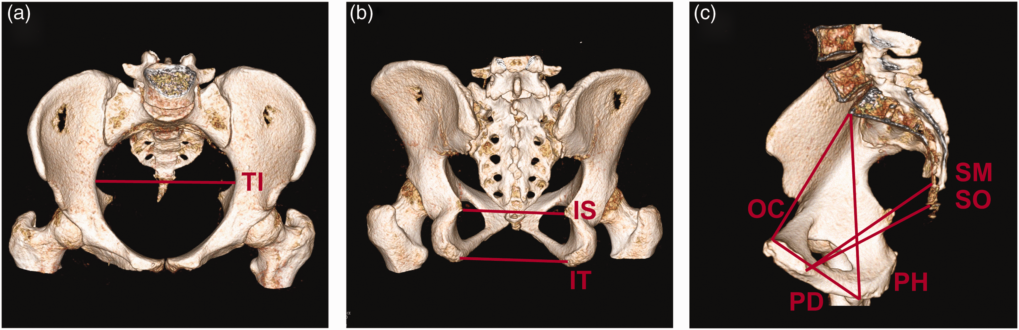
Statistical analysis
All continuous variables are displayed as mean and standard deviation (SD). Pearson’s correlation coefficient between a summation of pelvic diameters and the pelvic volume was calculated using IBM SPSS statistic software (IBM Corp. Released 2013. IBM SPSS Statistics for Macintosh, Version 22.0. IBM Corp., Armonk, NY, USA). The correlation was considered significant if the P value was less than 0.05.
Results
In total 142 patients (91 men, 51 women) with 64.8 ± 10.6 years of age at surgery were included in the study. Mean calculated pelvic volume of all patients was 1031.13 ± 180.06 cm3. Men had a mean calculated pelvic volume of 996.57 ± 172.43 cm3; women showed a significantly (P < 0.005) higher calculated pelvic volume of 1093.34 ± 178.39 cm3.
In the second approach, the 2D parameters transverse inlet, interspinous distance, intertuberous distance, obstetric conjugate, pelvic height, pelvic depth, sagittal outlet, and sagittal midpelvic were determined as described in the methods section. The mean transverse inlet was 130.03 ± 8.80 mm, with significantly (P < 0.005) higher values in women (133.27 ± 9.32 mm) than in men (128.21 ± 7.99 mm). Interspinous distance (97.48 ± 11.92 mm) and intertuberous distance (106.85 ± 14.99 mm), obstetric conjugate (109.99 ± 10.20 mm), sagittal outlet (110.54 ± 8.51 mm), as well as sagittal midpelvic (121.50 ± 8.63 mm) have also shown significantly higher values in women. Biggest gender-related differences were determined for interspinous distance (women, 108.71 ± 9.75 mm; men, 91.19 ± 7.64 mm). Men, however, turned out to have significantly higher (158.12 ± 10.62 mm vs. 152.80 ± 9.58 mm) and deeper (99.32 ± 7.24 mm vs. 87.84 ± 7.14 mm) pelvis.
Two to four 2D parameters were then summed up and correlated with the calculated pelvic volume. We restricted calculations to the reasonable parameter combinations. In total, 46 different combinations were calculated. All of the correlations were highly significant (P < 0.001). Best overall correlation result, i.e. best approximations to the pelvic volume, was found for the quadruple combination transverse inlet + obstetric conjugate + interspinous distance + pelvic height with a correlation coefficient of r = 0.798 (P < 0.001). The double combination with best correlation result was transverse inlet + pelvic height (r = 0.767, P < 0.001), best triple combination was transverse inlet + interspinous distance + pelvic height (r = 0.793, P < 0.001). Considering gender, best correlation results for men were achieved for the double combination transverse inlet + pelvic height (r = 0.799, P < 0.001), for the triple combination transverse inlet + sagittal outlet + pelvic height, and the quadruple combination transverse inlet + obstetric conjugate + interspinous distance + pelvic height (r = 0.768, P < 0.001, respectively). In women, best results were found for the following combinations: transverse inlet + pelvic height (r = 0.790, P < 0.001), transverse inlet + obstetric conjugate + pelvic depth (r = 0.836, P < 0.001), and transverse inlet + obstetric conjugate + interspinous distance + pelvic depth (r = 0.855, P < 0.001).
The mean duration to perform a 3D volumetry was 16.66 ± 0.87 min, whereas the 2D approach took 2.33 ± 0.37 min on average. Time analysis was restricted to only five datasets as variance turned out to be small.
Discussion
Our study is the first describing a radiological method to determine the surgically experienced inner pelvic volume. Thin-layered multi-slice CT scans, as performed in clinical routine, can be easily used for multiplanar reconstructions, realignment to the pelvic inlet plane, and slice by slice measurement of the inner pelvic surface. The inner pelvic volume is then automatically compounded based on the single inner pelvic surface measures.
Use of magnetic resonance imaging (MRI) would avoid ionizing radiation, but is time-consuming and susceptible to motion. The technicians who routinely acquire the MRI data frequently lack experience with pelvimetry, and an inaccurate planning of the sequences can result in gross measurement errors (15,16). MRI scans of the pelvis are routinely performed with a slice thickness of 5–8 mm, which might limit the accuracy of pelvic volumetry calculations. On the contrary, CT scans are acquired with continuous z-axis coverage and thus offer excellent postprocessing possibilities. Furthermore, CT is frequently performed for diagnostic reasons anyway and can be used retrospectively for pelvimetry. Thus, patients usually do not have to receive additional radiation for volumetric measurements. We showed that CT-based pelvic volumetry achieves very accurate volume measures without requiring special acquisition settings or specific pelvimetry training of the evaluating radiologist. A single pelvimetry set can be done within 15–20 min which presumably exceeds feasibility in clinical routine, but might be applicable on single patients in special cases. A less time-consuming, good approximation to the pelvic volume, though, can be achieved by the summation of 2D diameters of the bony pelvis performed on 3D volume rendered reconstructions. The combination of the parameters transverse inlet, obstetric conjugate, interspinous distance, and pelvic height showed best gender comprehensive approximations. Better results can be achieved using gender specific combinations like transverse inlet and pelvic height for men and transverse inlet, obstetric conjugate, interspinous distance, and pelvic depth for women. The gender specific differences might be explained by the sex specific geometry of the pelvis with a wider pelvis in women and a narrower and higher pelvis in men. In our study the inner pelvic volume measurements are biased, though, as primarily those patients who had surgery complications have undergone a CT scan of the pelvis. Thus, it might be possible that this group has a smaller pelvic volume compared to patients with good surgery outcome.
Whether the pelvic volume serves as a predictive factor for intra- or postoperative complications still needs to be evaluated. Some studies already proved a significant correlation of single pelvic 2D diameters and operating time, intraoperative blood loss or recurrence rate (5,7,8,12). The fact that other studies could not prove these results suggests that single 2D dimensions of the bony pelvis do not describe its size sufficiently (3,10,11). As best correlation results were found for combinations of pelvic dimensions (13), a correlation with pelvic volumetric measurements might be promising. Similar CT- or MRI-based volumetric approaches like organ volumetry already found broad acceptance in clinical routine, e.g. in presurgical planning of liver resections or transplantations (17–19).
In conclusion, pelvic volumetry is a feasible, precise, and reproducible method to determine the inner pelvic volume respecting not only bony, but also visceral boundaries. In some clinical institutions, however, pelvic volumetry might not yet be feasible due to the time effort or missing software features. Very good approximation can be reached by the summation of two to four specific 2D diameters. These measurements, though, do not regard visceral structures. To assess the clinical impact of pelvic volumetry, correlation analysis of the inner pelvic volume and surgery outcome still has to be performed. This might be of special interest for urologists and gynecologists, as well as visceral surgeons.
Footnotes
Declaration of conflicting interests
The author(s) declared no potential conflicts of interest with respect to the research, authorship, and/or publication of this article.
Funding
The author(s) received no financial support for the research, authorship, and/or publication of this article.