



Abstract
Background
The identification and subsequent management of liver diseases in children is challenging due to the lack of non-invasive imaging biomarkers. Ultrasound shear-wave elastography (US-SWE) is an emerging imaging technique which can quantitatively assess liver stiffness and may be useful as a tool in the management of liver disease in overweight and obese children.
Purpose
To evaluate US-SWE velocities of the liver in normal-weight and obese children, to correlate US-SWE findings with age and body-mass-index (BMI), and to compare US-SWE values with qualitative assessment (i.e. normal versus abnormal echogenicity) of the liver by conventional US.
Material and Methods
A cohort of 300 children (mean age, 9.9 ± 5.3 years; age range, 0.06–18.9 years) were studied, comprising 176 normal-weight and 124 obese participants. In each patient, both US-SWE and conventional US of the liver were obtained. Three pediatric radiologists individually and in consensus determined whether liver parenchyma was of normal or abnormal echogenicity.
Results
US-SWE velocities differed between normal-weight and obese children (1.08 ± 0.14 versus 1.44 ± 0.39 m/s; P < 0.001), but not by gender. Multivariate linear regression demonstrated US-SWE velocity to be primarily associated with age in normal-weight children (P < 0.05) and with BMI in obese children (P < 0.001). In the obese group, mean US-SWE velocity was statistically higher in participants with abnormal echogenic livers than in those with normal-appearing livers (1.53 ± 0.38 vs. 1.17 ± 0.27). The difference was not significant in the normal-weight group.
Conclusion
US-SWE provides a useful quantitative imaging biomarker for evaluating liver stiffness in children.
Keywords
Introduction
The increasing incidence of childhood obesity in the United States is paralleled by the rising prevalence of liver disease in pediatrics. In particular, non-alcoholic fatty liver disease (NAFLD) is one of the most common liver disorders in children and adolescents (1). The progressive accumulation of fat can cause inflammation and lead to scarring, a condition referred to as non-alcoholic steatohepatitis (NASH) (2–5). Consequences of NAFLD include liver fibrosis, cirrhosis, malignancy, hepatocyte damage, and eventual organ failure. The identification and subsequent management of liver diseases in children remains challenging due to the limited availability of non-invasive imaging biomarkers that can characterize disease onset and progression, such as in NAFLD and NASH. Current guidelines from the American Academy of Pediatrics recommend biannual measurement of liver enzymes such as serum alanine transaminase to screen overweight and obese children for NAFLD (6–8). Conventional ultrasound (US) is often subsequently recommended in children with abnormal liver function tests to determine whether the liver is abnormally echogenic and whether fatty infiltration is present or absent. However, the degree of liver fibrosis cannot be readily detected by conventional US. While liver biopsy with histological assessment remains the reference standard for definitive diagnosis of hepatic diseases, it is nevertheless limited by risks associated with the invasive procedure and small tissue sample size. Needle biopsies are also impractical for routine disease screening and monitoring of disease progression (9).
Recently, US-based shear-wave elastography (SWE) has emerged as a non-invasive imaging techniques that shows promising reliability and clinical utility in the quantitative assessment of liver tissue stiffness (10–14), particularly in children (15–21). US-SWE has been demonstrated with clinically acceptable repeatability and reproducibility (10,21) and statistically significant correlations between US-SWE measurements and semi quantitative liver stiffness categorical scores (i.e. METAVIR F scores) have also been demonstrated (11,16,21). The basic underlying premise in quantitative US-SWE measurements is that shear-wave velocity through the liver increases as liver parenchyma becomes stiffer under pathologic conditions, such as fibrosis (22–27). To date, the potential utility of US-SWE in obese children has not been reported.
In this work, we evaluate US-SWE measurements of the liver in normal-weight and obese children and additionally compare US-SWE measurements with qualitative assessments of the liver by conventional US. We hypothesize that obese children have stiffer livers (i.e. higher shear-wave velocities), that US-SWE measurements are positively correlated with age and body mass index (BMI), and that livers that appear abnormally echogenic exhibit greater US-SWE velocities.
Material and Methods
Participant recruitment
This prospective study was approved by our hospital’s institutional review board and was compliant with the United States Health Insurance Portability and Accountability Act. Written informed consent, and when appropriate, assent, were obtained from all participants and their parents and legal guardians. Between November 2013 and November 2015, a total of 300 children were recruited and enrolled in the study. At the outset, the children were separated into either normal-weight or obese groups, using age- and sex-specific BMI percentiles of < 95th and => 95th from World Health Organization (WHO) growth charts, respectively.
Approximately three-fourths of the obese children were referred to the Department of Radiology from the Departments of Endocrinology or Hepatology for liver ultrasound evaluation. These patients were suspected of having hepatic steatosis. The remaining one-fourth of the obese children and all normal-weight children were recruited from the general patient population pool at our tertiary care institution. Many were referred to the Department of Radiology for an unrelated ultrasound imaging exams unrelated to the liver. Children with a history of liver disease, other than hepatic steatosis, were excluded from the study. Anthropometric measurements such as weight and height were collected to compute BMI. In addition to the BMI percentile, BMI Z-scores were calculated according to growth charts from the Centers for Disease Control and Prevention.
SWE examination details
SWE measurements were obtained using the IU-22 ElastPQ system (Philips Healthcare, Best, The Netherlands). The procedures were performed in an US suite by a team of US trained technologists familiar with US-SWE techniques and under the supervision of a pediatric radiologist. A 5 MHz broadband (C5-1) curved array transducer was used to interrogate the liver at a minimum of 1 cm depth below the liver capsule. The transducer’s region-of-interest (ROI) sample box is a small sector with a fixed height of 12 mm that can be moved from the skin surface up to 8 cm deep using the trackball. As the sample box is moved from shallow to deep tissues, the width of the box changes automatically from 5 mm near surface of transducer to 9.3 mm at 8 cm depth. The sample box was placed with care to avoid vascular structures.
Briefly, in US-SWE, an acoustic pulse is used to generate and propagate ultrasonic waves through the tissue of interest. The velocity of the generated shear waves (m/s) is calculated by monitoring tissue displacement over time and is the direct output of the IU-22 system. The measured velocities can be subsequently converted to a measure of stiffness (kilopascals) using two constants, the Young modulus and the tissue density. Therefore, a high velocity measurement of the acoustic waves proportionately implies greater tissue stiffness.
In all patients, the ultrasound exam was performed either with light breathing or with a short breath-hold. In each patient, a total of 15 separate velocity measurements were obtained across the liver using subcostal and intercostal approaches, ten in the right lobe and five in the left lobe. A majority of the measurements were obtained at a depth of 3–5 cm. The measurements were then averaged to yield a global estimate of liver stiffness (velocity) and heretofore will be referred to as the mean US-SWE velocity. The entire US-SWE exam took 10–15 min and no sedation was required in any of the cases. There were no dietary restrictions prior to the exam.
Radiologists qualitative ratings
Three board-certified pediatric radiologists (years of post-fellowship experience: Rater 1, 16 years; Rater 2, 5 years; Rater 3, 12 years) independently reviewed conventional liver US data from all 300 patients. During the review, the radiologists were blinded to quantitative US-SWE data. Each radiologist determined whether the liver was of normal or of abnormal echogenic appearance. The liver was deemed abnormal if one of the following criteria were met: (i) the echogenicity of the hepatic parenchyma exceeded that of the neighboring renal cortex and spleen; and (ii) there was an evident loss of sharp demarcation of the diaphragm and poor delineation of the intrahepatic vessels. After the independent evaluation by each individual radiologist, the three readers convened and discussed discordant cases and a consensus assignment of normal or abnormal echogenic appearance was reached.
Data analysis
Data analysis was performed using STATA statistical software (Version 13, Stata Corp., College Station, TX, USA). A P value of 0.05 was set for statistical significance. Two-sample two-tailed t-tests were used to compare US-SWE measurements between the normal-weight and obese groups. Within the normal-weight and obese groups, comparisons of velocities between boys and girls were also performed using t-tests. Additional comparisons of velocities between cases that were labeled normal-appearance versus abnormal echogenic-appearance on conventional US were also performed. Multivariate linear regressions, with age and BMI as the independent variables, and mean US-SWE velocity as the dependent variable, were separately performed in the normal-weight and obese groups, and co-linearity was tested. Coefficients of determination (r2), the regression slope, the 95% confidence of the slope, and the associated P value were used to assess the presence of statistically significant trends in the linear regressions. The Fleiss’ Kappa coefficient was computed for the qualitative radiologist evaluations. Additionally, a pairwise Cohen’s Kappa scores were also computed between the three radiologists.
Mean US-SWE velocity data from the normal-weight group were further stratified into several age ranges—<1 year; 1–5 years; 6–10 years; and >11 years—to establish a look-up table of normative values. Within each age group, the 5th, 10th, 25th, 50th, 75th, 90th, and 95th percentiles for mean US-SWE velocity were computed.
Results
Demographics of the study cohort.
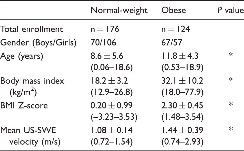
Mean US-SWE velocity measured in the liver for each group is reported in the last row.
For age, BMI, and BMI Z-score, range is given in parentheses along with the mean and standard deviation.
Statistically significant differences between normal-weight and obese groups (P < 0.001).

Box-and-whisker plots of US-SWE velocity measurements of the liver as a function of boys and girls and normal-weight and obese groups. Within each weight group (normal-weight, obese), there were no statistically significant differences between boys and girls. Independent of gender, the obese group are characterized by higher velocity measurements (P < 0.001) than the normal-weight group. Mean, standard deviation, and range are shown for each sub-group.
Fig. 2 summarizes the linear correlations between mean US-SWE velocity (dependent variable) and BMI and age (independent variables). The 95% confidence intervals (CI) of the regression slope for BMI versus SWE was 0.0045–0.018 for the normal-weight group and 0.018–0.029 for the obese group. The 95% CI of the regression slope for age versus SWE was 0.0037–0.011 for the normal-weight group and 0.024–0.053 for the obese group. Note that none of these CIs of slope contain zero, suggesting statistically significant, albeit very minute, positive linear trends. When BMI and age were jointly regressed against mean US-SWE velocity in a multivariate analysis to test for co-linear effects, age became the dominant parameter in the normal-weight group (95% CI of the slope: 0.001–0.01; P < 0.05) and BMI was not a statistically significant factor. Conversely, in the obese-group, BMI became the dominant independent variable (95% CI of the slope: 0.015–0.028; P < 0.05), while age was no longer a statistically significant factor.
Linear regression plots of US-SWE velocity measurements of the liver versus (top) BMI and (bottom) age for the normal-weight (left) and obese (right) groups. The individual correlations with BMI are statistically significant (P < 0.01) in both groups. Likewise, the correlations with age are statistically significant (P < 0.05) in both groups. With multi-variable linear regression of SWE velocity vs. BMI and age after co-linearity adjustment, age is the more statistically significant (P < 0.05) determinant in the normal-weight group, whereas BMI is not. Conversely, in the obese group, BMI is the more statistically significant (P < 0.01) determinant, and age is not.
Percentiles of US-SWE velocities (m/s) in the liver for the normal-weight group separated by four age bins.

Summary of qualitative assessment by three radiologists.

US-SWE velocity distributions of the liver in participants who exhibited abnormal echogenic-appearing livers versus those who exhibited normal-appearing livers, as determined from consensus scores by three radiologists reviewing conventional ultrasound data.

Statistically significant differences (P < 0.05).
NS, not significant.

US-SWE images in (a) a normal-weight 10-year-old girl with normal SWE velocity and (b) an obese 16-year-old boy with a high SWE velocity reading. White sample box in each image denotes the region where SWE velocity is being measured.

Ultrasound images in two obese patients: (a, c) an 11-year-old boy and (b, d) a 14-month-old boy. (a, b) Conventional ultrasound images. (c, d) US-SWE images. Note that both patients have normal SWE velocity measurements but while the echogenicity is relatively normal in the 14-month-old child (b), the 11-year-old boy (a) has an abnormal diffusely hyperechoic liver which is potentially attributable to changes of NAFLD without fibrosis.
Discussion
In this work, we have demonstrated that US-SWE is capable of detecting statistically significant differences in liver stiffness between normal-weight and obese children. Additionally, we have shown that particularly in obese children whose livers appear abnormally hyperechoic on conventional US that are typically suggestive of NAFLD, their corresponding US-SWE velocities are statistically higher than those whose livers appear normal. Furthermore, we have shown with multi-variable linear regression that US-SWE velocity is significantly associated with age in normal-weight children, but is more strongly associated with BMI among obese children. There also appears to be no gender-based differences in US-SWE velocities within the liver across the age range evaluated in this work. Finally, we have shown that conventional US interpretation by three pediatric radiologists can be subjective, with moderate inter-rater agreement that is potentially attributable to years of experience, and that quantitative US-SWE is a potentially superior and complementary method to conventional US. The data from our large pediatric cohort study support the promising clinical utility of US-SWE in measuring liver stiffness. The early detection of liver disease in children is critical in the subsequent management of additional screening and monitoring procedures to establish appropriate therapy.
The present study contributes to the existing ultrasound elastography literature with new liver data from a large pediatric cohort of 300 children, including normative data from 176 normal-weight children (BMI < 95%) and new data in 124 obese children (BMI > 95%). In the previous study by Haquinet et al. that involved 103 healthy children (15), the investigators reported a mean liver US-SWE velocity of 1.12 m/s (range, 0.73–1.45 m/s). These values are in close agreement with the current findings in our normal-weight group (Table 1). Furthermore, our 90th to 95th percentile values from the normal-weight group (Table 2) suggests a US-SWE velocity range of 1.2–1.3 m/s as a reasonable upper threshold point. The highest US-SWE value measured in our obese cohort was 2.93 m/s. This was from a 13-year-old Hispanic girl, nearly threefold higher than the average in the normal-weight group. Conversely, the lowest US-SWE value measured in our obese cohort was 0.74 m/s. This was from an 11-year-old Hispanic boy, suggestive of the fact that childhood obesity is not necessarily accompanied by liver fibrosis and that the onset of fibrosis likely occurs at different time points in each individual.
The current work has several notable limitations. First, our cohort consisted of largely Hispanic (55%) and non-Hispanic-white (35%) children, with the remainder (10%) representing Native American, Asian, and African-American ethnicities. Therefore, we were not able to compare liver US-SWE measurements across multiple race groups in a systematic manner. Our data do not currently suggest a statistically significant difference between Hispanics and non-Hispanic-Whites in either the normal-weight or obese groups. Second, very few patients in our study’s obese cohort had liver function tests or liver biopsies. The availability of such data and positive correlations with US-SWE measurements would have strengthened the clinical value of our work, in alignment with previous findings by (11,16,21).
We believe that our demonstration of the potential clinical utility of US-SWE is timely, given that Hispanic children, in particular, are the fastest growing segment of the pediatric population in the southwestern region of the United States, and it is widely recognized that they experience a disproportionate burden of cardiometabolic and obesity-related abnormalities, including diabetes and liver diseases (28–31). The Hispanic population also represents a large proportion of the population that our tertiary-care pediatric hospital serves. One promising direction of future work will be to compare Hispanic and non-Hispanic-White children to other race groups, including Native Americans, Asians, and African-Americans. Another opportunity for research is to determine whether US-SWE measurements can serve as a useful imaging biomarker in routine screening of liver abnormalities and whether it is more sensitive to disease onset in particular race groups. Future work should consider comparing measurements of pediatric liver stiffness using US-SWE with those obtained by new magnetic resonance elastography techniques (20,32–34), as well as assessing liver stiffness in the context of MR-based quantitative liver fat and iron imaging. Also, additional work should investigate whether liver US-SWE measurements can be used to track the organ’s health longitudinally, in the context of lifestyle (e.g. diet, physical activity) and therapeutic interventions.
In conclusion, US-SWE is a reliable screening tool for quantifying liver stiffness and can potentially assist in the early diagnosis and management liver diseases such as NAFLD and NASH in overweight and obese children.
Footnotes
Acknowledgments
The Department of Radiology at Phoenix Children’s Hospital acknowledges research and funding support from the Leadership Circle and Phoenix Children’s Hospital Foundation. The authors thank the ultrasound technologists at Phoenix Children’s Hospital for their dedication and contributions to this study. The authors thank librarian Kathy Zeblisky, MLS, of Phoenix Children’s Hospital for assistance with literature references.
Declaration of Conflicting Interests
The author(s) declared the following potential conflicts of interest with respect to the research, authorship, and/or publication of this article: The Department of Radiology at Phoenix Children’s Hospital receives research and funding support from Philips HealthTech. Individual authors do not receive research and funding support and have no conflicts of interest to declare with respect to the content of this manuscript.
Funding
The author(s) received no financial support for the research, authorship, and/or publication of this article.