



Abstract
Background
Conventional magnetic resonance imaging (MRI) is adversely affected by thick slices, small intersection gaps, and the partial volume effect, leading to the missed diagnosis or misdiagnosis of pituitary micro-lesions.
Purpose
To evaluate the diagnostic yield of three-dimensional sampling perfection with application-optimized contrasts using different flip-angle evolutions (3D-T2 SPACE) sequences compared with a standard MRI protocol for the diagnosis of pituitary micro-lesions.
Material and Methods
The MRI findings of 664 patients with clinically suspected pituitary lesions were retrospectively analyzed. All patients underwent coronal 3D-T2 SPACE sequences followed by T1-weighted (T1W) imaging. Conventional scanning sequences included coronal and sagittal T1W imaging and post-contrast enhanced coronal and sagittal T1 imaging. All images were independently evaluated by two experienced neuroradiologists. The inter-observer agreement was analyzed using kappa statistics.
Results
Compared with conventional sequences, there was an increase in diagnostic confidence of 60.3% for the diagnosis of pituitary micro-lesions with the addition of 3D-T2 SPACE sequences. The lesion conspicuity scores of combined conventional and 3D-T2 SPACE sequences were significantly higher than those of conventional imaging (z = −6.403, P < 0.01) and 3D-T2 SPACE sequences (z = −4.243, P < 0.01). In addition, the inter-observer agreement of 3D-T2 SPACE sequences was good (κ = 0.826).
Conclusion
Combined with routine sequences, post-contrast enhanced 3D-T2 SPACE sequences effectively improve diagnostic confidence in the diagnosis of pituitary micro-lesions. Post-contrast enhanced 3D-T2 SPACE is suitable for detecting pico-adenomas, micro-lesions adjacent to the cavernous sinuses or sellar floor, lesions between the anterior and posterior lobes, and lesions with early phase enhancement.
Introduction
Magnetic resonance imaging (MRI) is a good modality to assess pituitary lesions, offering good tissue contrast and high spatial resolution. However, conventional MRI is adversely affected by thick slices, small intersection gaps, and the partial volume effect (1), leading to the missed diagnosis or misdiagnosis of micro-lesions. Increasing the number of excitations or decreasing the thickness of slices in two-dimensional (2D) sequences may achieve smaller voxels, but elevates the risk of artifacts, specific absorption rate (SAR), and acquisition time and reduces the signal-to-noise ratio (SNR). Three-dimensional (3D) sequences reduce these effects through their ability to capture thin continuous slices and minimize the partial volume effect between small lesions and surrounding tissues (2). Regarding the pituitary, some artifacts are caused by differences in magnetic susceptibility between adjacent tissues such as bone and sinus. Gradient echo (GRE) sequences are fast scans that are very sensitive to flow; however, GRE sequences are very sensitive to magnetic susceptibility, which makes them unsuitable for detecting pituitary lesions, especially micro-lesions. The scan time of spin echo (SE) sequences is long, but their sensitivity to magnetic susceptibility is lower and their resolution is high. Moreover, fast spin echo (FSE) and turbo spin echo (TSE) sequences offer even shorter scan times and are more suitable for detecting pituitary micro-lesions. Three-dimensional sampling perfection with application-optimized contrasts using different flip-angle evolutions (3D-T2 SPACE) sequences, widely used in many aspects of imaging, are TSE T2-weighted (T2W) 3D sequences that use variable flip angles for refocusing instead of the conventional 180° refocusing pulse. The resulting reduction in acquisition time enables isotropic thin-section imaging within an acceptable examination time (3). In addition, MRI of the pituitary for micro-lesions is common because endocrinologists and neurosurgeons continue to seek such lesions, observe changes in their size, and monitor complications such as hemorrhage or cavernous sinus or bone invasion. For hormone control, some functional adenomas unresponsive to medical management require surgical resection (4).
The cavernous sinuses are a pair of dural venous plexuses situated on either side of the sella region and traversed by major neurovascular structures (5). Given the location (artifacts are more common because they are adjacent to bone and sinus) and structure (mixed signal intensity on MRI as a result of traversing complex neurovascular structures) of the cavernous sinuses, the potential for missed diagnosis or misdiagnosis of lesions of the pituitary gland, which is situated adjacent to the cavernous sinuses, is high. For this reason, micro-lesion detection and characterization remain important and challenging. Previously, we reported that 3D-T2 SPACE sequences were suitable for detecting pituitary micro-lesions (6). It has also been shown that the signal intensity of the cavernous sinuses gradually increases, whereas that of the pituitary gland temporarily decreases, in contrast-enhanced 3D-T2 SPACE images, providing better contrast between the pituitary and cavernous sinuses (7,8).
In this study, we evaluated the ability of 3D-T2 SPACE sequences to enhance diagnostic confidence in the diagnosis of pituitary micro-lesions.
Material and Methods
Patients
This study was approved by and conducted under the ethics committee of our hospital. Written informed consent was provided by every participant.
Between January 2013 and October 2014, 664 patients (627 women, 37 men; mean age = 36 years; age range = 17–70 years) with clinically suspected pituitary lesions (hyperprolactinemia, acromegaly, hypogonadism, vision loss, or menstrual disorder) underwent conventional MRI scanning of the pituitary, including 3D-T2 SPACE sequences, at our hospital.
The exclusion criteria were as follows:
pituitary lesions with diameters > 10 mm; postoperative evaluations; suprasellar lesions; parasellar lesions; contraindications for MRI scanning such as claustrophobia or a pacemaker.
Magnetic resonance imaging technique
Sequence parameters.

3D-T2 SPACE, three-dimensional sampling perfection with application-optimized contrasts using different flip-angle evolutions; TR, repetition time; TE, echo time; FA, flip angle; FOV, field of view; TA, acquisition time.
Radiologic imaging assessments
The MR images were reviewed on a syngo fastView Workstation (version VX57H31; Siemens Healthineers). Pituitary images acquired by conventional and 3D-T2 SPACE sequences were evaluated on separate occasions by two experienced neuroradiologists and until consensus was reached. Pituitary glands with homogenous signal intensity without focal signal abnormalities in all sequences were considered negative, whereas pituitary glands with focal signal abnormalities (whether high, low, or mixed) were considered positive (Fig. 1). The images acquired by conventional sequences were evaluated first, whereas the images acquired by 3D-T2 SPACE sequences were evaluated after a six-week interval in a randomized order. After another six-week interval, the readers analyzed both the conventional and 3D-T2 SPACE images of patients whose initial conventional images were not diagnostic. According to the degree of lesion conspicuity on conventional and 3D-T2 SPACE sequences alone and in combination, the lesions were divided into three grades: 2 = definite; 1 = probable; and 0 = negative.
Lesions positive on 3D-T2 SPACE (arrows). (a) A focal high-signal intensity lesion located on the left side of the pituitary; (b) a focal mixed-signal intensity lesion located on the right side of the pituitary; (c) a focal low-signal intensity lesion located on the right side of the pituitary.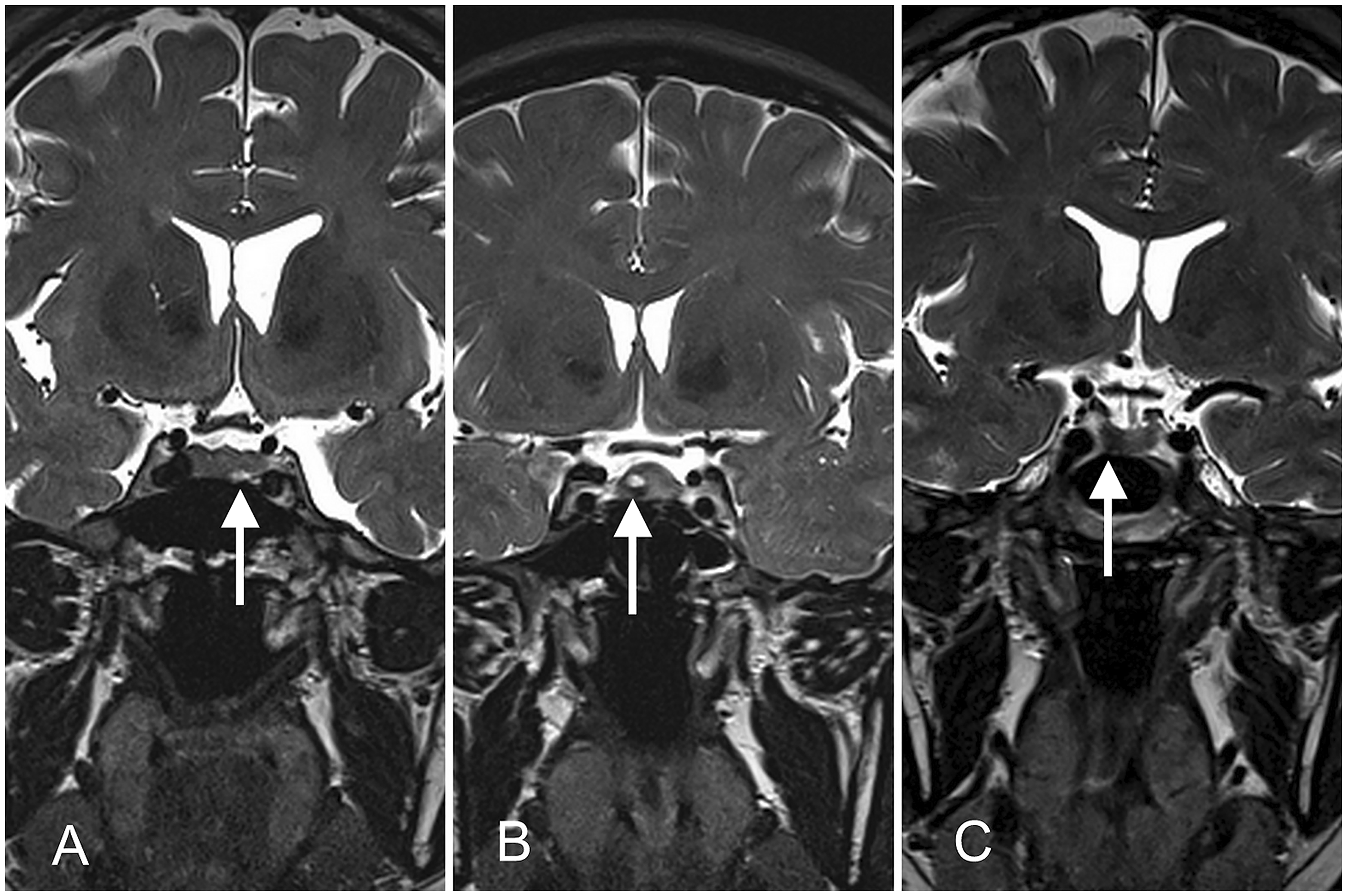
Statistical analysis
Statistical analyses were performed using SPSS version 19.0 (IBM Corp., Armonk, NY, USA). Inter-observer agreement was assessed using kappa statistics. Kappa values ≥ 0.8 were considered to indicate optimal consistency, values 0.4–0.8 were considered to indicate fair consistency, and values ≤ 0.4 were considered to indicate poor consistency.
The mean scores of the two readers were used to analyze the lesion conspicuity scores of conventional and 3D-T2 SPACE sequences alone and in combination. The data were compared using the paired Wilcoxon signed-rank test. A P value of < 0.01 was considered to indicate statistical significance.
Results
Patients
Between January 2013 and October 2014, 2000 consecutive patients underwent MRI pituitary examinations using this particular 3-T MRI system, excluding patients undergoing postoperative evaluations and those with suprasellar lesions, parasellar lesions, or pituitary lesions with diameters > 10 mm. Of them, 664 with clinically suspected pituitary lesions underwent imaging with conventional pituitary MRI and 3D-T2 SPACE sequences.
Radiologic assessments
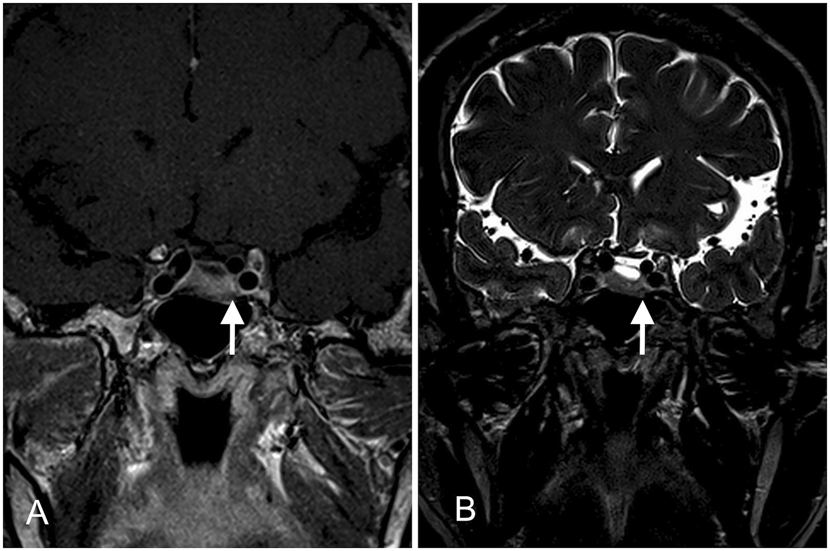
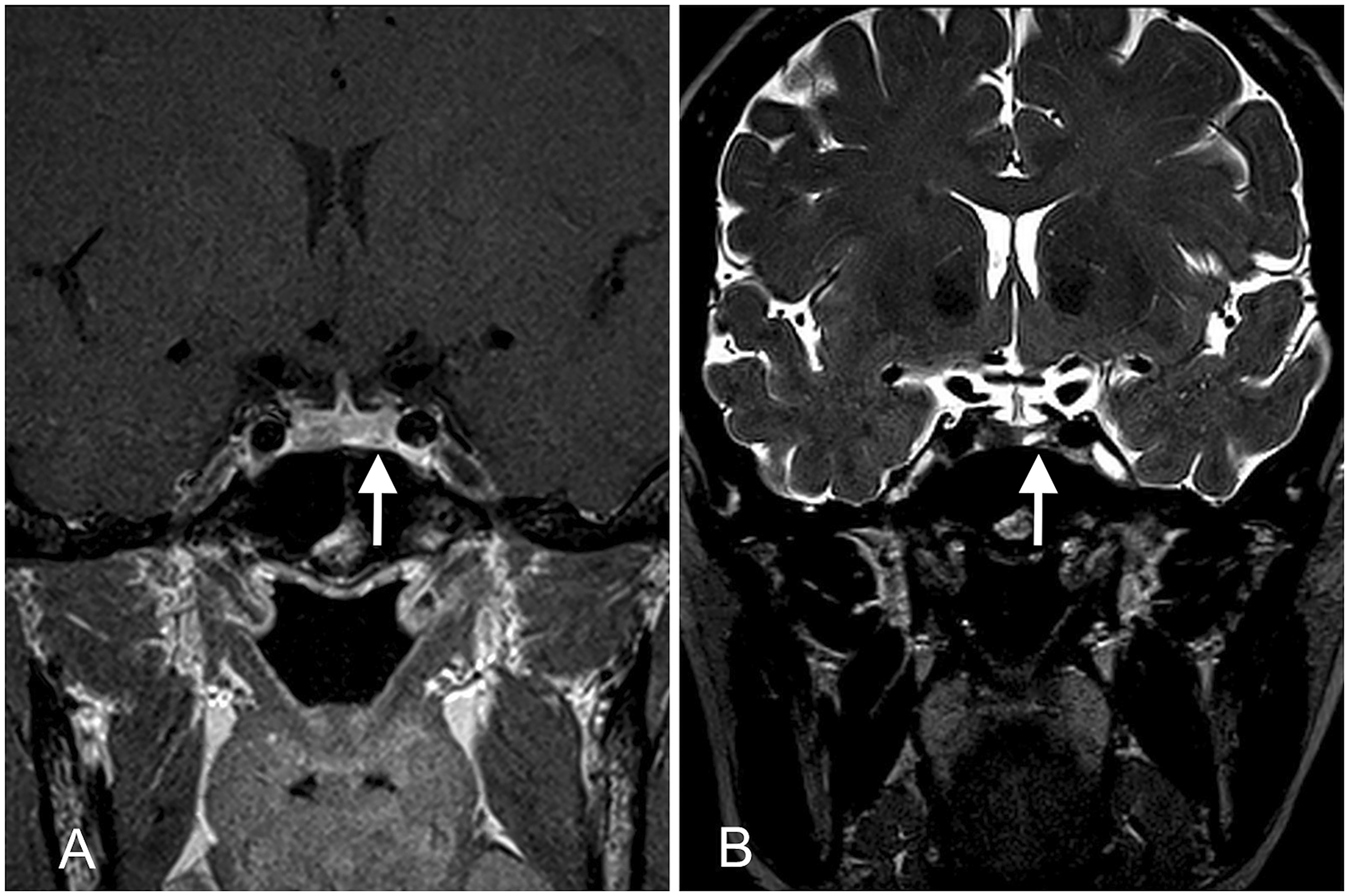
When the two readers evaluated the conventional images, they found that, on average, 61 of the images were non-diagnostic. The addition of 3D-T2 SPACE sequences increased diagnostic confidence by 60.7% on average in the diagnosis of pituitary micro-lesions (Figs. 2–4; Tables 2 and 3). The agreement between the two readers was optimally consistent (0.812 and 0.826 for the conventional and 3D-T2 SPACE sequences, respectively). The lesion conspicuity scores of conventional and 3D-T2 SPACE sequences both alone and in combination are shown in Table 4. The scores of combined conventional and 3D-T2 SPACE sequences were significantly higher than those of conventional (z = −6.403, P < 0.01) and 3D-T2 SPACE (z = −4.243, P < 0.01) sequences alone.
A pituitary lesion, along with an adrenal lesion, was found in a 33-year-old woman with Cushing’s disease. The tumors were confirmed to be a non-functional pituitary adenoma and an adrenocortical adenoma, respectively, by pathologic analysis. (a) The pituitary showed heterogeneous enhancement, particularly on the right side, on post-contrast enhanced coronal T1W imaging; (b) a focal low-signal intensity lesion (arrow) with a well-defined margin was evident on the left side of the pituitary on 3D-T2 SPACE. A growth hormone-secreting adenoma was found in a 56-year-old woman with acromegaly (growth hormone = 20.2 mU/L; Insulin-like growth factor-1 = 942 µg/L). (a) A suspicious lesion (arrow) was found on the left side of the pituitary on post-contrast enhanced coronal T1W imaging; (b) a focal moderately high-signal intensity lesion (arrow) with a circle of low signal intensity was visible on the left side of the pituitary on 3D-T2 SPACE. A prolactinoma was found in a 29-year-old woman with galactorrhea after giving birth and hyperprolactinemia (prolactin, 263 ng/mL). (a) The left side of the pituitary showed moderately high signal intensity (arrow) on post-contrast enhanced coronal T1W imaging; (b) A focal high-signal intensity lesion (arrow) with a well-defined margin was evident on the left side of the pituitary on 3D-T2 SPACE. Increased diagnostic confidence for uncertain lesions. 3D-T2 SPACE, three-dimensional sampling perfection with application-optimized contrasts using different flip-angle evolutions. Classification of lesions with increased diagnostic confidence. Lesion conspicuity scores. 3D-T2 SPACE, three-dimensional sampling perfection with application-optimized contrasts using different flip-angle evolutions.


Treatment and follow-up
One- and two-year follow-up assessments were conducted between 2015 and 2016 by telephone. Thirty-two patients with endocrine disturbances and pituitary lesions underwent surgery to remove pituitary micro-adenomas. Five patients with diabetes insipidus and pituitary lesions diagnosed as Rathke’s cleft cysts (RCCs) also underwent surgery. A total of 126 patients with increased prolactin levels and pituitary lesions were treated with bromocriptine. Two hundred and fifteen patients with headaches, normal endocrine function, and pituitary lesions were followed-up annually (of them, 23 patients were considered to have RCCs). In total, 286 patients with headaches or visual disturbances with clinically suspected pituitary lesions were found to be normal.
Discussion
Previous studies of the pituitary using MRI have focused on the detection of micro-lesions and found that a dynamic, time-dependent relationship exists between the gland and adenoma enhancement (4). Although dynamic contrast-enhancement can visualize most pituitary lesions, the diagnosis of some lesions is challenging using conventional sequences because of the similarity between the dynamic enhancement curves of the lesion and pituitary. Contrast-enhanced 3D-T2 SPACE imaging not only provides differential enhancement of the lesion and pituitary, but also reveals the different components of the lesion, because it is essentially a TSE T2W imaging sequence. The signal captured by 3D-T2 SPACE imaging is complicated. The different signal intensities are related to different components of the lesion, such as cystic changes, hemorrhage, or necrosis. Greater gland-to-lesion contrast equates to superior definition of the lesion border, allowing a more accurate depiction of the size, shape, and conspicuity of the lesion. However, some pituitary micro-lesions present iso-intensity on 3D-T2 SPACE imaging, resulting in a missed diagnosis. Therefore, the detection of pituitary micro-lesions using combined conventional and 3D-T2 SPACE sequences is superior to that using conventional or 3D-T2 SPACE sequences alone.
Recently, the development of 3-T MRI systems and techniques have yielded advantages such as an increased SNR, which reduces acquisition time and/or increases spatial resolution (9). Conventional sequences such as GRE sequences, which are very sensitive to magnetic susceptibility, especially during 3-T MRI, are easily affected by the skull, air in the sphenoid sinus, and blood flow in the vessels around the pituitary. Although SE sequences offer decreased magnetic susceptibility and high resolution, they require a long scan time. For the detection of pituitary micro-lesions, 2D sequences are adversely affected by thick slices. Decreasing the thickness of slices may achieve smaller voxels but elevates the risk of artifacts. In contrast, 3D-T2 SPACE sequences can provide thin-section images with high spatial resolution, thus reducing the partial volume effect between small lesions and surrounding tissues (10). In addition, the images can be obtained in a reasonable acquisition time (11). In our previously study, we found that one particular advantage of 3D-T2 SPACE sequences compared with conventional 2D sequences was their great reduction in magnetic susceptibility artifacts. Therefore, 3D-T2 SPACE sequences are suitable for detecting pituitary lesions (6).
The results of our large-scale study showed that 3D-T2 SPACE images improved the radiologist’s diagnostic confidence in diagnosing pituitary micro-lesions in the following ways. First, 3D-T2 SPACE sequences are helpful for detecting pituitary lesions of smaller size. Lesions < 3–4 mm in diameter, termed “pico-adenomas,” are usually MRI-negative and represent a major challenge in pituitary lesion diagnosis. Our findings showed that an additional 16.23% lesions < 3 mm in diameter were detected after the addition of 3D-T2 SPACE compared with conventional sequences. Determining the existence and location of such pico-sized lesions may be of significant value in guiding subsequent treatment, whether surgical or medical. Second, 3D-T2 SPACE imaging aids in the diagnosis of lesions in difficult locations. In this study, the existence of 28.36% and 24.34% of lesions adjacent to the cavernous sinuses and sellar floor, respectively, was confirmed by the addition of contrast-enhanced 3D-T2 SPACE sequences. Pituitary lesions adjacent to the cavernous sinuses or sellar floor are more easily missed or misdiagnosed because of the partial volume effect. However, it is significant to find a lesion in this region or determine that a lesion is invading the cavernous sinuses. In our previous study, because gadolinium decreases both the T1 and T2 relaxation time in tissues, we observed an increasing signal intensity in the cavernous sinuses and a decaying signal intensity in the pituitary after the injection of gadolinium during 3D-T2 SPACE imaging, which provided better contrast between the relatively hypointensity of the pituitary and the hyperintensity of the cavernous sinuses (8). Third, 3D-T2 SPACE imaging is suited to the diagnosis of abnormal enhancement lesions. Small numbers of pituitary adenomas exhibit an early phase enhancement pattern. For these lesions, dynamic contrast-enhanced imaging cannot definitively distinguish the lesion from normal pituitary gland. 3D-T2 SPACE imaging may provide more information in patients with these lesions; indeed, in our study, 13.53% more abnormal enhancement lesions were confirmed with the use of 3D-T2 SPACE sequences.
Therefore, our study demonstrates that, as a supplemental sequence, 3D-T2 SPACE is suitable for the detection of pituitary micro-lesions, especially those in difficult locations (involving the partial volume effect), that exhibit early phase enhancement (difficult to distinguish from the pituitary gland), or are of small size (necessitating high resolution). In general, 3D-T2 SPACE sequences increased diagnostic confidence for uncertain lesions by >60% in this study.
The 3D-T2 SPACE sequence is also suitable for detecting micro-RCCs. The persistence of Rathke’s cleft can result in an accumulation of fluid and cystic dilation, which leads to a purely intrasellar RCC (12,13). It is difficult to diagnose because of its location, especially when the lesion is small in size. By using parallel acquisition techniques, 3D-T2 SPACE sequences yield isotropic datasets, which enable the creation of multi-plane reconstructions in any orientation, such as the sagittal plane (Fig. 5) (14).
A Rathke’s cleft cyst found in a 37-year-old woman with hyperprolactinemia (prolactin = 91 ng/mL). (a) A low-signal intensity lesion (arrow) was evident in the middle of the pituitary on coronal 3D-T2 SPACE; (b) a reconstruction in the sagittal plane showed the lesion (arrow) in the posterior lobe of the pituitary; (c) the lesion (arrow) showed non-enhancement on post-contrast enhanced sagittal T1W imaging.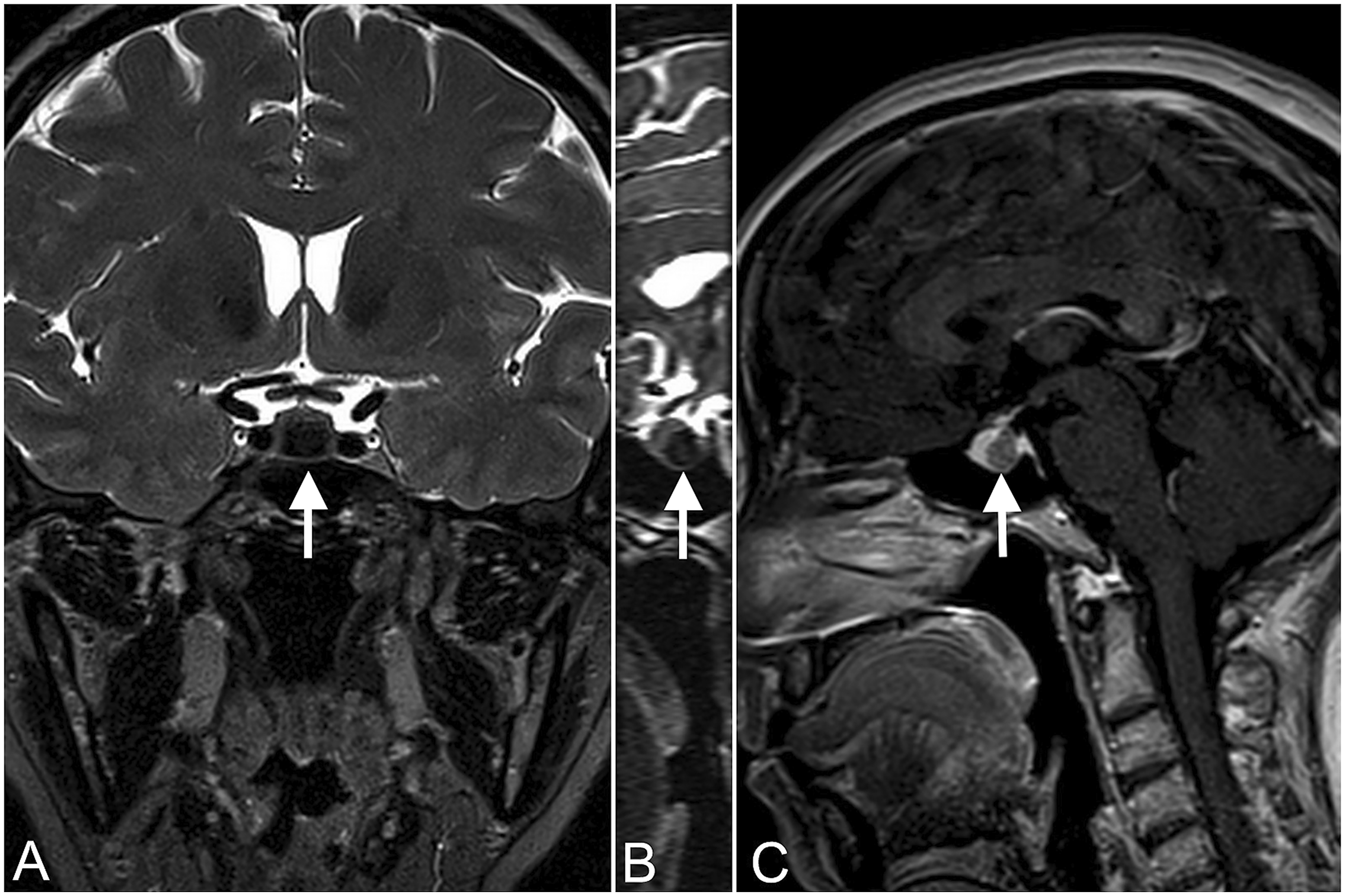
A major limitation of this study was that most of our patients did not undergo surgery. Thus, these lesions were not confirmed by pathologic analysis and the true-positive rate could not be accurately assessed. Further investigations with a longer-term follow-up are needed in future to determine the sensitivity, specificity, false-negative rate, and false-positive rate of 3D-T2 SPACE sequences.
In conclusion, 3D-T2 SPACE sequences can significantly enhance diagnostic confidence in the diagnosis of pituitary micro-lesions, especially those in difficult locations, that exhibit early phase enhancement, or that are small in size. 3D-T2 SPACE imaging should become part of the routine protocol for the detection of pituitary micro-lesions.
Footnotes
Declaration of Conflicting Interests
The author(s) declared no potential conflicts of interest with respect to the research, authorship, and/or publication of this article.
Funding
The author(s) received no financial support for the research, authorship, and/or publication of this article.