



Abstract
Background
Fistulas are serious complications of splenic and perisplenic fluid accumulations, which are often difficult to detect by routine imaging methods.
Purpose
To evaluate the occurrence of spontaneous fistulas detectable during computed tomography-guided percutaneous drainage placement (CTGDP) with contrast filling of splenic or perisplenic fluid collections and to assess characteristics in comparison with perihepatic or peripancreatic fluid accumulations, also being treated with CTGDP.
Material and Methods
In 127 CTGDP-procedures, pre-interventional CTs conducted with intravenous contrast agent were compared to post-interventional CTs including contrast filling of the drain to identify spontaneous fistulas. Patient and case characteristics were evaluated, and therapeutic consequences of fistula identification were analyzed.
Results
A total of 43 perisplenic, 40 peripancreatic, and 44 perihepatic drains were evaluated; 13 (30.2%) perisplenic, 7 (17.5%) peripancreatic, and 10 (22.7%) perihepatic fistulas were observed. Concerning the frequency of fistulas, no significant difference was found between the patient groups (P = 0.39). All fistulas were solely proven in CT scans including contrast filling of the drain. Seven fistulas (23.3%) required additional interventions. Perihepatic drains were significantly more often associated with recent surgery (P < 0.001). The mean size of peripancreatic drains was significantly greater (11.8 ± 3.9 F; P < 0.001) than in perihepatic or perisplenic fluid collections.
Conclusion
Spontaneous fistulas detected during CTGDP of splenic or perisplenic fluid collections are common. Post-interventional contrast filling of the drain drastically improves the detection rate of perisplenic, peripancreatic and perihepatic fistulas simultaneously initiating appropriate follow-up interventions.
Introduction
Splenic and perisplenic fluid collections are usually detected by ultrasound or computed tomography (CT) and may have different causes. One of the most common reasons is a splenic abscess, which is defined as an infectious process involving filling defects in the parenchyma of the spleen or in the subcapsular space (1–3). With the spleen representing the most frequently damaged organ during abdominal blunt trauma (4–7), traumatic abdominal injuries represent another commonly observed cause of splenic or perasplenic fluid collections. In both cases, an early diagnosis followed by immediate therapy is indispensable. In the past, the treatment was mainly based on splenectomy. Especially traumatic splenic injuries were mostly treated with surgery based on the concept that the spleen does not have an essential function for life and that the high vascularization of splenic parenchyma may cause uncontrollable bleeding (8–11), without surgery.
Nowadays, based on the developing knowledge of the immunological functions of the spleen (12,13) and the increasing role of interventional radiology, minimally invasive techniques such as the use of CT-guided perisplenic drains represent a viable alternative in the therapy of a large number of splenic and perisplenic fluid collections of diverse origin.
A non-negligible complication associated with splenic or perisplenic fluid collections is the presence of spontaneous fistulas (14–22), which can be difficult to detect using routine imaging methods. The current literature offers only little information about the occurrence of these fistulas. Moreover, not much is known about the occurrence of spontaneous fistulas associated with splenic or perisplenic fluid collections compared to other abdominal fistulas.
The aim of the present study was to evaluate the occurrence of spontaneous perisplenic fistulas detected during CT-guided percutaneous drainage placement (CTGDP) of splenic or perisplenic fluid collections by contrast filling. The results and associated clinical and material parameters were compared to two reference groups experiencing perihepatic and peripancreatic fluid collections, which were also treated with CTGDP. Furthermore, the therapeutic consequences of spontaneous fistulas were analyzed.
Material and Methods
Using a full-text search in our radiology information system (RIS), we identified all patients undergoing CTGDP in the last five years in our radiology department, where diluted contrast agent (Solutrast 300; Bracco Imaging, Konstanz, Germany or Accupaque 350; GE Healthcare, München, Germany) was applied via the drain to identify spontaneous fistulas. In none of the evaluated cases the existence of fistulas was expected before the intervention. All detected fistulas represented incidental findings during CTGDP. To identify the fistulas, the pre-interventional CT scan, which was performed with intravenous contrast agent, and the post-interventional CT scan including contrast filling of the drain were evaluated and compared. Patient characteristics, recent surgeries (<14 days before CTGDP), the maximum diameter of addressed fluid collection, the anatomic proximity to either spleen, liver, or pancreas, and drain diameter were noted.
Another important aspect of this study was the management of the detected fistulas. For this purpose, the study compiles an overview of all radiological and surgical interventions, which were directly associated with the detected fistulas and were performed within the first 21 days after their first detection. Moreover, the occurrence of peri-interventional complications was observed. Complications that required an additional intervention or caused further or prolonged hospitalization were defined as major complications. All other complications were regarded as minor complications.
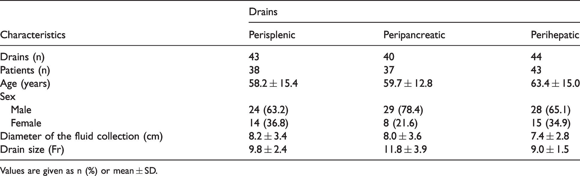
In total, we evaluated 127 drains in 118 patients (81 men; 37 women). In total, 43 drains (38 patients) were perisplenic, 40 drains (37 patients) were peripancreatic, and 44 drains (43 patients) were perihepatic. An overview of the patient, disease, and drain characteristics is given in Table 1. The size of the drains was individually chosen by the interventionalist depending on the size of the fluid collection and the composition of the punctured secretion. In cases of large accumulations containing secretion with high viscosity, larger drains were used.
Patient, disease and drain characteristics.
Values are given as n (%) or mean ± SD.
The study was approved by the local independent ethics committee of the University Regensburg (approval number: 20-1906-104) and conducted according to national and state law as well as the WMA Declaration of Helsinki – Ethical Principles For Medical Research Involving Human Subjects and the EMA Good Clinical Practice Directive. Written informed consent for the CTGDP was obtained from each patient before the intervention. Written informed consent for the anonymous use of the patients’ data was not required, as all data had been acquired during clinical routine and because of the retrospective design of the study.
Statistical analysis was performed using standard contingency calculations and one-way ANOVA with Bonferroni’s multiple comparison test where appropriate. Statistically significant differences were assumed for P < 0.05. Calculations were made with GraphPad Prism 5.0a for Mac (GraphPad Software, San Diego, CA, USA).
Results
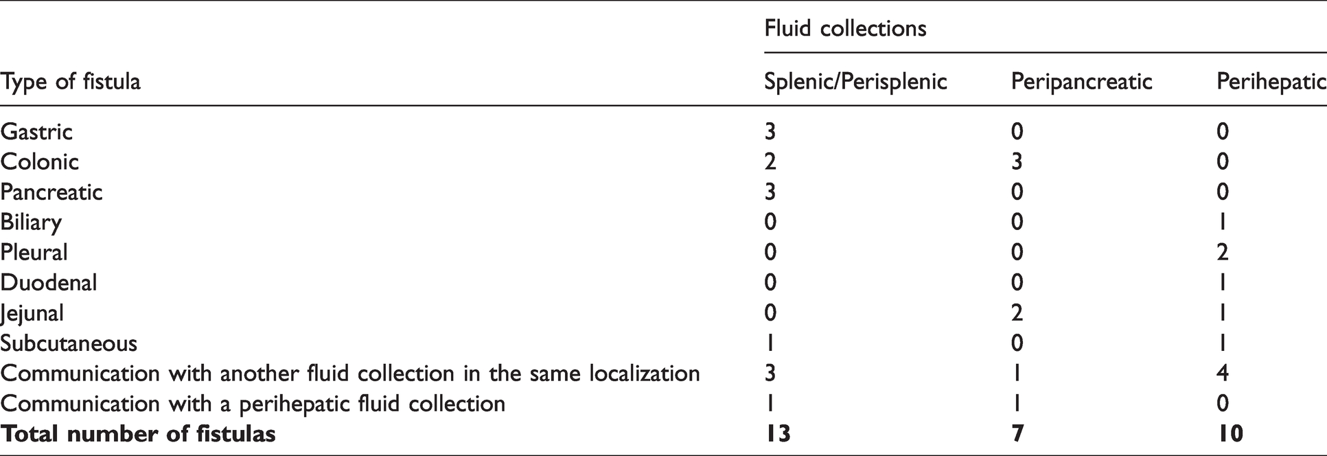
In total, we evaluated 43 perisplenic, 40 peripancreatic, and 44 perihepatic drains. Spontaneous fistulas were detected in 13 of all 43 cases (30.2%) with perisplenic drains. Furthermore, 7 (17.5%) peripancreatic fistulas and 10 (22.7%) perihepatic fistulas were observed. Fig. 1 summarizes the mentioned results. None of the detected spontaneous fistulas could be reliably proven in the pre-interventional examination, which was performed by a CT scan with intravenous contrast agent. Using a chi-square calculation, no statistically significant difference concerning the frequency of spontaneous fistulas was found between the three patient groups (chi-square = 1.89; P = 0.39). Table 2 presents an overview of the different types of fistulas in every patient group. In 20 of 43 cases of perisplenic drains a recent surgery had been performed. The same result applies to 19 of the 40 peripancreatic drains and 39 of the 44 perihepatic drains (Fig. 2). Perihepatic drains were significantly more often associated with a recent surgery (88.6%; chi-square = 21.0; P < 0.001) than perisplenic (46.5%) and peripancreatic (47.5%) cases. There was no statistical difference in the diameter of the fluid collections in perisplenic, peripancreatic, and perihepatic cases with 8.2 ± 3.4 cm, 8.0 ± 3.6 cm, and 7.4 ± 2.8 cm, respectively (P = 0.51). The mean size of drains was significantly greater in peripancreatic cases (11.8 ± 3.9 F) compared to 9.8 ± 2.4 in perisplenic and 9.0 ± 1.5 F in perihepatic fluid collections (P < 0.001). Fig. 3 presents the distribution of age, tube size, and the diameter of fluid collections in the observed patient groups. Of the 30 patients with spontaneous fistulas, an additional radiological or surgical intervention was necessary in 7 (23.3%) within the first 21 days after their first detection. In four cases of perihepatic fluid collections associated with biliary leaks, a percutaneuos transhepatic cholangiodrainage (PTCD) placement had to be performed. Two patients experiencing perisplenic accumulations with associated fistulas had to undergo splenectomy combined with a resection of the left colon flexure. In one case of a peripancreatic fluid accumulation accompanied by a fistula, a distal pancreatectomy and a resection of the left colon flexure were unavoidable. No major peri-interventional complications occurred. A total of four minor complications (pneumothorax, n = 3; initially dislocated perihepatic drain, which could be replaced immediately, n = 1) required no intervention and caused no further or prolonged hospitalization.
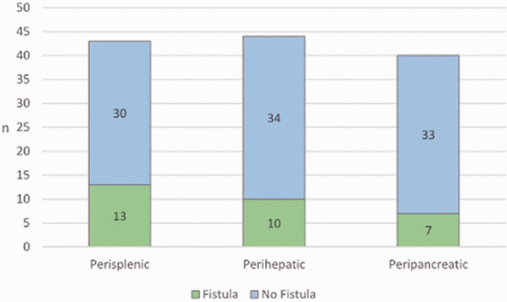
Number of fistulas in splenic or perisplenic, perihepatic, and peripancreatic fluid collections.
Types of fistulas.
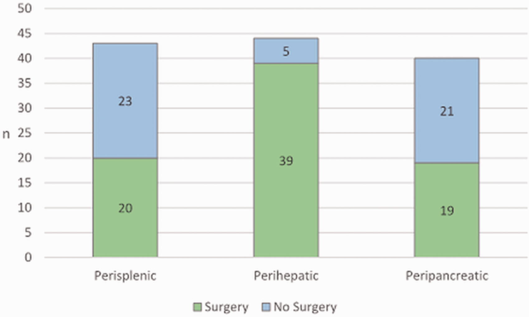
Number of previous surgeries in splenic or perisplenic, perihepatic, and peripancreatic fluid collections.
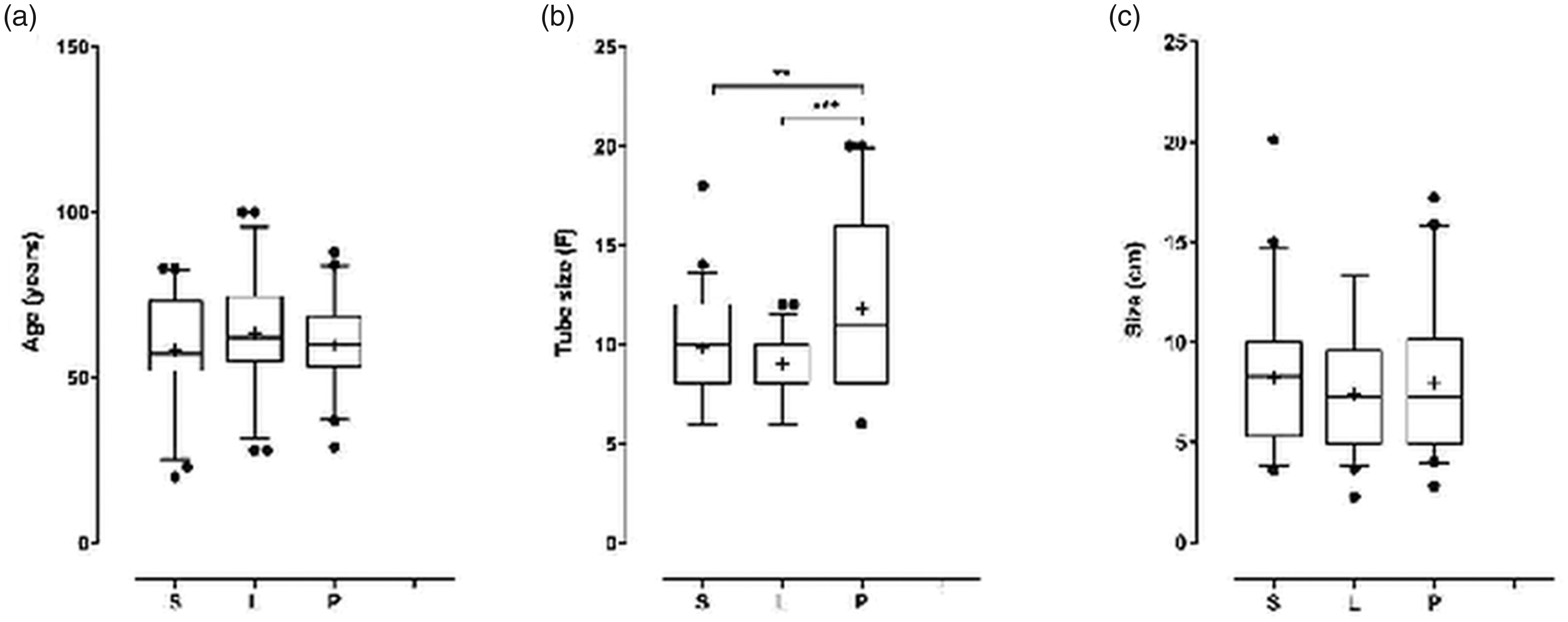
(a–c). Distribution of age (a), tube size (b), and size of the fluid accumulation (c) in splenic or perisplenic (S), perihepatic (L), and peripancreatic (P) fluid collections.
Discussion
Being a minimally invasive therapeutic option, CTGPD represents a viable alternative to surgery in the therapy of splenic and perisplenic fluid collections, not least because of its safety and effectiveness (23). Its application in the therapy of splenic or perisplenic accumulations combined with post-interventional contrast filling can simplify the identification of associated fistulas. The current literature offers almost exclusively individual cases of perisplenic fistulas (14–22). So far, this is the first trial evaluating the incidence of spontaneous perisplenic fistulas while simultaneously presenting a comparison to reference groups, who had perihepatic and peripancreatic fluid collections, respectively.
In 30.2% (n = 13) of the splenic or perisplenic fluid collections spontaneous fistulas were observed (Figs. 4 and 5). Although no significant difference concerning the occurrence of spontaneous fistulas could be proven between the patients experiencing splenic or perisplenic fluid collections and the two reference groups (peripancreatic fistulas, n = 7, 17.5%; perihepatic fistulas, n = 10, 22.7%), the large number of spontaneous fistulas indicates that, regardless of the abdominal localization of the drain, a post-interventional contrast filling should be considered to be routinely included into the procedure of placing CT-guided drains. In the whole study population, there was not one single case, in which post-interventionally detected fistulas could be reliably proven in the pre-interventional CT scan, which was performed by a CT scan with an intravenous contrast agent. This fact also highlights the importance of post-interventional contrast filling of the drain, as otherwise the majority of the observed fistulas in all three patient groups would probably never have been detected at such an early stage of the disease, potentially leading to further complications (24,25).
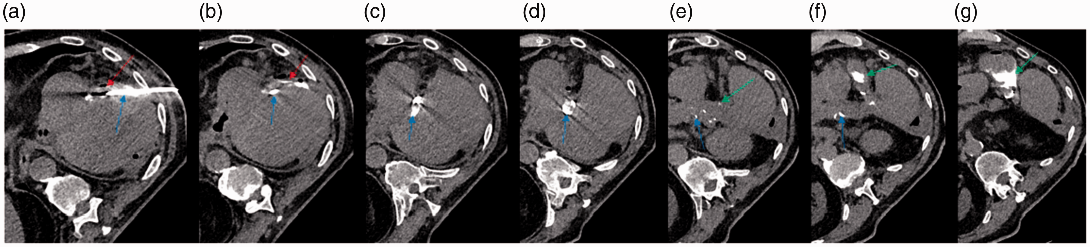
(a–g) Axial unenhanced CT images show a perisplenic fluid collection and a colonic fistula in a 58-year-old man treated with a CT-guided drain. After post-interventional contrast filling of the drain (blue arrow), the perisplenic fluid collection (red arrow) and a colonic fistula (green arrow) can be identified. CT, computed tomography.
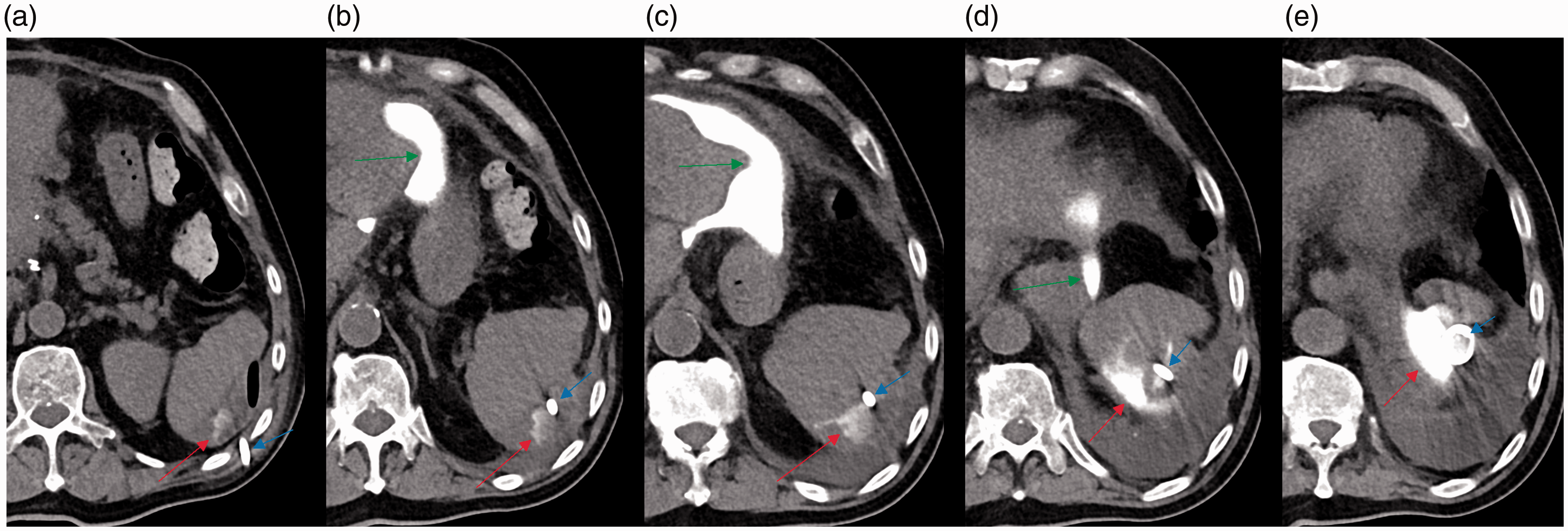
(a–e) Axial unenhanced CT images show a perisplenic fluid collection and a perihepatic fistula in a 55-year old man treated with a CT-guided drain. After post-interventional contrast filling of the drain (blue arrow), the perisplenic fluid collection (red arrow) and a perihepatic fluid accumulation including a fistula (green arrow) can be identified. CT, computed tomography.
Regarding the organs that were afflicted with splenic and perisplenic fistulas (Table 2), the pancreas (n = 3) and the stomach (n = 3) were affected most frequently, followed by the colon (n = 2). The results are consistent with the findings of other authors. Kato et al. (26), who focused on the postoperative occurrence of pancreatic fistulas after splenectomy as part of cytoreductive surgery for the treatment of ovarian cancer, reported six pancreatic fistulas in 21 female patients after postoperative day 3, which were detected by elevated amylase levels in the drainage fluid obtained by a surgically placed drain. Moreover, the current literature offers several individual reports of gastrosplenic fistulas occurring in association with splenic abscesses (14–17,19). Splenocolonic fistulas associated with splenic abscesses have been described before in case reports by McCrystal et al. (20), presenting a 16-year-old boy with blunt abdominal trauma and splenic injury, and by Pappalardo et al. (22), reporting on a 31-year-old woman with Crohn’s disease. In contrast to single case reports by other authors (18,21), splenothoracic or splenobronchial fistulas could not be observed in the present study. As a potential reason for the evolution of perisplenic fistulas, iatrogenic injuries of the organ, which could result from a previous intervention (27), have to be considered. Moreover, perisplenic fistulas can be associated with splenic lymphoma (28).
Focusing on the types of fistulas detected in the two reference groups (Table 2), the findings indicate that perihepatic fluid collections particularly developed fistulas affecting further perihepatic accumulations (n = 4) and that peripancreatic collections primarily showed a pathological intestinal communication, especially colonic (n = 3) (Fig. 4). The results are in accordance with the findings of other authors. Kochhar et al. (29) identified intestinal fistulization as a rare complication of pancreatitis, describing fistulas in 12 of 289 patients with acute pancreatitis, of which four were colonic. Furthermore, several single cases of pancreaticocolonic fistulas associated with pancreatitis and peripancreatic fluid collections are reported in the current literature (30–32). An important fact that must be considered in this context is prior pancreatic resection. With postoperative pancreatic fistula representing one of the most common complications after pancreatic surgery (33), it remains the main source of major morbidity and mortality after pancreatic resection, affecting 13%–41% of all patients (34,35). The fact that pancreatic secretions include several digestive enzymes may cause erosive processes in the adjacent organs possibly provoking the evolution of fistulas. Therefore, especially in these cases, the early evaluation of potential associated fistulas is of great importance.
Abdominal abscesses or fluid collections are a frequently observed complication after abdominal surgery (36,37) requiring postoperative drainage. The current study indicates that perihepatic drains are significantly more often associated with a recent surgery (P < 0.001) than perisplenic or peripancreatic drains. These findings are similar to the results of Shahnazi et al. (37), who evaluated 41 patients with abdominopelvic abscesses or fluid collections. In 31 cases (75.6%) a previous surgery was reported, with the liver most frequently affected (n = 17; 41.5%). Due to its relatively high vascularization, the liver seems to be at high risk of postoperative hematoma. Moreover, an insufficient bile duct located at the resection margin can cause postoperative bilioma potentially even evolving into an abscess by superinfection. The fact that all these pathomechanisms may make the liver more vulnerable to fluid accumulations than other abdominal organs provides a possible explanation for the results of this trial. Thus, the results may heighten awareness of the postoperative course in patients treated with hepatic surgery.
Compared to perisplenic and perihepatic drains, the average size of peripancreatic drains was significantly greater (P < 0.001), which coincides with the outcomes of other studies reporting a range of 8–12 Fr in splenic drains (23,38), 7–12 Fr in hepatic drains (39,40), and 14–30 Fr in peripancreatic drains (41,42). Baudin et al. (41) described an average drain diameter of 24.4 ± 4.4 Fr in 48 patients with peripancreatic fluid collections treated with CT-guided drainage (41). This difference of size might be due to the commonly observed diffusely distributed peripancreatic fluid collections, for example in cases of pancreatitis, which are often hard to manage by using smaller sized drains.
In 7 of 30 patients (23.3%) experiencing spontaneous fistulas an additional radiological or surgical intervention had to be performed, as the patients’ state of health became increasingly precarious due to the consequences of the detected fistulas. In four cases of small biliary leaks associated with perihepatic accumulations, a surgery could be avoided by placing a PTCD, as recommended by Popat et al. (43). In three cases a surgery, which is considered to be one of the last therapeutic opportunities (44), was indispensable. Although the fistulas had been detected in a very early stage of the disease by post-interventional contrast filling of the drain after CTGDP, almost one-quarter of all affected patients had to undergo another intervention to limit the fistulas` consequences. As fistulas can potentially result in a severe state of illness, such as fulminant sepsis (44), one must assume that a delayed or even lacking detection of those fistulas would probably have worsened the patients’ outcome, emphasizing the importance of post-interventional contrast filling of the drain after CTGDP.
The present study has some limitations. The first is the retrospective nature of the study. The second limitation is the small sample size of the observed study population and the reference groups. Third, the trial does not include further controls to evaluate the further development of the detected fistulas. Fourth, the study groups consisted of a heterogenous patient population concerning number of patients, number of drains, sex, and age.
In conclusion, the presented findings demonstrate that the occurrence of spontaneous perisplenic fistulas detected during CT-guided drainage of splenic or perisplenic fluid collections is not rare. Moreover, the study illustrates two important advantages of post-interventional contrast filling of the drain after CTGDP in abdominal fluid collections. On the one hand, post-interventional contrast filling of the drain drastically improves the detection rate of perisplenic, peripancreatic, and perihepatic fistulas without requiring any extra time or financial effort. On the other hand, the application of contrast agent via the placed drain can hereby initiate necessary follow-up interventions preventing severe fistula-associated complications. Compared to peripancreatic and perihepatic fluid accumulations, no significant difference concerning the occurrence of spontaneous fistulas could be proven. Perihepatic drains were more often associated with recent surgery, while peripancreatic fluid collections required larger drains.
Footnotes
Acknowledgements
We thank Esther Kiszler for the linguistic revision of the manuscript.
Declaration of conflicting interests
The author(s) declared no potential conflicts of interest with respect to the research, authorship, and/or publication of this article.
Funding
The author(s) received no financial support for the research, authorship, and/or publication of this article.
