



Abstract
Background
Iterative reconstruction techniques (IRTs) are commonly used in computed tomography (CT) and help to reduce image noise.
Purpose
To determine the minimum radiation dose while preserving image quality in head CT using IRTs.
Material and Methods
The anthropomorphic phantom was used to scan nine head CT image series with varied radiation parameters. CT dose parameters, including volume CT dose index (CTDIvol [in mGy]) and dose length product (DLP [in mGy/cm]), were recorded for each scan series. Different noise levels (iDoseL1-6) were used in IRT reconstructions for soft and bone tissues. In total, 15 measurements were taken from five regions of interest (ROI) with an area of 10 mm2. The signal-to-noise ratio (SNR) and noise values obtained at different ROIs were compared among various reconstruction methods with repeated measures of statistical analysis.
Results
In the head CT scan, applying IRT iDoseL5 had the lowest noise and highest SNR for soft tissue (P < 0.05), and increased iDose can decrease CT dose by 54.6% without compromising image quality. While for bone tissue reconstruction, no clear association was found between the level of iDose and noise. However, when CTDIvol is >20 mGy, iDoseL4 is slightly superior to other reconstruction methods (P < 0.065).
Conclusion
Using IRTs in head CTs reduces radiation dose while maintaining image quality. IDoseL5 provided optimal balance for soft tissue.
Introduction
Computed tomography (CT) imaging is one of the most frequently performed radiological examinations providing valuable diagnostic information (1–3). Examinations performed using CT methods are associated with a risk of oncogenesis depending on the radiation dose levels received (4–6). According to the main principle of radiation protection, as low as reasonably achievable (ALARA), it is advised to use the lowest possible dose for radiological examinations (7). Image noise may usually be increased by reducing the ionizing radiation dose; however, it is possible to keep the adequate diagnostic quality of the images. The main goal of examinations in radiology procedures is to obtain a “diagnostic image” rather than a “perfect image,” so in patients, it is common to work with “noisy” images (8–10).
To decrease radiation exposure from CT scans, several methods have been implemented, such as automatic regulation of tube current and voltage, and adjustable beam collimation; however, they must not be examined on patients in clinical practice. Therefore, different tissue-equivalent phantoms are commonly used in radiology for research purposes (11). Notably, a variety of CT image reconstruction algorithms for quality enhancement are developed (10). These algorithms were applied to CT examinations in 1972, but their use had been limited by low computer reconstruction power as well as high computing time and poor quality.
The reconstruction of images in modern CT scans is mainly performed using the filtered back projection (FBP) algorithm. This method, which involves filtering and back projecting the data, is fast and efficient. However, there is a limit to the amount of dose reduction possible with the use of the FBP algorithm. Despite having been the standard for CT image reconstruction for a long time, the FBP approach has a drawback of increasing image noise if the radiation dose is greatly reduced.
One drawback of reducing radiation dose excessively using the FBP algorithm is the potential for streak artifacts and a noticeable increase in image noise. However, the recent revival of iterative reconstruction techniques (IRTs) in CT scans provides a potential solution that enables a reduction in radiation exposure while maintaining image quality (12–14). Multiple studies have demonstrated that the implementation of IR algorithms effectively decreases radiation dose and enhances image quality in CT exams compared to the FBP method (15–18).
In 2009, IRTs, which are mathematical algorithms, were commercially available and started to be used instead of FBP (19).
The IRT works by modeling and comparing the resulting signal projection data with the theoretical calculation based on the signal mentioning (guess) principle (7,13). The ultimate goal of all IR algorithms is to reduce image noise and improve resolution while preserving edges and minimizing artifacts. This ability of IR techniques enables the use of low-dose CT scans with decreased scanning parameters, such as lower tube current or voltage. Numerous studies have demonstrated the potential to decrease radiation exposure with the use of various IR techniques on various CT scanners (20–22).
However, there are also several limitations to improving the suboptimal image with IRT. If the image quality is not optimal, the IRT may render the image more granular or excessively confusing by changing the representation of a real tissue, which may result in an incorrect interpretation (12).
Given the nature of the IRT and the need to reduce radiation doses, it is necessary to understand the impact of IRT on image quality and obtain CT images with adequate diagnostic quality. The aim of the present study was to assess the impact of IRT reconstruction with five different noise strengths/levels, and find the lowest dose and exposure parameters with reasonable image quality in the head CT examinations.
Material and Methods
This single-institution experimental study was conducted at the Diagnostic Radiology department of Children's Clinical University Hospital in Riga, Latvia.
CT examinations
A 64-slice scanner (Ingenuity; Philips Medical Systems, Cleveland, OH, USA) with the manufacturer software (Extended Brilliance Workspace) was used to obtain raw phantom images. Radiological imaging postprocessing with the IntelliSpace Portal version 5 (Phillips, Best, The Netherlands) was used to reconstruct the CT images with IRT algorithms. The images were obtained from the head of a tissue-equivalent phantom (PIXY, MIRION Technologies (Capintec), Florham Park, NJ, USA). The phantom length and weight were 156 cm and 48 kg, respectively (Fig. 1).
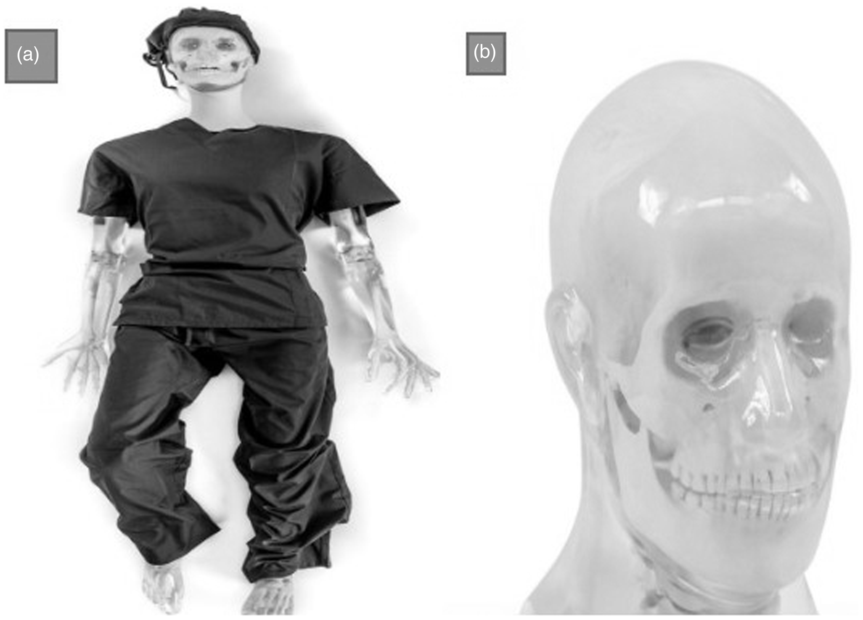
(a) Take-Apart Pixy Skeleton. (b) From head to toe. The human skull with the Take-Apart Pixy Head Phantom (https://rsdphantoms.com/product/take-apart-ct-pixy). Made of tissue-equivalent materials and lifelike articulations, Take-Apart CT Pixy is more realistic than a cadaveric skeleton with radiographs that are optically equivalent in density and contrast to human patients.
Radiographs of PIXY are optically equivalent to density and contrast to human patients https://rsdphantoms.com/product/take-apart-pixy/).
The phantoms’ head was imaged with a matrix of 512 × 512 pixels. The exposure parameters were changed for each scan as kilovolts (kV) = 80–120 and tube current seconds (mAs) = 55–227.
The whole combinations of exposure parameters were as follows:
We selected a fixed window width (WW) and window level (WL) for bone, 500 and 2000, and soft tissue, 40 and 80, respectively, for all combinations.
The scan length, in spiral mode with a pitch of 1, of 15.4 cm was selected with the 153 transverse images. The scan and reconstructed slice thickness of 2 mm was selected.
For each scan series, CT dose parameters calculated using the Philips image acquisition software, including volume CT dose index (CTDIvol [in mGy]) and dose length product (DLP [in mGy/cm]), were recorded. Five regions of interest (ROI) with an area of 10 mm2 were selected to evaluate the signal (average of CT HU) and noise (standard deviation of CT HU) values. A total of 15 measurements were obtained in each reconstruction series. The mean was calculated for a set of scores.
Image reconstruction
The images of every CT examination were reconstructed from the raw data separately in soft tissue and bone gray-level windows. A single FBP reconstruction was performed for all scan series, as well as several IRT Philips iDose reconstructions at all levels offered by the program. For each exam, six reconstruction series (one FBP, iDoseL1, iDoseL2, iDoseL3, iDoseL4, and iDoseL5) were performed for soft tissue image reconstruction. In addition to the mentioned reconstruction series, an excessive iDose (level 6) was performed for bone tissue reconstruction. Two scan series with the highest exposure parameters were used for IRT with iDose 6. Furthermore, two scan series with the lowest exposure parameters were allowed by the Philips program to be reconstructed with iDoseL4. The other exam images were reconstructed by iDoseL1-5.
Signal and noise assessment in IRT reconstructions
In consultation with an experienced medical physicist and a radiologist, five ROIs with an area of 10 mm2 (center, right, left, top, and bottom of a transverse image) were selected to evaluate the signal (average of CT HU) and noise (standard deviation of CT HU) values. Measurements were made in three slices (located in the middle, inferior, and superior of the topogram) for each exam and each reconstruction series. A total of 15 measurements were obtained in each reconstruction series. An average of the results was calculated based on a total of 15 measurements obtained in each series of reconstructions.
To estimate image quality, the mean noise value and the corresponding signal-to-noise ratio (SNR) were calculated for each reconstruction series. Reconstructions with the highest SNR and lowest SD values were considered to be the most optimal.
The SNR and noise (SD) values obtained at different ROIs were compared among various reconstruction methods with repeated measures of statistical analysis.
The statistical test was repeated for various image series obtained by different exposure parameters. The level of statistical significance was set at P < 0.05 and all the statistical tests were performed in SPSS version 18 (SPSS Inc., Chicago, IL, USA).
Results
Exposure and CT dose parameters
Exposure and CT dose parameters are presented in Table 1. All exams were arranged in ascending order based on CTDIvol values.
DLP depending on the exposure parameters applied in the examination.

CT, computed tomography; CTDIvol, volume CT dose index; DLP, dose length product.
Signal and noise assessment in soft tissue reconstructed images
The mean ± SD of noise values in soft tissue are illustrated in Table 2. In most reconstructed image series, noise values were gradually decreased with increasing the CTDIvol values. The greatest noise value was observed in the FBP reconstruction series in all exposure settings (P < 0.035).
Noise values and SNR in soft tissue window reconstructions.

Values are given as mean ± SD.
CTDIvol, volume CT dose index; FBP, filtered back projection; SNR, signal-to-noise ratio.
iDoseL5, within all CTDIvol values, provided the lowest image noise value in comparison to other iDose reconstructions (P < 0.035). The best reconstruction with the lowest image noise (SD = 2.9) is associated with the iDose 5 series 9 (Fig. 2)

Soft tissue window reconstructions in image series no. 5 (exposure parameters = 120 kV, 146mAs, CTDIvol 18, 6). Among all CTDIvol values tested, iDoseL5 exhibited the least image noise for soft tissue reconstructions compared to other iDose settings. CTDIvol, volume CT dose index; FBP, filtered back projection.
The mean ± SD of SNR values in soft tissue reconstructed images are provided in Table 2. The iDoseL5 has the highest SNR values for all the CT dose parameters (P < 0.031). Within comparable image quality levels (SNR = 8), iDoseL5 in series 3 (CTDIvol = 14.3 mGy) and iDoseL1 in series 8 (CTDIvol = 26.2 mGy), it is possible to acquire similar quality images and reduce the dose by 54.6% (P < 0.05). The values for the other iDose reconstructions are as follows: iDoseL2 in series 3 (CTDIvol = 14.3 mGy) and iDoseL6 in series 8 (CTDIvol = 26.2 mGy).
The largest noise and SNR difference within a single scan (equal CTDIvol) was observed in series 8 and 9 (noise difference = 37.5%; SNR difference = 50%). The smallest SNR difference is observed in series 1–0.8.
Signal and noise assessment in bone window reconstructed images
The mean ± SD of noise and SNR values in bone tissue reconstructed images are illustrated in Tables 3. For scan series 1 and 2 with the lowest dose, bone reconstructions were performed up to the iDose 4 level, while iDose 6 was also reconstructed for series 8 and 9.
Noise values and SNR in bone window reconstructions.

Values are given as mean ± SD.
CTDIvol, volume CT dose index; FBP, filtered back projection; SNR, signal-to-noise ratio.
In the bone reconstructed image series, the noise was not associated with the increasing iDose level. In contrast to soft tissue reconstructed images, the noise values for bone reconstructed image series show that no reconstruction method is convincingly superior to anyone else (P > 0.05). The lowest noise (20.9) and the highest SNR (47.7) values were observed for the reconstruction of the scan series 9 with iDoseL4 due to higher CT exposure parameters (Fig. 3).

Bone window reconstructions in image series 9 (exposure parameters = 120 kV, 225 mAs, CTDIvol 29.4). The panels labeled FBP show the noise characteristics without using any IRT algorithm. IDose1- iDose5 represent the noise reduction achieved by progressively more advanced IRT algorithms. The lowest noise and the highest SNR values were observed for the reconstruction with iDoseL4. CTDIvol, volume CT dose index; FBP, filtered back projection; IRT, iterative reconstruction technique; SNR, signal-to-noise ratio.
IRT iDoseL6, compared to iDoseL5, provided nearly the same noise and SNR results at the same CTDIvol values for all the imaging with different CT dose parameters (P > 0.05). It can also be observed from the noise and SNR results that when CTDIvol is >20 mGy, iDoseL4 is slightly superior (but not statistically significant) to other reconstruction methods.
For the noise and SNR values of the bone tissue reconstructed images, the noise is directly dependent on the CDTIvol exam, which depends on the exposure parameters applied in the exam.
Discussion
Our findings suggest that the choice of reconstruction method significantly impacts image quality, and the optimal choice depends on the imaging protocol and the specific tissue of interest. The findings showed that the iDoseL5 reconstruction method provided the lowest noise values in soft tissue images across all CT dose parameters, and the highest SNR values for all CT dose parameters in both soft and bone tissue images.
In addition, the study revealed that within comparable image quality levels, iDoseL5 in series 3 and iDoseL1 in series 8 could produce similar quality images while reducing the dose by 54.6%. Interestingly, the study also found that the noise values in bone window reconstructed images were not associated with increasing iDose levels. This is in contrast to the findings in soft tissue images, which showed a gradual decrease in noise values with increasing iDose levels. The authors suggested that this discrepancy may be due to differences in the anatomy and attenuation properties of soft and bone tissues, which may require different reconstruction algorithms and CT dose parameters. Furthermore, the results of the present study showed that when CTDIvol is >20 mGy, iDoseL4 is slightly superior to other reconstruction methods in terms of noise and SNR values in bone tissue reconstructed images.
In soft tissue reconstructed images, we observed that the iDoseL5 method consistently provided the lowest noise values compared to other iDose reconstructions. This is in line with previous studies that have shown the effectiveness of iDoseL5 in reducing image noise and improving image quality (23,24). In addition, our study found that within comparable image quality levels, such as SNR = 8, it is possible to acquire similar quality images with a dose reduction of 54.6% using iDoseL5 in series 3 and iDoseL1 in series 8. This finding is in agreement with previous studies that have also demonstrated the potential of dose reduction without sacrificing image quality when using IRTs (25,26).
We also observed a significant difference in noise and SNR values between different exposure settings, with the greatest noise value observed in the FBP reconstruction series at all exposure settings. This finding is consistent with previous studies that have shown that FBP produces higher levels of image noise compared to IRTs (27,28). In addition, we found that the largest noise and SNR difference within a single scan (equal CTDIvol) was observed in series 8 and 9, while the smallest SNR difference was observed in series 1.
In bone window reconstructed images, we found that the noise values were not associated with the increasing iDose level, and there was no reconstruction method that was convincingly superior to others. However, we did observe that higher CT exposure parameters resulted in lower noise values and higher SNR values. This finding is consistent with previous studies that have also reported higher SNR values in bone tissue with increased CT exposure parameters (29,30). In addition, our study found that iDoseL4 provided the lowest noise and the highest SNR values in the bone reconstructed images of series 9 due to higher CT exposure parameters.
Our findings suggest that the choice of reconstruction method significantly impacts image quality in CT imaging, and the optimal choice depends on the imaging protocol and the specific tissue of interest. These findings are in agreement with previous studies that have also reported the effectiveness of IRTs in reducing image noise and improving image quality while allowing for dose reduction (31,32).
Overall, the IRT provides an alternative means of using lower radiation doses in CT scans. By enhancing the SNR, this technique can effectively reduce the noise level and photon beam starvation artifacts, ultimately leading to a reduction in dose. Our current research has examined the impact of IRT on a head phantom using five distinct noise levels to identify the optimal dose parameters while maintaining acceptable image quality in CT scans.
Several investigations have been conducted in this area, including studies by Niu et al. (33), Tozakidou et al. (13), and Southard et al. (14). However, these studies have primarily focused on CT exams of the abdominal, thoracic, and temporal bones in patients or cadavers using various noise strengths/levels. For instance, Niu et al. (29) evaluated the effect of IRTs on image quality and radiation dose in temporal bone CT scans using multiple iDose levels and FBP. They found that the combination of 100 mAs/section and iDoseL5 yielded the lowest dose while maintaining diagnostic image quality, with CNR slightly higher than their routine institution protocol of 200 mAs/section with FBP reconstruction. Tozakidou et al. (13) assessed the impact of IR (iDoseL4 and 6) and FBP on image quality in CT scans of clavicular epiphysis in 19 patients due to forensic reasons. They reported that quantitative noise was lower in iDoseL6 reconstructed images compared to FBP, and the qualitative assessment showed an enhancement of image quality on both iDose reconstructed images in comparison with FBP. In a retrospective study, Southard et al. (14) compared non-contrast head CT protocols reconstructed with FBP and the iterative model in children. They found that CTDIvol was significantly reduced by 22% in studies reconstructed with IRTs, while the SNR and CNR improved twofold. In the present study, we directly compared the image quality of FBP and iDose reconstructions on the same head soft tissue CT study. We found that increasing levels of IR significantly improved subjective image quality for all assessed parameters, consistent with the findings of these studies.
There are several limitations related to our study. First, the appearance of iDose images (particularly at the higher levels) can readily differentiate from the traditional FBP images based on the radiologist's experience; in this regard, although iDose with higher levels (>5) are available, they were not utilized due to the easier recognition by a radiologist. Second, we performed our investigation in a head phantom, due to the repetition of CT examination, and removed the extra dose received to the real patients based on the ALARA principles. Third, human heads are all different sizes, while our study was performed on a specific phantom. Further research can be carried out to assess whether IRTs offer a diagnostic advantage regarding the specific pathology sensitivity detection for iDose compared to FBP.
In conclusion, these findings suggest that the choice of CT dose parameters and reconstruction methods can significantly impact image quality, and that IRTs such as iDoseL5 can provide significant improvements in SNR and noise reduction without sacrificing image quality. These findings have important implications for clinical practice, as they highlight the potential for optimizing CT protocols to improve patient outcomes while reducing exposure to radiation.
Footnotes
Declaration of conflicting interests
The author(s) declared no potential conflicts of interest with respect to the research, authorship, and/or publication of this article.
Funding
The author(s) received no financial support for the research, authorship, and/or publication of this article.
