



Abstract
Background
The neurophysiological mechanisms underlying manifestations of bulbar paralysis in acute thyrotoxic myopathy (ATM) and the afflicted brain areas are unclear.
Purpose
We used resting-state functional magnetic resonance imaging (rs-fMRI) to evaluate the regional brain activities in patients with ATM.
Material and Methods
In total, 16 patients with ATM, 16 patients with hyperthyroidism without ATM, and 16 healthy controls underwent functional MRI scans. By calculating the fractional amplitude of low-frequency fluctuation (fALFF), regional homogeneity (ReHo), and functional connectivity (FC), we assessed variations in resting-state cerebral activity. The correlation between the resting-state functional indexes and clinical assessments was also explored.
Results
Compared to the hyperthyroid patients, patients with ATM had stronger ReHo in the left precentral gyrus, reduced ReHo in the left orbitofrontal gyrus (OFG), and decreased FC in the left precentral gyri, left superior frontal gyrus (SFG), and left middle frontal gyrus (MFG). Patients with ATM showed reduced fALFF and ReHo in the right SFG and decreased ReHo in the bilateral supplementary motor area (SMA). A significantly decreased FC in the left SFG and left MFG, right precentral gyrus, and the orbital part of the right interior frontal gyrus was observed in patients with ATM compared to healthy controls. Additionally, fALFF and ReHo values were positively correlated with serum thyroid-related hormones and antibodies.
Conclusion
The findings of rs-fMRI demonstrate that particular brain regions’ functional activity was aberrant in individuals with ATM, especially in SFG area. This finding may help with better understanding of underlying pathophysiology of patients with ATM.
Keywords
Introduction
Hyperthyroidism can occasionally cause a dangerous complication called acute thyrotoxic myopathy (ATM). Patients with ATM present with dysphagia, nasal regurgitation, dysarthria, lalopathy, hoarse voice or disappearance of pharyngeal reflexes, or even lethargy and coma, in addition to the typical manifestations of hyperthyroidism (1). Moreover, bulbar paralysis with binocular diplopia and hyperreflexia may be observed among patients with ATM. The disease is easy to be misdiagnosed, is often mistreated, and progresses rapidly. Patients with rare ATM have nerve fiber degeneration in the vagus (2). Pyramidal tract symptoms and bulbar palsy may also occur with ATM (1,3). These symptoms indicate functional changes in brain regions in patients with ATM.
ATM in patients manifests with different neurological symptoms. Some previous case reports found no structural or pathological changes in brain magnetic resonance imaging (MRI) among patients with ATM, and medullary paralysis has been significantly improved after clinical treatment (3,4). Resting-state functional MRI (rs-fMRI), an automatic method for unbiased assessment of brain function, has been used extensively to examine brain activity. Compared to task-state fMRI, rs-fMRI is a simpler test that does not require a stimulus to be formed on the participant, nor does it require the participant to respond to the stimulus. It is also easier to image certain patient populations, such as the very young or elderly or those with lower literacy levels, because they do not need to perform movements that may be difficult (5). Rs-fMRI can capture brain trends that task-state fMRI cannot or does not capture. For example, one study used rs-fMRI to classify individuals’ social and neurocognitive performance based on sensorimotor network (SMN) connectivity. That study also used any-state-based fMRI, but it was less sensitive at detecting brain connectivity and the finding was not well replicated in another independent test sample, whereas rs-fMRI was able to accomplish the above tasks (6).
Emerging evidence has shown that patients with thyroid dysfunction demonstrate morphological and functional changes that are associated with particular cerebral activities based on rs-fMRI (7,8). Our recent study employed an independent component analysis of rs-fMRI in patients with ATM and suggested that the left frontoparietal network and sensorimotor network of rs-fMRI changed in several brain regions in these patients compared with patients without ATM (9). This study implies that rs-fMRI is a possible choice to indicate the detailed brain changes among patients with ATM. ATM is mainly a symptom of medullary palsy, and several brain regions are involved in the regulation of functions, such as articulation and swallowing, and the mechanism of the abnormal brain activity in ATM is not clear and cannot be anchored to a specific brain region (10). This study focuses on the changes in functional activity of the whole ATM brain, and the choice of rs-fMRI is more in line with the purpose of the study. Moreover, the reports about the correlation of ATM patients and the MRI image changes (rs-fMRI) are very rare.
The aim of the present study was to use the fractional amplitude of low-frequency fluctuation (fALFF) (11), regional homogeneity (ReHo) (12), and functional connectivity (FC) (13) metrics to evaluate the spontaneous neural activity changes in patients with ATM based on rs-fMRI.
Material and Methods
Participants
All patients provided their written, voluntarily informed consent. This study was approved by the Medical Ethics Committee at Guangxi Medical University First Affiliated Hospital (reference no. 2017KY-E-086), and followed the Declaration of Helsinki. Recruitment criteria were first determined, potential participants were approached through the outpatient and inpatient departments of our hospital, and the study was advertised through social media. During the recruitment process, it was ensured that participants’ privacy and rights were protected, detailed information about the study was provided, participants were made aware of the purpose, risks, and benefits of the study, and voluntary participation was ensured.
Patients with newly diagnosed ATM and those with newly diagnosed hyperthyroidism without ATM from the Endocrinology Department of our hospital between January 2018 and June 2022 were enrolled. In addition, healthy controls without any physical or mental illness who were matched by age, sex, and education were enlisted. Participant demographics are shown in Fig. 1.
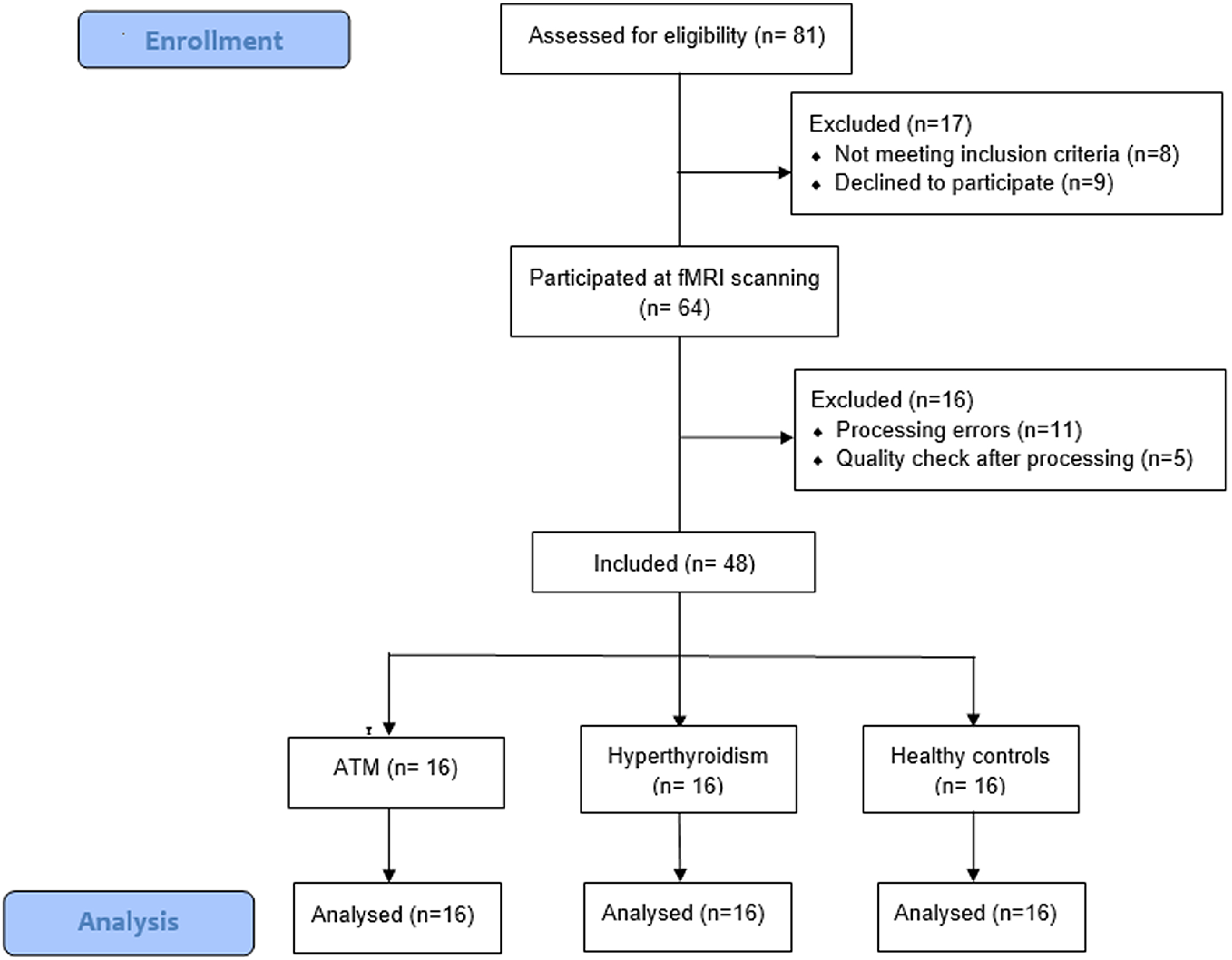
The flow chart of participants.
According to the 2016 edition of ‘Guidelines for Diagnosis and Management of Hyperthyroidism and Other Causes of Thyrotoxicosis’ compiled by the American Thyroid Association (14), all patients with ATM and hyperthyroidism should satisfy the diagnostic criteria for hyperthyroidism. All participants in this study were subject to the following exclusion criteria: (i) a history of cardiovascular or pulmonary conditions that could affect blood oxygenation level dependent (BOLD) fluctuations; (ii) abnormal thyroid function secondary to other serious physical diseases; (iii) serious dysfunction of liver, kidney, and other organs; (iv) neurological illness or psychiatric disorders; (v) pregnancy or lactation; (vi) acetylcholine receptor antibodies, creatine kinase; and (vii) contraindications to MRI.
The inclusion and exclusion criteria for the ATM group were as follows: (i) satisfied the diagnostic criteria for hyperthyroidism; (ii) the patient with hyperthyroidism exhibits at least one symptom of bulbar paralysis (dysphagia, dyspnea, or hoarseness); and (iii) the patient ruled out other illnesses that might induce bulbar paralysis, such as myasthenia gravis (excluded by neostigmine experiment), central disease (excluded by MRI), and pharyngeal disease (consultation of otolaryngologist and laryngoscopy).
Before starting antithyroid medication, the blood levels of free triiodothyronine (FT3), free thyroxine (FT4), suppressed thyroid-stimulating hormone (TSH), TSH receptor antibody (TRAb), thyroglobulin antibody (TGAb), and thyroid peroxidase antibody (TPOAb) of the patients were tested. An MRI scan was conducted the same day the blood values were gathered. The blood test results from patients with ATM were gathered throughout the course of their acute illness of bulbar palsy. The individuals in the healthy control group did not get any therapy and all completed the blood tests and thorough evaluations at the start of the trial.
The medical personnel responsible for blood collection were all working nurses in our hospital who have received professional training in specimen collection and mastered the correct collection methods and operation procedures. The blood collection tubes and anticoagulants and other reagents were regularly checked and updated to ensure their effectiveness. Blood specimens were stored and transported at room temperature after collection and sent for testing within 2 h. The testing of blood specimens was carried out by the same medical staff, who regularly checked and maintained the testing equipment to ensure its normal operation. The electrochemiluminescence used were automatic biochemistry (MAGLUMI 4000 PLUS; Shenzhen New Industry Biomedical Engineering Co., Ltd., Shenzhen, PR China)
The normal ranges for TPOAb levels were as follows: FT3 = 3.60–6.00 pmol/L; FT4 = 7.86–14.41 pmol/L; TSH = 0.34–5.65 mIU/L; TGAb = <30%; TPOAb = 0.00–30.00 IU/mL.
MRI data acquisition and preprocessing
The medical staff who performed the MRI scans were the same radiographers from our hospital who had received professional training in fMRI acquisition to ensure the reliability of the data. Once the data acquisition was completed, two specialized radiographers usually check the data individually and eliminate data of substandard quality. In case of inconsistency, a chief radiographer with more than 10 years of experience was invited to make a judgment.
Each patient underwent an fMRI scan using a 3.0-T MRI scanner (Achieva 3.0 T; Philips, the Netherlands). Axial registration of the fMRI images was accomplished by employing an echo planar imaging (EPI) sequence with the following parameters: 31 slices; slice thickness = 5 mm; no slice gap; TR/TE = 2000/30 ms; flip angle (FA) = 90°; field of view (FOV) = 220 × 220 mm; and matrix = 64 × 64. The duration of the fMRI scan for each participant was 374 s and 240 volumes were acquired. High-resolution structural images were acquired using a T1-weighted three-dimensional fast field echo sequence (T1W-3D FFE) with the following scan parameters: no slice gap; 156 slices; slice thickness = 1.0 mm; TR/TE = 20/3.5 ms; FA = 15°; FOV = 240 × 240 mm; and matrix = 512 × 512.
The SPM12 software program was used to preprocess the data on the MATLAB R2013b platform (https://www.fil.ion.ucl.ac.uk/spm/). The first 10 volumes were eliminated to avoid infection or confounding. According to A previous study (15), slice timing, realignment, and normalization were conducted using these samples. Instead of ReHo, smoothing was then performed using a 6-mm full-width at half-maximum (FWHM) Gaussian kernel for fALFF. All data were also filtered, detrended, and normalized to the Montreal Neurological Institute (MNI) template.
fALFF calculations (11) were carried out using the Resting-State fMRI Data Analysis Toolkit plus V1.22 (RESTplus V1.22). RESTplus software was applied to calculate ReHo values (12). FC calculations (13) were conducted with RESTplus software.
Statistical analysis
SPM12 software was devoted to analyze the fALFF, ReHo, and FC maps. After adjusting for covariates like age, sex, and duration of education (years), two-sample t-tests were performed on a voxel-by-voxel basis to characterize the substantial variation in fALFF, ReHo, and FC between the ATM, hyperthyroidism, and control groups (n = 16 in each group); the statistical significance should be set at cluster level P < 0.05/3. The signal values of fALFF, ReHo, and FC in the differential brain regions were first extracted by restplus (V1.22) software, and then Pearson's correlation analysis was carried out between the signal values and the clinical data based on SPSS software (IBM Corp., Armonk, NY, USA) (P < 0.05, two-tailed). For a further FC analysis, we used the peak locations of brain regions defined in the fALFF and ReHo results from patients with ATM as seed regions. The above-mentioned maps were all adjusted using RESTplus software's AlphaSim technique (P < 0.001) on a voxel-by-voxel basis. For the clinical data, normality tests were performed, using the Kolmogorov-Smirnov test. All statistical analyses were applied using SPSS version 23.0 (IBM Corp., Armonk, NY, USA).
Results
Demographic and clinical outcomes
A total of 19 patients with ATM were included, three of whom were excluded due to diagnostic inconsistency and poor image quality (ATM group, n = 16). In total, 16 patients with newly diagnosed hyperthyroidism without ATM were included (hyperthyroidism group, n = 16). In addition, 16 healthy controls (control group, n = 16) without any physical or mental illness who were matched by age, sex, and education were enlisted.
Age, sex, and educational attainment did not significantly differ among all groups (P > 0.05) (Table 1). The control group's TH levels were within the usual limits. (FT3 = 3.60–6.00 pmol/L, FT4 = 7.86–14.41 pmol/L, TSH = 0.34–5.65 mIU/L, TGAb = <30%, TRAb = 0.00–1.50 IU/L, TPOAb = 0.00–30.00 IU/mL). Compared to individuals with hyperthyroidism, the patients with ATM had considerably higher blood FT4 and TPOAb levels (P < 0.05) (Table 1). The 16 patients with ATM exhibited a statistically significant rise in thyroid-related hormones and antibodies compared to the healthy controls (P < 0.001) (Table 1). We did not collect TSH levels because these values exceeded the reference ranges in several patients. All brain MRI scans of the patients and the control group were normal and confirmed by T1W sequence.
Demographic and clinical data of the participants.

Values are given as n (%), mean ± SD), or median (range).
*One-way ANOVA (equal variance) or Kruskal–Wallis test (heterogeneity of variance).
Two independent sample t-test; comparison between patients with ATM and those with hyperthyroidism.
Independent sample t-test; comparison between patients with ATM and healthy controls.
Chi-square test.
ATM, acute thyrotoxic myopathy; BMI, body mass index; FT3, free triiodothyronine, FT4, free thyroxine; TGAb, thyroglobulin antibody; TRAb, thyrotropin receptor antibody; TPOAb, thyroid peroxidase antibody.
fALFF analysis
There were no significant differences in fALFF values between patients with ATM and those with hyperthyroidism (P > 0.05). As opposed to healthy controls, individuals with ATM had lower levels of fALFF in the right superior frontal gyrus (SFG) (P < 0.001, AlphaSim corrected) (Table 2 and Fig. 2).

Brain regions showing significantly different fALFF and ReHo values (P < 0.001, AlphaSim corrected). Broadman areas were also indicated. (a) Decreased fALFF in the right SFG between ATM patients and healthy controls. (b) Abnormal ReHo in the left precentral gyrus and orbital part of left IFG between ATM and hyperthyroidism patients. (c) Decreased ReHo in the right SFG and bilateral SMA between ATM patients and healthy controls. fALFF, fractional amplitude of low-frequency fluctuation; IFG, inferior frontal gyrus; ReHo, regional homogeneity; SFG, superior frontal gyrus; SMA, supplementary motor area.
Brain regions showing significantly different fALFF and ReHo between patients with ATM, HPs, and HCs (P < 0.001, AlphaSim corrected).

ATM, acute thyrotoxic myopathy; BA, Brodmann area; fALFF, fractional amplitude of low-frequency fluctuation; HC, healthy control; HP, hyperthyroid patient without ATM; MNI, Montreal Neurological Institute; ReHo, regional homogeneity.
ReHo analysis
In the patients with ATM, the ReHo values were greater in the left precentral gyrus but lower in the orbital region of the left inferior frontal gyrus (IFG) compared to the patients with hyperthyroidism (P < 0.001, AlphaSim corrected). In addition, compared to the healthy controls, the patients with ATM exhibited lower ReHo values in the right SFG and bilateral supplementary motor area (SMA) (P < 0.001, AlphaSim corrected) (Table 2 and Fig. 2).
FC results
There was substantially aberrant FC between seed areas and the overall brain based on the seed sites revealed by the abnormal fALFF and ReHo regions (P < 0.001, AlphaSim corrected) (Table 3 and Fig. 3). FC varied significantly between patients with ATM and hyperthyroidism. Based on the peak position (−27, −18, 60) of the left precentral gyrus, we discovered a decreased FC in the left SFG and left middle frontal gyrus (MFG) (P < 0.001, AlphaSim corrected) (Table 3 and Fig. 3). However, with the peak coordinate (−42, 27, −15) in the orbital part of the left IFG, there were no notable changes in FC results.

Brain regions showing significantly different FC values (P < 0.001, AlphaSim corrected). Broadman areas were also indicated. (a) Decreased FC in the left precentral gyrus, left SFG and left MFG with the seed region (−27, −18, 60). (b) Increased FC in the right SFG, right precentral and opercular part of right IFG with the seed region (18, 42, 39) based on the fALFF values. (c) Increased FC in the right SFG, right precentral and opercular part of right IFG with the seed region (18, 45, 39) based on the ReHo values. fALFF, fractional amplitude of low-frequency fluctuation; FC, functional connectivity; IFG, inferior frontal gyrus; MFG, middle frontal gyrus; ReHo, regional homogeneity; SFG, superior frontal gyrus.
Brain regions showing significantly different FC between patients with ATM, HPs, and HCs (P < 0.001, AlphaSim corrected).

The seed regions refer to the brain regions that are abnormal regions confirmed by fALFF and ReHo in the previous section. and the time-series correlation coefficients between this seed point and the whole brain are analyzed, i.e. the functional connectivity between the seed point and the whole brain; and the peak area in the table refers to the brain regions that are abnormally functionally connected to the seed point in the whole brain, and is followed by the MNI coordinates voxels and the BAs to describe the peak area.
ATM, acute thyrotoxic myopathy; BA, Brodmann area; fALFF, fractional amplitude of low-frequency fluctuation; HC, healthy control; HP, hyperthyroid patient without ATM; MNI, Montreal Neurological Institute; ReHo, regional homogeneity.
Obvious abnormal FC was also found in patients with ATM compared with healthy controls (P < 0.001, AlphaSim corrected) (Table 3 and Fig. 3). Using the peak location (18, 42, 39) in the right SFG as the seed coordinate, ATM groups exhibited stronger FC in right precentral gyrus and the orbital part of the right IFG (P < 0.001, AlphaSim corrected). It is noteworthy that the fALFF and ReHo consequences for these two groups (ATM and the healthy control) show the same brain regions with only slight changes in MNI coordinates. Therefore, when the peak point (18, 45, 39) of the right SFG was defined as seed points for FC calculations, the results were consistent with the other peak point (18, 42, 39).
Correlation among abnormal fALFF and reHo values, aberrant FC and clinical data
Comparing patients with ATM to healthy controls, the fALFF values in the right SFG showed a strong positive correlation with serum TRAb (P = 0.035, r = 0.529) (Fig. 4) and TPOAb levels (P = 0.034, r = 0.531) (Fig. 4). In addition, there was a significant association between the lowered ReHo in the right SFG and the serum FT4 (P = 0.03, r = 0.542) and TRAb levels (P = 0.042, r = 0.514) (Fig. 4).
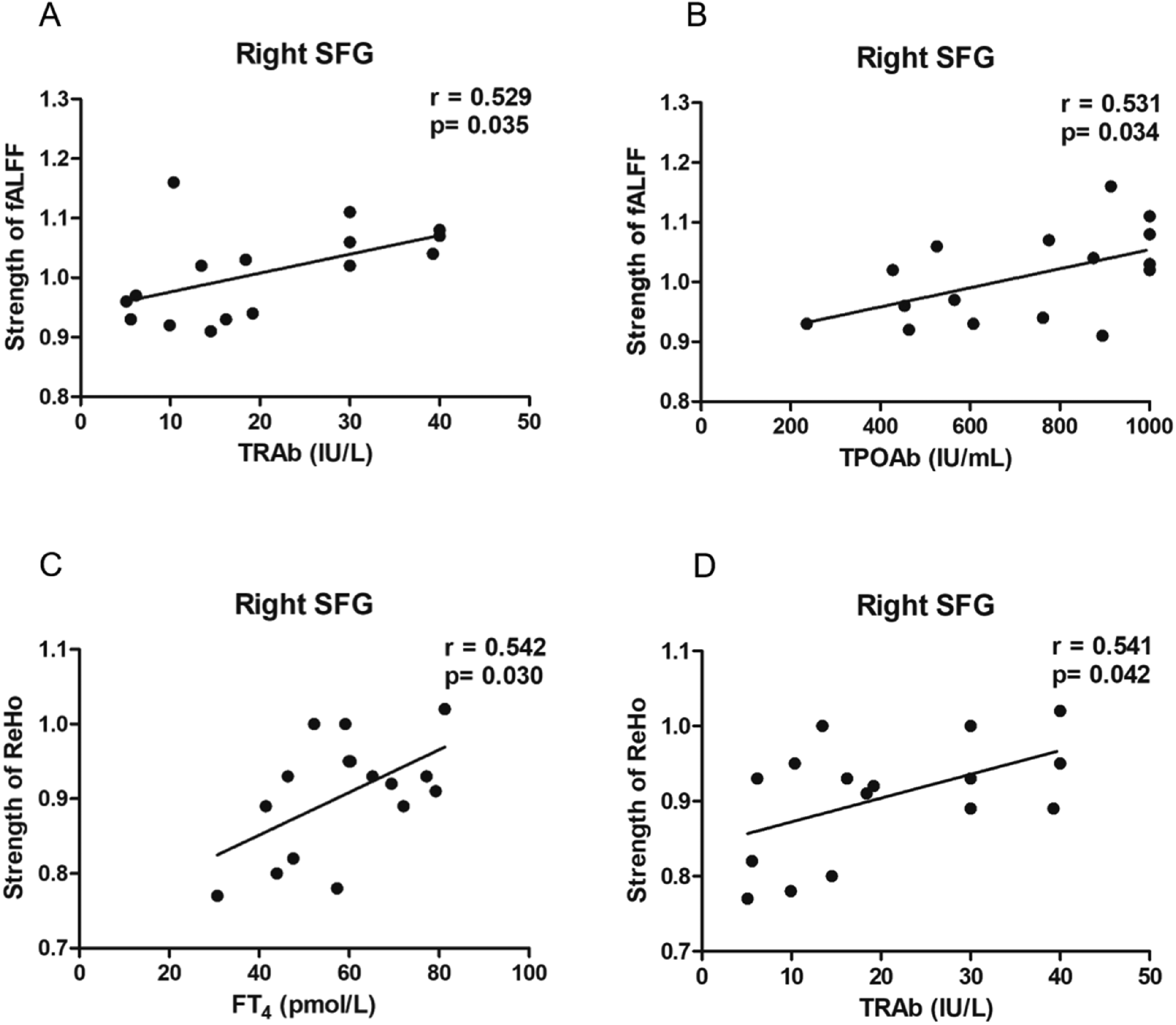
Scatter diagrams show the significant correlations between the clinical indexes and the fALFF, and ReHo values in the ATM group. (a, b) Decreased fALFF values in the ATM patients compared with the healthy controls in the right SFG were positively correlated with the serum TRAb (P = 0.035, r = 0.529) and TPOAb levels (P = 0.034, r = 0.531). (c, d) Decreased ReHo values in the ATM patients compared with the healthy controls in the right SFG were positively correlated with the serum FT4 (P = 0.03, r = 0.542) and TRAb levels (P = 0.042, r = 0.514). fALFF, fractional amplitude of low-frequency fluctuation; FC, functional connectivity; ReHo, regional homogeneity; SFG, superior frontal gyrus; TPOAb, thyroid peroxidase antibody; TRAb TSH receptor antibody.
Discussion
This study assessed variable functional brain activities by computing fALFF, ReHo, and FC values among the patients with hyperthyroidism and the healthy controls. Our results revealed that brain functional activities changed between those with and without ATM in several brain regions (especially in the SFG) and were possibly related to the medulla oblongata's innervation of the vital center's functions. The anomalous functional networks were distributed primarily in the brain’s swallowing and language networks. Furthermore, there was a correlation between biochemical indicators of disease severity and functional activity detected by rs-fMRI in some of the brain regions in patients with ATM.
Rs-fMRI computational methods generally study brain activity in terms of both functional segregation and functional integration. Functional segregation focuses on the characteristics of individual rs-fMRI signals, while functional integration emphasizes the interactions between different brain regions. The functional segregation includes ALFF, fALFF, ReHo, and DC, while the functional integration includes FC and ICA (16,17). The fALFF response is the mean intensity of the low-frequency portion of the BOLD signal for each voxel, with high sensitivity and specificity. fALFF measures the concordance between the temporal signals of a voxel and the surrounding voxels, and is a parameter calculated based on the Kendall coefficient of concordance (KCC). ReHo measures the temporal signal concordance between a voxel and the surrounding voxels, and is a parameter calculated based on the KCC. Seed-based FC calculates the BOLD time-series correlation coefficient between a given voxel and all other voxels or regions of interest in the brain to derive the functional connectivity between brain regions (16,17).
To date, there have been studies in the literature selecting fALFF, ReHo, and FC for use in hyperthyroidism-related disorders (7–9). ATM belongs to one of the complications of hyperthyroidism, which can also be used with the above metrics. In addition, a study described the cross-regional correlation of resting-state fALFF and ReHo with voxel-based hemodynamic and metabolic variables, suggesting that the hemodynamic characteristics of voxel-based BOLD signals in the resting state reflect underlying metabolic demands, and further uncover the immobile coupled metabolic information in fALFF and ReHo (18). The results of this study support the idea that fALFF and ReHo can be considered to be metabolic markers for MRI, and that ATM belongs to autoimmune metabolic diseases for which the selection of fALFF and ReHo is compatible. The seed points selected for FC analysis in this study were based on the brain regions obtained from the identification of fALFF and ReHo analyses, which is a more effective and reliable method than the traditional seed localization method based on a priori assumptions for selecting specific brain regions (7).
In the study by Li et al., the ICA method was used to analyze specific resting-state networks of the brain; the two main functional networks, SMN and LFPN, were analyzed, while other regions of the brain, other functional networks, were not analyzed (9). Different from the study by Li et al. (9), this study used fALFF values to analyze the frequency intensity of each voxel in the brain to find abnormal areas, using ReHo to explore the coherence of temporal signals between each voxel and surrounding voxels in the brain. The seed-based FC index was used to explore the time-series correlation coefficients between the seed points and the whole brain by taking the regions with abnormalities in fALFF and ReHo as seed reigons and exploring the brain regions with abnormalities in functional connectivity with the seed points. In this study, two major FMRI indicators, namely functional integration and FC, were combined to analyze the brain function of the whole brain of ATM patients, and the indicators used and the scope of the study were more comprehensive. This study found that the right SFG showed lower fALFF values in the ATM groups compared to the healthy controls. Previous studies reported that patients with thyrotoxic status exhibited attenuation of ALFF values in particular brain regions, which are located in the frontal lobe (7,19), demonstrating consistency with our research. fALFF activity could be used as one of the indicators to assess the severity of disease in aphasia (20). Brodmann areas (BA) 44 and 45, the essential components of Broca's area, which has been proposed as a crucial brain area for linguistic computations, was localized in the left triangular IFG (21). In this study, the reduction of ALFF in the left triangular IFG of ATM patients indicates that the spontaneous neural activity in this brain area is reduced, which leads to abnormal language function.
Reduced ReHo values were found in the left SMA of patients with ATM in contrast to the healthy controls in our study. In addition, the right medial SFG showed lower ReHo between the patients with ATM and those with hyperthyroidism. SMA was located in the medial part of BA 6, which corresponds to parts of the posterior subregion of the SFG (22). A previous study showed that one subregion of the SFG, which is crucial to language function, corresponds to BA 6, 8, 9, and 32 (23). BA 6 is mainly associated with somatic motor control, and most patients with ATM have symptoms of myopathy, including proximal muscle weakness, involuntary tremor of muscle fibers, and even generalized floppy paralysis in severe cases (24), presumably due to abnormal functional activities in this brain region. BA 8, 9, and 32 are mainly activated in cognitive-related tasks and are involved in the cognitive control processes of the body (25). Furthermore, an ER-fMRI study found that the four Brodmann areas mentioned above were involved in asymmetric language switching in second-language learners, especially BA 32, indicating that the SFG plays a role in monitoring and controlling the use of languages (26). The functional anomalies of the SFG would lead to muscle tremors, dysfunction of linguistic performance, and impaired cognitive function in ATM (22–26).
Moreover, our study found that the lower ReHo of the right SFG was significantly positively linked with serum FT4 and TRAb levels. The previous study by Liu et al. found that the thyrotropin receptor (TSHR) in microglial cells could react specifically to TSAb (one form of TRAb) stimulation, indicating the effect of TRAb on brain regional function (27). The ReHo values of the bilateral frontal lobe are significantly reduced in patients with hyperthyroidism, speculating that excessive thyroid hormones (THs) may cause abnormal brain regional activities (19). Microglia are found to be an important part of the cortical tissue that makes up the brain, and TRAb can cross the blood–brain barrier and bind to TSHR in brain tissue to promote further microglia polarization (28). Together, these findings implicate that SFG plays a crucial role of regional brain function in ATM, which may be regulated by the THs and thyroid-related antibodies together.
Anomalous ReHo levels were also discovered in the left precentral lobe and orbital region of the left IFG in patients with ATM in contrast to the hyperthyroid patients without ATM. Dysphagia is one of the most characteristic manifestations of medullary palsy in patients with ATM. In previous fMRI studies, precentral motor cortical regions in humans have been shown to become activated after swallowing water and saliva spontaneously (29), and stronger activation emerged in the left precentral cortex of all participants during verbal cue swallowing (30). These data indicated that the precentral gyrus may be involved in impairing the swallowing function of patients with ATM and may also partake in the process of dysfunctions to pronunciation. The orbitofrontal cortex (OFG), the smallest part of the frontal lobe in the brain, includes a host of distinct subregions (BA 10, 11, and 47) according to the Broadman division. A magnetoencephalographic study showed aberrant activation of the OFG in one participant with stuttering, and that unsuccessful speech onset was associated with weaker left BA 47 stimulation, in particular (31). Further, BA 10 and 11 might contribute to the processes of high-level integration, regulation, and execution as well as the detection of emotions and sensory input (32,33). According to these findings, we speculated that the abnormal functional variations in OFG, which were related to language execution and processing, may also lead to impaired neurocognitive function in the patients with ATM.
The left triangular IFG showed reduced regional FC with the left ITG and left MFG in ATM compared with healthy controls. However, we found that patients with ATM exhibited significantly stronger FC in the left SMA to the right medial SFG in contrast to patients with hyperthyroidism, but weaker FC occurred in the bilateral SFG to the left SMA compared with the healthy controls. There are anatomical white matter fiber connections between the IFG, MFG, and ITG, suggesting that these brain regions with fibrous structural connections exhibited functional connectivity damage and contribute to the abnormal brain functional network of patients with ATM (34–37). Chen et al. reported that when reading Chinese characters and pinyin, the function of certain brain regions, including the ITG, IFG, middle temporal gyrus (MTG), and parietal lobes can be activated, indicating that the ITG is involved in the processing of the language function (30). The MFG is mainly involved in the execution of language, working memory, and a variety of brain information, and the functional impairment of this brain area can lead to the disorder of language and execution (36). In summary, we believe that dyspraxia, unclear articulation and inattention in patients with ATM may be related to abnormal activities in the ITG and MFG. Furthermore, the SFG and SMA all localized in the frontal lobe, whose function was modulated by the dopaminergic neuron of fronto-striatal circuitry (38). A previous study reported that the THs played a crucial role in dopaminergic neuron development (39). Liu et al. (7) also showed that increased FT3 could lead to increased excitability in some brain regions and enhanced the FC between brain regions of the default mode network (DMN) and other brain networks, indicating that the gradually increased serum FT3 can partially antagonize the influence of abnormal brain functional activities in the pathogenesis and development of ATM. Literature with independent component analysis showed brain regions (precentral gyrus, SFG, cerebellum, and cingulate gyrus) with typical FC alterations in the SMN and LFPN of patients with ATM, which might lead to dysphagia and dysarthria in patients with ATM (9), demonstrating consistency with our research. We speculated that the afore-mentioned mechanism might account for the aberrant FC values between the SFG and SMA in our study. It was speculated that the symptoms of medullary paralysis, myopathy, and cognitive dysfunction in ATM patients may be the result of joint involvement in several different brain regions, of which SFG may be considered to play a leading role.
In our study, abnormal functional activities were mainly observed in the SFG, SMA, MFG, IFG, and precentral gyrus, and only the functional abnormalities of SFG were correlated with serum THs and thyroid-related antibodies. ATM is a rare complication of hyperthyroidism and the mechanisms remain unclear. Prior research has confirmed that the expression of the myosin heavy-chain gene in skeletal muscle is changed with thyroid disease states but reversed with restoration of normal serum concentrations of THs (24), which suggests that ATM may be a neuromyopathy. It was reasonable to then examine the abnormal activity in various brain regions to identify corresponding differences in clinical symptoms of ATM. A large proportion of the ATM patients who participated in our study exhibited TH disorder and excessive serum levels of TGAb and TPOAb. The two thyroid antibodies have been associated with psychiatric and motor disturbances (40). Our current research found that the ReHo values in the SMA were negatively correlated with the serum TGAb and TPOAb levels, implying the importance of these thyroid antibodies in ATM.
Our study discovered a favorable correlation between the serum TRAb and TPOAb values and the reduced fALFF in the right SFG. It is suggested that the higher the serum antibody levels, the more neuron activities in SFG were inhibited, implying the abnormal effects of thyrotoxicosis on regional brain function. Moreover, TRAb is an autoantibody of thyroid gland and indicates thyroid autoimmune. TPOAb, which is thought to occur owing to a secondary response to thyroid injury, forms part of a dysregulated autoimmune response in autoimmune thyroid diseases (41). According to the above mechanism, we consider that ATM may be related to autoimmunity, and TRAb and TPOAb could be the important detection indexes in this disease. However, it should be considered that there were no fALFF differences between patients with or without ATM. At present, the pathogenesis of ATM has not yet been elucidated, while excessive THs were reported to be the primary reason (42). Excessive THs could directly accelerate the oxidation process in mitochondria of muscle cells and abnormally regulate energy metabolism. Nerve corpuscles sustain more damage than other tissues when oxidative stress is out of balance. In addition, the body's tolerance to THs is reduced and its responsiveness to stimulation is altered, leading to abnormal muscle contraction and abnormal neuromuscular junctions, and may interfere with medullary function (43). The clinical data of all the participants showed that TH levels in ATM patients were highest among the three groups, but the discrepancy through TH levels between the ATMs and the hyperthyroid cohort was smaller than the difference between the ATMs and the healthy controls, which may not be sufficient to cause aberrant activity in specific brain areas in either group.
The present study has some limitations. First, the correlation analysis results should be carefully considered because the sample size is rather small due to the exceedingly rare incidence of ATM. In a follow-up study, we would enlist additional ATM patients to boost the sample size. Second, to date, there have been more case reports of ATM patients instead of a unified treatment process for this disease. Perhaps it is controversial to claim that the participants in our study matched the criteria based on standardized data. Clinical heterogeneity was not significant. Third, the correlation analysis between MRI alterations and patients’ clinical symptoms could not be completed due to the small sample size and the multiple symptoms of bulbar paralysis in patients. Third, a non-hyperthyroid control group should be provided to attribute observed differences specifically to ATM. Fourth, the relationship between serum thyroid antibodies and regional brain activity should be further analyzed. Finally, the absence of a detailed characterization of neuropsychological dysfunction due to abnormal brain activity weakens the comprehensiveness of our findings. This type of characterization needs to be evaluated in the future.
In conclusion, abnormalities in regional brain function can be demonstrated in ATM by calculating the fALFF, ReHo, and FC values with the rs-fMRI technique, especially in the SFG area. Our findings provide light on the neurological processes behind brain dysfunction caused by ATM, and the fALFF, ReHo, and FC values in rs-fMRI could be potential markers for better understanding of the underlying pathophysiology of ATM. However, the potential of the rs-fMRI indicators should be validated by a large sample size with better grouping (adding the non-hyperthyroid control group), deeper analysis on the relationship between serum thyroid antibodies and regional brain activity.
Footnotes
Data availability
All data analyzed during this study are available from corresponding author upon reasonable request.
Declaration of conflicting interests
The authors declared no potential conflicts of interest with respect to the research, authorship, and/or publication of this article.
Funding
The authors disclosed receipt of the following financial support for the research, authorship, and/or publication of this article: This study was supported by the National Natural Science Foundation of China (grant nos. 81860146 and 82260159).
