



Abstract
Background
68Ga-labled fibroblast activating protein inhibitor (68Ga-FAPI) represents a new and exciting positron emission tomography-computed tomography/magnetic resonance (PET-CT/MR) radiotracer.
Purpose
To compare the diagnostic efficacy of 68Ga-FAPI PET CT/MR and 18F-fluorodeoxyglucose (18F-FDG) PET/CT in metastatic lesions of gynecological cancers (GCs).
Material and Methods
The PubMed, Embase, and Web of Science databases were thoroughly investigated from inception until 22 December 2023. A head-to-head contrast between 18F-FDG PET/CT as well as 68Ga-FAPI PET CT/MR for the assessment of GCs was presented by the included studies. A random variable model was employed to examine the sensitivity in detection of lymph node (LN) and peritoneal metastases (PM).
Results
The pooled sensitivity for 68Ga-FAPI PET CT/MR and 18F-FDG PET/CT in lymph node metastases (LNM) of GC were 0.98 (95% confidence interval [CI] = 0.86–1) and 0.85 (95% CI = 0.65–0.98), respectively, while the results about peritoneal metastases in ovarian cancer were 0.98 (95% CI = 0.93–1) and 0.71 (95% CI = 0.55–0.86). Compared with 18F-FDG PET/CT, 68Ga-FAPI PET CT/MR exhibited a better sensitivity in peritoneal involvement of ovarian cancer with a relative risk of 0.24 (95% CI = 0.09–0.40) and P = 0.002.
Conclusion
68Ga-FAPI PET CT/MR displayed a superior sensitivity over 18F-FDG PET/CT in detecting metastatic lesions of ovarian cancer. However, there was insufficient evidence to favor the superiority of 68Ga-FAPI PET CT/MR in LNM of CC. Further studies are needed for evaluating primary and metastatic lesions of 68Ga-FAPI PET CT/MR in different GC.
Introduction
Gynecological cancer (GC) encompasses a range of cancers originating in the female reproductive system. The most prevalent types are cervical cancer (CC), uterine corpus cancer, and ovarian cancer (OC), which represent 3.1%, 2.2%, and 1.6% of new cases, respectively, and 3.4%, 1.0%, and 2.1% of new deaths of all cancer sites globally in 2020. Notably, cervical cancer has the second-highest mortality rate behind breast cancer, which continues to be the main trigger of cancer-related fatalities for women (1). The outlook for individuals with metastatic cancers remains impoverished, underscoring the need for a reliable staging tool for effective treatment planning (2,3).
18F-FDG positron emission tomography/computed tomography (PET/CT) is pivotal in diagnosing, tracking, as well as following up on GC. It may be an effective method for evaluating a possible extraperitoneal or extra-pelvic metastases and suspicion of recurrence and is considered to be a valuable diagnostic imaging method (4–6). However, it has limitations, including false-positive uptake of 18F-FDG in endometriosis cysts, uterine fibroids, and the uterine endometrium during menstrual, ovulatory, and premenopausal periods, as well as in lesions with intense inflammation (7). Moreover, false-negative results have been observed in urinary tract tumors, scirrhous stomach cancer, and hepatocellular cancer (8). The fibroblast activation protein inhibitor (FAPI) variants have emerged as promising tracers for future PET imaging applications (9–11). 68Ga-FAPI PET/CT has displayed high diagnostic efficiency and detection rates in various tumors, particularly gastric cancers, lung cancers, and other malignant tumors, and has proven effective even in cases challenging for 18F-FDG PET/CT (12–15).
68Ga-FAPI PET-CT/magnetic resonance (MR) might surpass 18F-FDG PET/CT in detecting gynecological tumors (16). However, a comprehensive comparison of these two tests in assessing metastatic gynecological malignancies has still not been conducted. The goal of the research is to determine and compare the efficacy of 18F-FDG PET and 68Ga-FAPI PET for individuals with gynecological tumors.
Material and Methods
The Preferred Reporting Items for Systematic Reviews and Meta-Analyses (PRISMA) guidelines were followed within this meta-analysis (17).
Literature search
Electronic literature databases, including PubMed, Embase, and Web of Science, were looked over on 22 December 2023. The search algorithm incorporated terms such as: (i) “women's” OR “gynecological” OR “cervical” OR “uterine” OR “uterus” OR “endometrial” OR “ovarian” OR “ovary” OR “tubal” AND (ii) “cancer” OR “neoplasm” OR “tumor” OR “tumour” AND (iii) “FAPI-04” OR “68Ga-FAPI-04” OR “FAPI” AND (iv) “fluorodeoxyglucose” OR “FDG” AND (v) “Positron Emission Tomography” OR “PET” OR “PET/CT” OR “PET/MRI.” The search did not impose any starting date or language restrictions.
Inclusion and exclusion criteria
The studies that made up this evaluation satisfied these requirements: (i) the primary focus was a head-to-head contrast of the diagnostic accuracy of 18F-FDG PET/CT and 68Ga-FAPI PET CT/MR for individuals with GC; (ii) the reference standard was based on histopathological outcomes as well as combination of histopathological and follow-up outcomes; and (iii) enough information to reevaluate sensitivity.
The exclusion criteria were as follows: (i) articles no relevant to our field of research; (ii) case reports, conference, meeting abstract, and review articles; (iii) non-comparative trials; and (iv) articles not in English.
Two researchers examined each manuscript to determine if they qualify for inclusion. The examination was needed to repeat twice.
Extracting data and evaluating quality
The first author's name, publication date, country, research structure, individual's features, along with technical information were gathered as basic information. We utilized the Quality Assessment of Diagnostic Accuracy Studies 2 (QUADAS-2) tool to assess the studies’ quality (18). All the publications were independently evaluated by two researchers, and any disagreements were settled in a consensus meeting.
Statistical analysis
A random variable model was employed to evaluate the two imaging modalities’ sensitivity. Pooled data and risk variance, along with 95% confidence intervals (CIs), were derived at a lesion-based level. Forest plots were used to visually represent the statistical pooling of data. I2 index (I2) statistics were employed to gauge between-study statistical heterogeneity. Funnel plots were utilized to test the publication bias. An additional sub-analysis compared the sensitivity of the two examinations for ovarian cancer and cervical cancer. The Stata program version 15.1 and Review Manager 5.4 were used to conduct statistical evaluations. If a value's two-sided P value was <0.05, it was deemed statistically significant.
Results
Literature search
The flow diagram of eligible studies is displayed in Fig. 1. A total of 147 papers were found using our search approach; of these, 28 were via PubMed, 83 were via Embase, and 36 were via Web of Science. Of these, 136 studies that did not fit the inclusion criteria were eliminated. After a thorough reading of the remain 11 full-text publications, eight trials were eventually included in this meta-analysis (19–26).
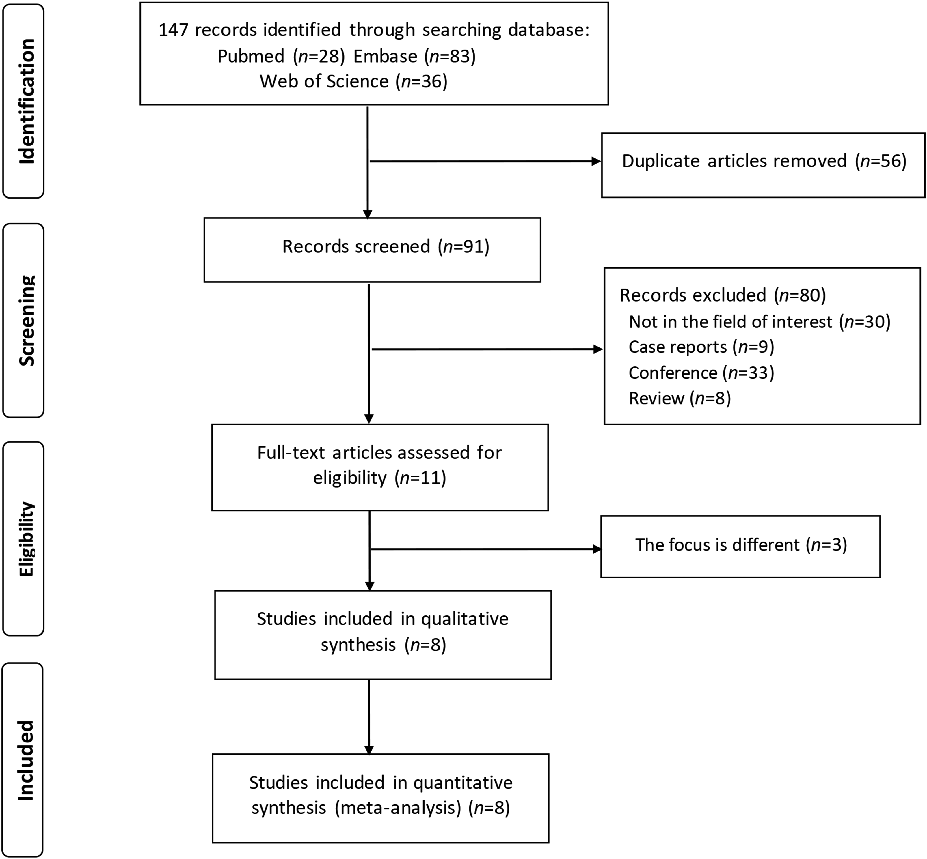
Flow chart of eligible literature.
Characteristics of the included research
Tables 1 and 2 lists the patient and study characteristics as well as specific technical details. All eight studies were analyzed based on a lesion level. Among the eight studies, five focused on ovarian cancers, while the remaining three consisted of cervical cancer patients. Of the eight included studies, six had a prospective design and two were retrospective. There were two studies utilizing 68Ga-FAPI PET/MR as the imaging modality and one study performing PET/CT using 68Ga-FAPI-46 as the radiotracer.
Characteristics of included studies.
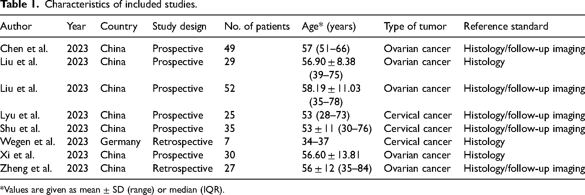
*Values are given as mean ± SD (range) or median (IQR).
Technical features.

*Unless otherwise indicated.
SUVmax, maximum standardized uptake value; TBR, tumor-to-background ratio; TLR, tumor-to-liver ratio.
Quality assessment
The findings of the methodological quality analysis are collected in Fig. 2, which included the risk of bias (patient selection, index test, reference standard, and flow timing) and applicability (patient selection, index test, and reference standard).

Risk of bias based on QUADAS-2 tool.
Sensitivity of 68Ga-FAPI PET and 18F-FDG PET in metastatic lesions
The pooled sensitivity of 68Ga-FAPI PET CT/MR and 18F-FDG PET/CT for lymph node metastases (LNM) of GC were 0.98 (95% confidence interval [CI] = 0.86–1) as well as 0.85 (95% CI = 0.65–0.98), with I2 values of 74.2% and 80.7%, respectively (Fig. 3).
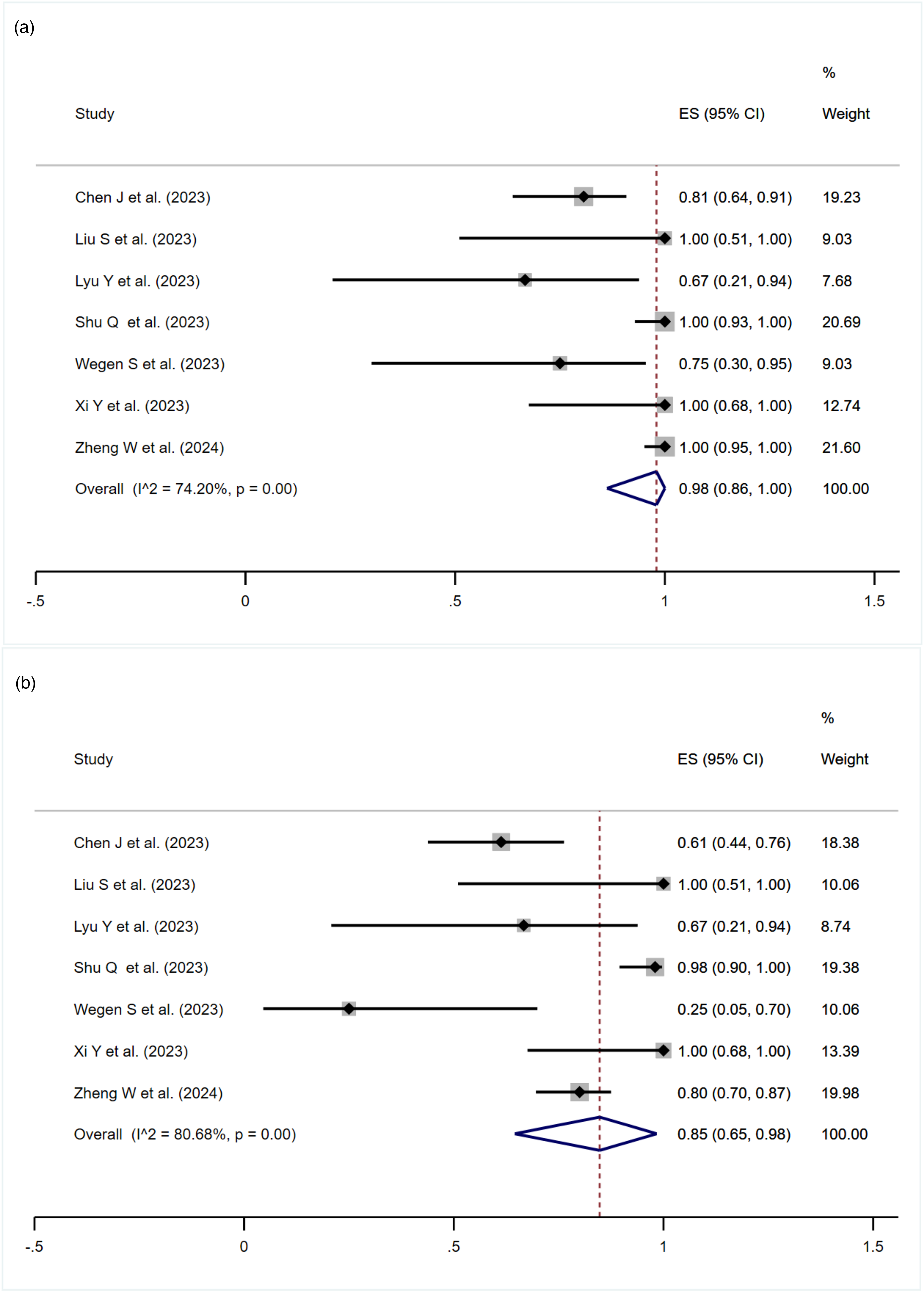
Forest map of the pooled sensitivity of (a) 68Ga-FAPI PET CT/MR and (b) 18F-FDG PET/CT in lymph node metastases of gynecological malignances.
Five studies focusing on OC provided data on peritoneal carcinomatosis (PC). The forest plot analysis (Fig. 4) revealed that the sensitivity of 68Ga-FAPI PET CT/MR and 18F-FDG PET/CT for diagnosing peritoneal carcinomatosis were 0.98 (95% CI = 0.93–1) as well as 0.71 (95% CI = 0.55–0.86), with I2 values of 12.2% and 67.9%, respectively.
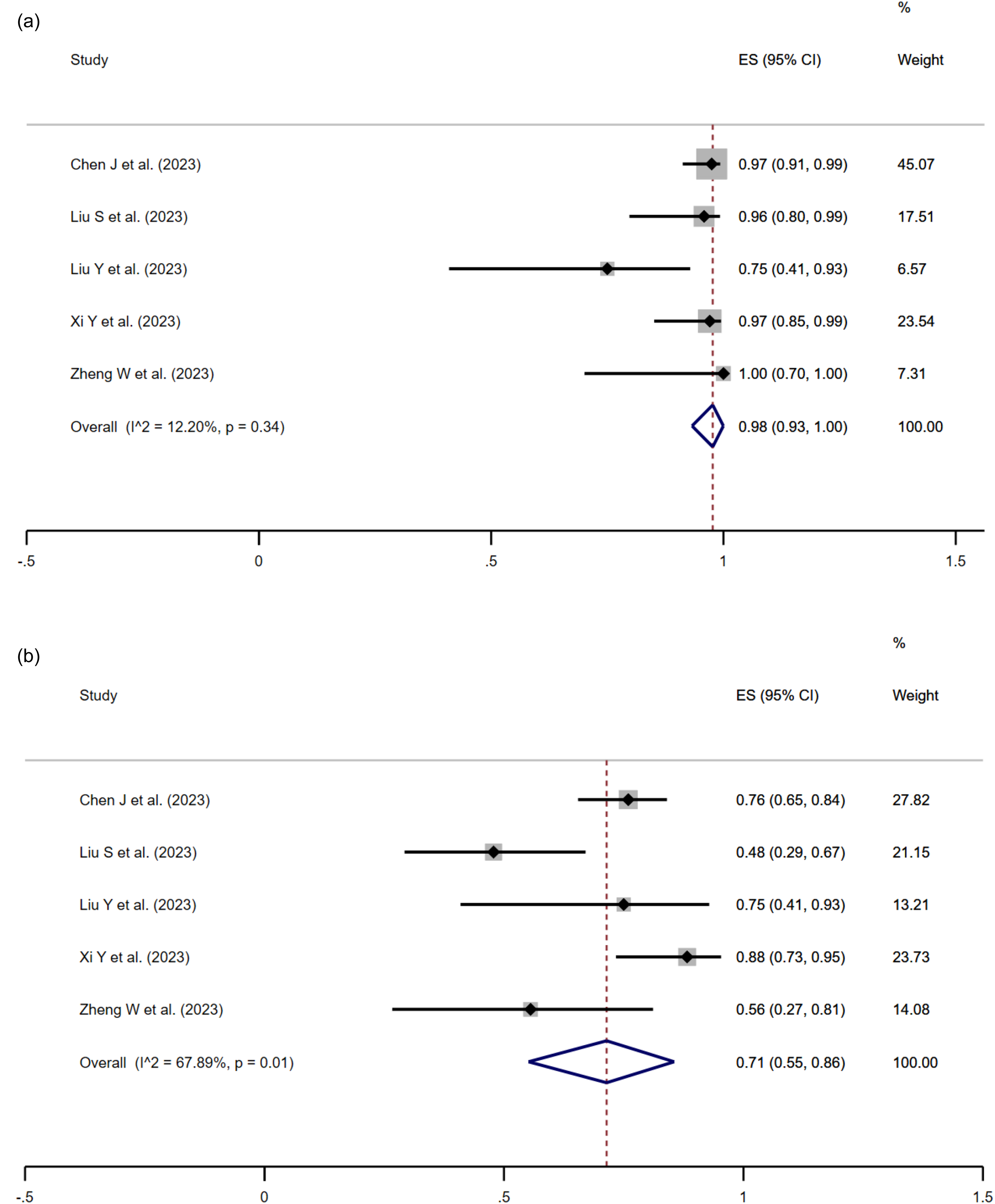
Forest map of sensitivity in assessment peritoneal carcinomatosis of (a) 68Ga-FAPI PET CT/MR and (b) 18F-FDG PET/CT.
Sensitivity of 68Ga-FAPI CT/MR imaging versus 18F-FDG PET/CT
The risk difference for LNM of GCs using 68Ga-FAPI PET CT/MR versus 18F-FDG PET/CT was in the range of 0–0.50, giving a pooled statistic of 0.11 (95% CI = −0.03–0.25) (Fig. 5). The Z value was 1.52 with a P value of 0.13. Subgroup analysis of LNM by different type of GC revealed the risk difference of 0.15 (95% CI = 0.04–0.25) for ovarian tumor with Z value of 2.78 as well as P value of 0.005, respectively. The risk difference was 0.12 (95% CI = −0.22–0.46) for cervical cancer with Z value of 0.69 as well as a P value of 0.49.

Comparison forest map of sensitivity in lymph node metastases of 68Ga-FAPI PET CT/MR and 18F-FDG PET/CT.
The risk difference for peritoneal carcinomatosis of GCs using 68Ga-FAPI PET versus 18F-FDG PET was in the range of 0–0.48, giving a pooled statistic of 0.24 (95% CI = 0.09–0.40) (Fig. 6). The Z value was 3.08 with a P value of 0.002.
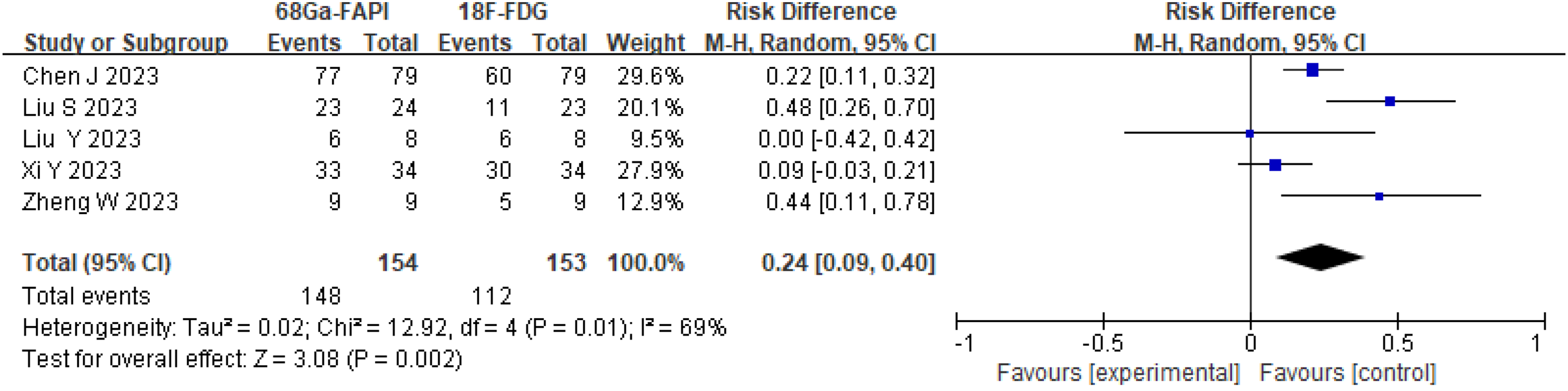
Comparison forest map of sensitivity in assessment peritoneal carcinomatosis of 68Ga-FAPI PET CT/MR and 18F-FDG PET/CT.
Publication bias
The funnel plots and egger tests regarding 68Ga-FAPI PET CT/MR versus 18F-FDG PET/CT in LNM and peritoneal carcinomatosis were shown in Fig. 7. The P values of the egger test were 0.61 and 0.54, correspondingly, indicating no publication bias. In addition, funnel plots did not display obvious asymmetry.

(a, b) Egger tests and (c, d) funnel plots of lymph node metastases and peritoneal involvement.
Discussion
Accurate diagnosis, staging and restaging in GC, are crucial for patient clinical management. In addition, it may improve survival rates. For patients with cervical cancer, lymphatic spreads are closely related to prognosis. In other words, nodal staging plays a decisive role in management selection. Once pelvic lymphatic metastasis was demonstrated by 18F-FDG PET/CT or CT/MR, patients would be recommended to receive concurrent chemoradiotherapy (CCRT), which has a deep influence on fertility (27). Apart from LNM, peritoneal involvement is another major metastatic and recurrent mechanism in OC. Thus, detecting metastatic lymph nodes and peritoneal lesions was essential for staging and restaging. 18F-FDG PET/CT has been recommended as a radiological biomarker and routine examination for pre- and postoperative assessment (28). However, there were several diagnostic pitfalls of 18F-FDG PET/CT hampering its application, such as physiological 18F-FDG uptake in intestines, difficult identification in inflammation and malignancies as well as false-negative 18F-FDG uptake in tiny, mucinous, and necrotic lesions (29). Recently, 68Ga-FAPI PET CT/MR has been gradually utilized to evaluate various tumors and non-tumor lesions owing to its advantages of increased tracer uptake in metastatic lesions and decreased background activity (16,30–34).
To our knowledge, this is the first meta-analysis comparing the diagnostic efficacy of 68Ga-FAPI PET CT/MR and 18F-FDG PET/CT for metastatic lesions of GCs. It included information for 254 participants across eight independent comparative studies who underwent both tests. The meta-analysis revealed 68Ga-FAPI PET CT/MR is usually superior to 18F-FDG PET/CT in terms of diagnostic efficacy for LNM and peritoneal carcinomatosis of OC, with higher pooled sensitivity (0.88 vs. 0.69 and 0.97 vs. 0.74). However, the detection rate of 68Ga-FAPI PET CT/MR and 18F-FDG PET/CT were comparable in LNM of cervical cancer.
In terms of the diagnostic efficiency of an LNM diagnosis of OC, 68Ga-FAPI-04 PET outperformed 18F-FDG PET/CT, with an extremely short axis size of less than 1 cm (19). The superior detection rate of 68Ga-FAPI PET might be attributed to the higher tracer uptake in OCs as well as lower tumor-to-background ratio (TBR) (35,36). However, tiny LNs, with an axis size smaller than 0.4 cm, were missed easily in both modalities. This might be because of the limited FAP expression in tiny LNs and confusable distinguishment from peritoneal involvement (37). Of note, lymphatic inflammation might also lead to false-positive LNs on 68Ga-FAPI-04 PET. Regarding LNM of cervical cancer, the sensitivity of the two examinations was not satisfactory. However, only three studies reported related data. The results should be interpreted with caution, and further research is needed to investigate the diagnostic efficiency of 68Ga-FAPI-04 PET and 18F-FDG PET/CT.
Peritoneal implant is a main metastatic mode of OC and considered to be a terminal stage of the disease (38,39). Kim et al. revealed that 18F-FDG PET/CT exhibited an excellent diagnostic efficiency in detection of PC in different cancers with a pooled sensitivity of 0.87 and specificity of 0.92 (40). Tsili et al. reported that compared to MRI and MDCT, FDG PET/CT has a higher combined sensitivity (93.7% vs. 82.7% vs. 79.7%), and the diagnostic accuracy of FDG PET/CT and MRI were comparable in detection of PCs in OC (41). Those findings seemed to indicate a high detection rate of PC for 18F-FDG PET/CT. Nevertheless, a recent meta-analysis showed a contrasting view. The diagnosis of FDG PET/CT in PC was poor with sensitivity of 59.9%, while 68Ga-FAPI PET/CT exhibited excellent sensitivity (98.2%) (42). This finding was in accordance with our research (sensitivity of 0.98 vs. 0.71). 68Ga-FAPI PET CT/MR showed an obviously higher tracer uptake ratio than FDG PET/CT in detecting PCs. Xi et al. (25) speculated this may depend on high density NAFs in the mesentery and omentum, which can cause increased FAP expression indirectly when presenting metastasis. Moreover, 68Ga-FAPI PET CT/MR always has advantages of higher volumetric parameters, and lower background activity (43). In addition, surgery-induced fibrous tissue hyperplasia should not be overlooked, which was the major reason for false-positive findings. The drawback of both modalities was the influence of abdomen movement and small lesions.
The present study has some limitations. First, there are insufficient data to evaluate the specificity for ovarian and cervical cancer, respectively, since limited articles reported the true-negative findings. Moreover, the diagnostic value of 68Ga-FAPI PET CT/MR and FDG PET/CT for LNMs of cervical cancer needs more exploration. Second, not all the studies focused on newly diagnosed cancer; two focused on tumor recurrence and metastasis (20,21). Finally, imaging examinations and radiotracers are not unified. One study performed PET/CT using 68Ga-FAPI-46 and two trials utilized PET/MR instead of PET/CT to detect primary and metastatic lesions.
In conclusion, when evaluating metastatic GC, 68Ga-FAPI PET MR/CT displayed superior sensitivity to 18F-FDG PET/CT in detecting LNM and PC of ovarian cancer. There was no significant difference between the two tests in evaluating the LNM of cervical cancer. Nevertheless, as the findings are based on limited sample sizes, further investigation is needed to confirm the conclusion and explore the diagnostic ability of 68Ga-FAPI PET MRI/CT for different types and stages of GC.
Footnotes
Declaration of conflicting interests
The authors declared no potential conflicts of interest with respect to the research, authorship, and/or publication of this article.
Funding
The authors received no financial support for the research, authorship, and/or publication of this article.
