



Abstract
Background
The Bosniak classification is designed to standardize evaluation of cystic renal masses and to communicate the risk of malignancy.
Purpose
To determine whether radiologists vary in their communication of Bosniak class III and IV cystic renal masses.
Material and Methods
This retrospective study included 186 patients with CT or MRI reporting a Bosniak class III or IV mass. Radiology reports were evaluated to determine the noun representing the mass, the modifier to convey the likelihood of cancer, and recommendations for urologic referral. Electronic medical records were reviewed to determine if the patient saw a urologist.
Results
Of the patients, 112 (60%) had a class III mass and 74 (40%) had a class IV mass. Class III masses were more likely to be represented by the noun “lesion” rather than a “mass” (61/112 [54%] vs. 31/112 [28%]). Class IV masses are more likely to be represented as a “mass” (36/74 [59%] vs. 28/74 [38%]; P < 0.015). Cancer was described in 100/186 (54%) cases: 38/112 (35%) class III masses and 62/74 (72%) class IV masses (P < 0.001). The cancer terminology used included “renal cell carcinoma” (n = 57), “neoplasm” (n = 12), and “malignancy/malignant” (n = 86). Most radiology reports (n = 133, 72%) did not recommend urologic referral but 183 (98%) patients were referred and 181 (97%) ultimately saw a urologist.
Conclusion
Radiologists vary in their communication of class III and IV masses, reflecting historical terminology usage, nuanced interpretations, and an evolving understanding of renal cell carcinoma biology. This variance had minimal impact on urologic referral rates.
Introduction
Cystic renal masses are extremely common and found in over one-quarter of the population. The prevalence increases with age; more than half of men aged over 70 years have cystic renal masses (1). Cystic renal masses range from completely benign simple cysts of no clinical consequence to renal cell carcinoma with variable aggressiveness, with the latter requiring urologic management (2).
Radiologists and urologists use the Bosniak classification system to risk stratify the likelihood of cancer in a cystic renal mass. Within the Bosniak classification system, class I and II masses are benign and do not require any further follow-up. Class IIF masses have a low but non-zero likelihood of malignancy and are thus followed by imaging. Class III and IV masses have approximately 50% and >90 likelihoods of malignancy, respectively (2,3). Given their higher likelihoods of malignancy, these two latter classes are included in the most recent American Urological Association (AUA) guidelines on evaluation, management, and follow-up of localized renal masses, which may include active surveillance or nephron-sparing surgery (4,5).
The Bosniak classification is designed to standardize the evaluation of cystic renal masses using computed tomography (CT) or magnetic resonance imaging (MRI) and to communicate the risk of malignancy to the ordering provider in a consistent manner (2,6). Despite standardization, radiologists may vary in how they communicate the risk of malignancy. The aim of the present study was to determine whether radiologists vary in their communication of Bosniak class III and IV cystic renal masses and if this affects urologic referral rates.
Material and Methods
Study population
This single-center study was approved by the institutional review board. Due to the retrospective nature of the study, the requirement for informed consent was waived. We searched our informatics database for patients with a mention of “Bosniak” paired with “3,” “4,” “III,” and “IV” in a CT or MRI report between January 2017 and December 2022. The original search generated 231 patients. Baseline CT and MRI reports were then individually evaluated to confirm the presence of a Bosniak class III or IV cystic renal mass described in the findings and impression. This excluded 45 patients who had a Bosniak class III or IV mentioned in the study indication by the ordering provider but actually had a different diagnoses, e.g. Bosniak IIF or lower, solid renal mass, or a resected Bosniak class III or IV cystic renal mass (without baseline imaging). Among the patients with multiple qualifying renal masses, the mass with the higher class was recorded.
Image report analysis
As above, the impression section from baseline CT or MRI was evaluated and recorded. The impressions were aggregated and analyzed using a word cloud. In addition, the impressions were evaluated to determine the noun to represent the mass (e.g. lesion, mass, cyst, cystic renal mass), if cancer or other terminology was explicitly used (e.g. cancer, neoplasm, renal cell carcinoma, malignancy), and the modifier to convey the likelihood of cancer (e.g. highly suspicious for, highly concerning for, consistent with, not excluded, until proven otherwise). These modifiers were then grouped by approximate certainty of cancer based on prior studies (7–12). Finally, the impressions were reviewed to determine if urologic referral was recommended, and the electronic medical record was reviewed to determine if the patient saw a urologist.
Statistical analyses
Categorical variables are expressed as their integer value with percentages and compared using Fisher’s exact test. A statistical threshold of P < 0.05 was used to determine significance. Statistical analyses were performed using Prism version 10.2.1 (GraphPad, Boston, MA, USA).
Results
A total of 186 patients (116 men [62%], 70 women [38%]; median age = 67 years) were included. There were 112 (60%) patients with class III masses and 74 (40%) patients with class IV masses. The masses were assessed at baseline with CT in 99 (53%) patients and MRI in 87 (47%) patients. Most (n = 162, 87%) patients were outpatients, while the remaining patients obtained imaging while hospitalized. Baseline characteristics are summarized in Table 1.
Baseline characteristics of the patient cohort.

Values are given as n (%) or median (range). The sum of the percentages may not add up to 100% due to rounding.
CT, computed tomography; MRI, magnetic resonance imaging.
Table 2 summarizes the basic terminology used to describe the cystic renal mass and urologic outcome. Cancer was described in 100/186 (54%) of radiology report impressions: 38/112 (35%) of class III masses and 62/74 (72%) of class IV masses (P < 0.001). The terminology used to describe cancer included “renal cell carcinoma” (n = 57 across all masses), “neoplasm” (n = 12), and “malignancy” or “malignant” (n = 86). No report used the term “cancer.”
Basic terminology used to describe the cystic renal mass and urologic outcome.

Values are given as n (%). The sum of the percentages may not add up to 100% due to rounding.
*Comparison of whether cancer was mentioned in the report.
Comparison of “lesion” vs. “mass” as the noun to represent the cystic renal mass.
The nouns used to represent the class III and IV masses included “lesion” (n = 89/186; 48%), “mass” (n = 67; 36%), “cyst” (n = 25; 13%), “structure” (n = 1; 1%), and “neoplasm” (n = 4; 2%). Class III masses were more likely to be represented as a “lesion” rather than a “mass” (61/112 [54%] vs. 31/112 [28%]) whereas class IV masses are more likely to be represented as a “mass” rather than a “lesion” (36/74 [59%] vs. 28/74 [38%]; P < 0.015).
Most radiology reports (n = 133, 72%) did not have a formal recommendation for urology referral. Nevertheless, 183 (98%) patients had a urology referral to address the Bosniak cystic renal mass and 3 (2%) patients did not. Of the patients, 181 (97%) ultimately saw a urologist. All five patients who did not see a urologist had a class III mass (P = 0.278) and all were undergoing systemic therapy for a widely metastatic cancer of a non-renal primary.
Table 3 summarizes the modifier used by the radiologist and grouped by approximate diagnostic certainty based on prior publications. Class III masses were most commonly described by phrases typically associated with a likelihood of approximately 50% or 75%, such as “intermediate” probability and “concerning for,” respectively. Class IV masses were more likely to be described using terms associated with a likelihood of approximately 75% or 90%, such as “concerning for” and “large majority is malignant.” Fig. 1 represents word clouds generated from aggregate reports for class III and IV masses.
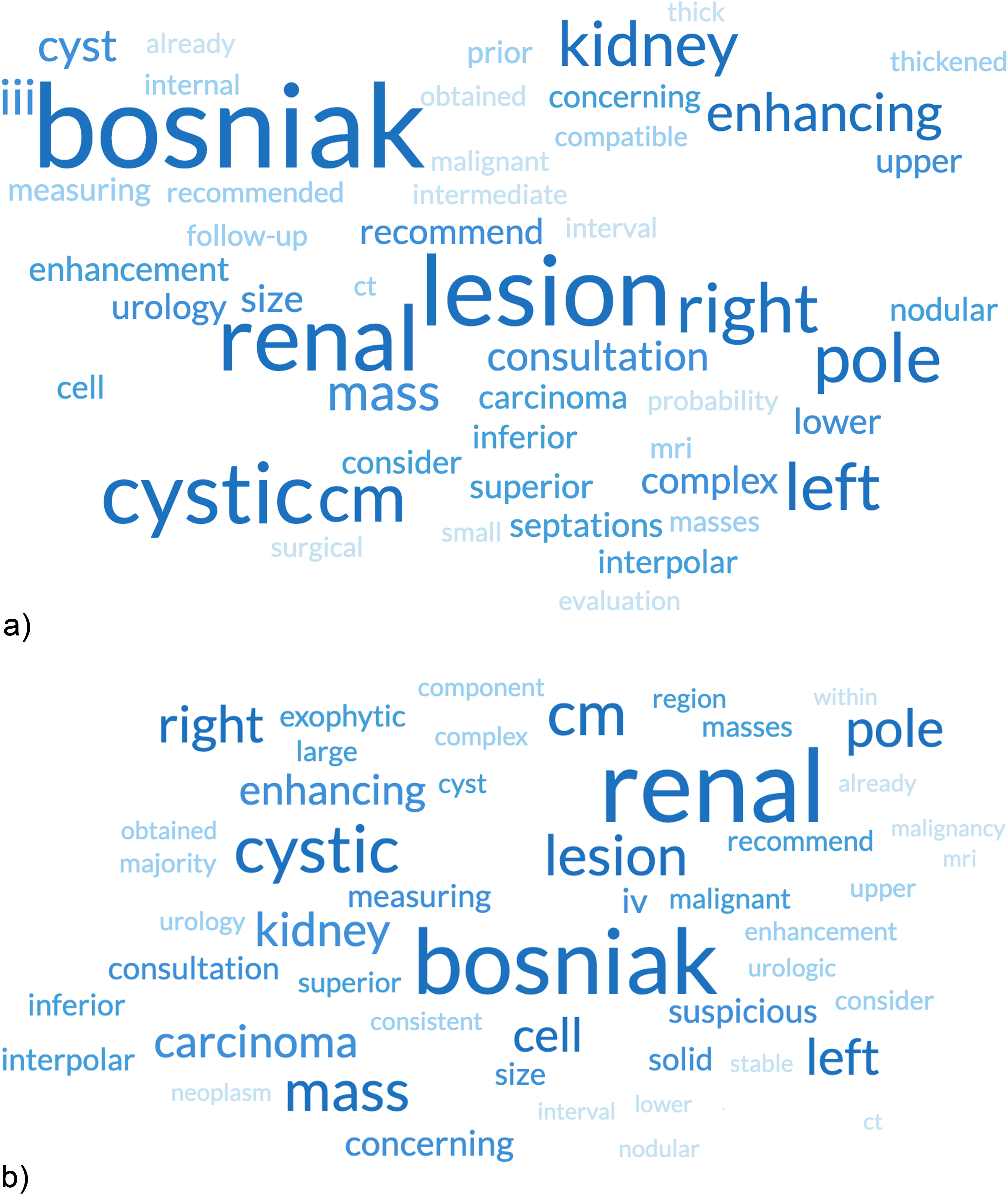
Word cloud of the 50 most common terms used in the radiology report impression for (a) class III masses and (b) class IV masses.
Modifiers used by radiologists and grouped by approximate diagnostic certainty based on prior publications.

Values are given as n or n (%). The sum of the percentages may not add up to 100% due to rounding.
Discussion
The Bosniak classification system and its related lexicon standardize the evaluation and reporting of cystic renal masses. In our study, we found that radiologists varied in their communication of class III and IV masses. Specifically, radiologists were more likely to refer to a class III mass as “lesion” rather than a “mass,” and a cancer synonymous term was not explicitly stated in most class III masses. The opposite was observed when describing a class IV mass. Nevertheless, this did not ultimately affect whether a patient saw a urologist.
There are several potential reasons why the term “lesion” was preferred to “mass” for class III masses. First, the formal lexicon to define any space-occupying abnormality as a “mass” was not published until 2021 (6). Even then, most radiologists continued to use the term “lesion” as this terminology is extensively used in both radiology and urology literature, including the most recently updated 2021 AUA guidelines (4,5,13–15). Second, “mass” in the context of other organ systems has a stronger connotation of malignancy. For example, a solid lung mass by definition measures least 30 mm and would be highly concerning for malignancy (16). A liver mass distorts or destroys local anatomic structures, whereas a lesion can also include benign processes that do not destroy local anatomic structures (17). Thus, radiologists may have opted to describe a class III mass as a “lesion” as a relatively neutral term to suggest that benign processes are just as likely as malignant processes. Furthermore, there is emerging research that class III masses are increasingly overtreated, less likely to grow, and less likely to exhibit aggressive biology even when malignant, of which the latter two drive extirpative therapy (18,19). Renal cell carcinomas are increasingly viewed as a spectrum of aggressiveness in which some histologic subtypes are known to be indolent despite being classified as a carcinoma (20,21). The dichotomy of malignant versus benign is insufficiently nuanced to represent the spectrum of class III masses. Similarly, simply stating the probability of malignancy does not truly risk stratify class III masses as the vast majority do not grow or metastasize. This may explain why a cancer synonymous term was not explicitly stated in most class III masses and the modifiers to describe the likelihood of cancer varied.
Radiologists also uncommonly recommended a urology referral although nearly all patients were ultimately referred to and saw a urologist. The suggested template by the most recently updated Bosniak classification system includes a recommendation to consider urology consultation (2). There are several reasons for this discordance. First, as our institution is a standalone healthcare system, patients can be referred within the local network and may be less likely to be lost to follow-up. Second, as the Bosniak classification system has been used for several decades, it is possible that non-specialists already have some familiarity with the terminology, particularly as cystic renal masses are so common (22). Third, many radiologists report on clinical context, which may reflect our local practices. Notably, all patients who did not see a urologist had class III masses and were undergoing systemic therapy for metastatic cancer of a non-renal primary. This raises questions about the appropriate management of incidental renal masses in patients with advanced malignancies and highlights the importance of individualized treatment decisions based on patient-specific factors and overall prognosis (23).
A prior study evaluated radiologists’ communication of focal liver lesions in patients at risk for hepatocellular carcinoma (HCC) (24). The study found that hepatologists and radiologists differ in their interpretations of certain terminology. For example, radiologists used “consistent with HCC” to describe LR-4 lesions (i.e. lesions that probably but not definitely represent HCC) but also used “suggestive of HCC” or “could represent HCC” to describe LR-5 lesions (i.e. lesions that definitely represent HCC). The study did not assess referral rates to interventional radiology or transplant surgery. Nevertheless, among patients at risk for HCC, management pathways are more strictly defined (i.e. who is a transplant candidate, who requires downstaging, and so on). This is because HCCs are almost always aggressive and clinically significant; active surveillance for untreated HCCs would not typically be offered (25). On the other hand, renal cell carcinomas vary substantially in biologic behavior and thus treatment options, which may include renal mass biopsy, active surveillance, delayed intervention, expectant management, or immediate intervention (4,5). Thus, the absolute likelihoods of malignancy may be less relevant for class III and IV masses.
The present study has some limitations. First, this was single-center, retrospective study. Dictation styles vary by institution and geographic regions, and our tendencies may be specific to our hospital. Furthermore, as a tertiary hospital, patients may have easier access to a urologist or may have already been seeing a urologist, and thus an explicit recommendation for urologic referral was uncommon. Second, our study was underpowered to account for language shifts during the time period, as version 2019 and a formal lexicon for renal masses was published during the data collection time span. Finally, we did not evaluate whether patients underwent surgery or percutaneous intervention after urologic referral, as this was a personalized decision between the patient and urologist rather than a decision based purely on the radiology report.
In conclusion, our study sheds light on the variability in radiologists’ communication regarding Bosniak class III and IV cystic renal masses, highlighting a preference for the term “lesion” over “mass” for class III masses. This preference may stem from historical terminology usage, nuanced interpretations of mass characteristics, and evolving understanding of renal cell carcinoma biology. Despite this variation, the ultimate decision of whether a patient sees a urologist was not significantly influenced, suggesting that clinical context and patient-specific factors play a more important role in the referral process. Our findings contribute to the ongoing discussion surrounding the standardization of radiology reporting and emphasize the importance of contextualizing terminology within the broader clinical framework. Continued efforts to refine communication strategies and integrate evolving knowledge into clinical practice will be essential for optimizing patient care in the evaluation and management of cystic renal masses.
Footnotes
Declaration of conflicting interests
The authors declared no potential conflicts of interest with respect to the research, authorship, and/or publication of this article.
Funding
The authors received no financial support for the research, authorship, and/or publication of this article.
