



Abstract
Background
Higher-resolution magnetic resonance imaging sequences are needed for the early detection of pancreatic cancer.
Purpose
To compare the quality of our novel T2-weighted, high-contrast, thin-slice imaging sequence, with an improved spatial resolution and deep learning-based reconstruction (three-shot turbo spin-echo with deep learning-based reconstruction [3S-TSE-DLR]), for imaging the pancreas with imaging using three conventional sequences (half-Fourier acquisition single-shot turbo spin-echo [HASTE], fat-suppressed 3D T1-weighted [FS-3D-T1W] imaging, and magnetic resonance cholangiopancreatography [MRCP]).
Material and Methods
Pancreatic images of 50 healthy volunteers acquired with 3S-TSE-DLR, HASTE, FS-3D-T1W imaging, and MRCP were compared by two diagnostic radiologists. A 5-point scale was used for assessing motion artifacts, pancreatic margin sharpness, and the ability to identify the main pancreatic duct (MPD) on 3S-TSE-DLR, HASTE, and FS-3D-T1W imaging, respectively. The ability to identify MPD via MRCP was also evaluated.
Results
Artifact scores (the higher the score, the fewer the artifacts) were significantly higher for 3S-TSE-DLR than for HASTE, and significantly lower for 3S-TSE-DLR than for FS-3D-T1W imaging, for both radiologists. Sharpness scores were significantly higher for 3S-TSE-DLR than for HASTE and FS-3D-T1W imaging, for both radiologists. The rate of identification of MPD was significantly higher for 3S-TSE-DLR than for FS-3D-T1W imaging, for both radiologists, and significantly higher for 3S-TSE-DLR than for HASTE for one radiologist. The rate of identification of MPD was not significantly different between 3S-TSE-DLR and MRCP.
Conclusion
3S-TSE-DLR provides better image sharpness than conventional sequences, can identify MPD equally as well or better than HASTE, and shows identification performance comparable to that of MRCP.
Keywords
Introduction
Pancreatic cancer is often detected at an advanced stage, and the prognosis is poor (1). However, if pancreatic cancer is detected at a size of ≤10 mm, a 5-year survival rate of ≥80% can be expected (2).
Recent studies have shown that precancerous pancreatic intraepithelial neoplasia lesions cause slight changes in pancreatic morphology (1,3–5), i.e. local atrophy and mild dilation of the main pancreatic duct (MPD), while themselves being macroscopically invisible (5).
Local atrophy progresses on a yearly basis (mean of 4.6 years before a mass is detected) (5), and long-term imaging studies are advisable for patients at high risk of pancreatic cancer with a family history (4) or intraductal papillary mucinous neoplasm (6).
Pancreatic cancer has been reported to be detected 1.1 years after dilation of the MPD (5), and more frequent follow-up is recommended in the event of detection.
In past reports (4,5), contrast-enhanced computed tomography (CT) was used for evaluation, though the radiation exposure and administration of contrast media are not suitable for frequent long-term follow-up.
Unenhanced magnetic resonance imaging (MRI) is ideal for frequent long-term follow-up because of its non-invasive and high-contrast characteristics. However, in MRI, increasing the matrix to improve the resolution also increases the imaging time and causes motion artifacts. Furthermore, thinner slices increase noise. Therefore, it has been difficult to achieve a CT-like spatial resolution and thin-slice images with MRI. Reports on MRI (7–9) have been limited to the detection of visible masses and evaluation of MPD with magnetic resonance cholangiopancreatography (MRCP). Therefore, a new MRI sequence allowing detailed assessment of pancreatic morphology and MPD simultaneously is needed. In recent years, higher-resolution techniques with deep learning-based reconstruction (10) (DLR) have become available for commercial MRI scanners (11). Although there are restrictions on the sequences that can be used, high-resolution MR images, which were difficult to obtain in the past, can be expected. Thus, we have developed an improved-resolution, high-contrast, thin-slice MRI sequence for the pancreas using a combination of technologies available for commercial scanners. Before clinical use of the novel sequence, we evaluated image quality in healthy volunteers.
In this study, with the aim of investigating the quality of our novel sequence (three-shot turbo spin-echo with deep learning-based reconstruction [3S-TSE-DLR]), we performed a comparative reading experiment of images of the pancreas of healthy volunteers obtained via this novel sequence and images obtained via three conventional sequences (half-Fourier acquisition single-shot turbo spin-echo [HASTE], fat-suppressed 3D T1-weighted [FS-3D-T1W] imaging, and MRCP) at our institution. We chose these conventional sequences for comparison and contrast because they are the sequences that we actually use in clinical practice to evaluate the pancreas. Thus, the aim of the present study was to evaluate whether the novel sequence improves the image quality and stability of conventional sequences in the pancreas of healthy volunteers.
Material and Methods
Study population
This study was conducted with the approval of the Ethics Committee of our institution; we recruited healthy volunteers and obtained informed consent. A total of 61 participants were scanned with the novel sequence, and three conventional sequences were included in our protocol for the pancreas. All images were evaluated and discussed by two diagnostic radiologists (with 20 and 10 years of experience, respectively), and 11 participants’ images, a number deemed sufficient to present a sample of scores, were selected for evaluator training. Those of the remaining 50 participants (mean age = 43.4 ± 11.7 years) were used for comparison.
MRI acquisition
Imaging was performed with a 3-T MRI scanner (MAGNETOM Vida; Siemens Healthcare, Erlangen, Germany). The MRI scanner and the software used were all commercially available at the time of the study, and no prototype scanners and software were used.
The novel sequence (3S-TSE-DLR) was developed as a T2-weighted (T2W) imaging sequence without fat suppression to enhance the contrast between the pancreas, MPD, and surrounding fat. The novel sequence was created to balance the suppression of motion artifacts associated with the reduced imaging time with the suppression of blurring caused by the increased echo train length. Imaging sequences under development are described in Fig. 1; compared with HASTE (Fig. 1a), turbo spin-echo (TSE) with three shots (Fig. 1b) reduced blurring, clarified organ margins, and improved contrast. We further applied the currently available DLR to this three-shot TSE (Fig. 1c), which reduced noise and further clarified organ margins. For DLR, Deep Resolve Gain and Deep Resolve Sharp (Siemens Healthcare) (11) were applied after imaging. Deep Resolve Gain reduces noise and improves the signal-to-noise ratio. This is expected to reduce noise caused by thin-slice imaging. Deep Resolve Sharp is expected to increase the matrix size without increasing the imaging time, revealing fine structures. With our commercially available scanner, these DLR methods were available for 2D TSE imaging but not for HASTE or 3D sequences. Only the pancreas was targeted for imaging, and signal suppression was applied to the abdominal wall via a spatial saturation pulse to reduce motion artifacts (Fig. 2). Because the acquisition time was increased by increasing the number of shots, the respiratory synchronization method with navigator echo was selected to avoid breath-hold failure. Although this increased the imaging time, 60 images of 2-mm slices took approximately 4.5 min to acquire, which was judged to be acceptable as it was comparable with the conventional TSE T2W imaging sequence at our institution.
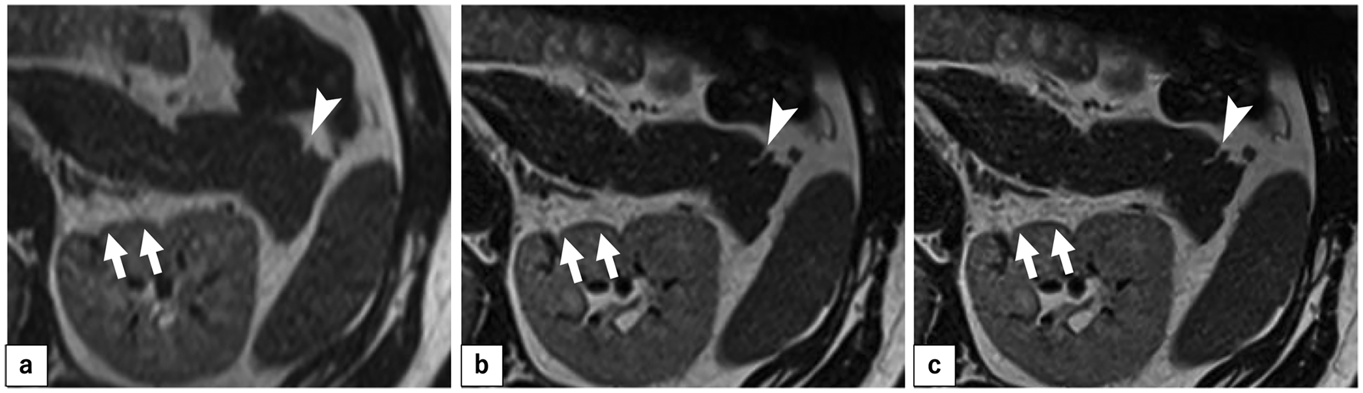
Imaging sequences under development. (a) HASTE. (b) 3S-TSE. (c) 3S-TSE-DLR. 3S-TSE (b) reduced blurring and delineated the margins of each organ more clearly compared with what was achieved with HASTE (a). When DLR is applied to 3S-TSE (c), the margins of organs are more clearly defined, fine structures are revealed, and noise is reduced compared with 3S-TSE alone (b). For example, the narrow fissures of the pancreas (white arrowheads) are invisible in HASTE (a), visible in 3S-TSE (b), and more clearly visible in 3S-TSE-DLR (c). Furthermore, the margin of the left kidney (white arrows) is noisy and irregular in HASTE (a), but it becomes sharper in 3S-TSE (b) and clearer with less noise in 3S-TSE-DLR (c). The low signal intensity region of the surface capsule is also clearer in 3S-TSE-DLR (c). 3S-TSE, three-shot turbo spin-echo; 3S-TSE-DLR, three-shot turbo spin-echo deep learning-based reconstruction; HASTE, half-Fourier acquisition single-shot turbo spin-echo

Signal suppression via a spatial saturation pulse was applied to the abdominal wall (square with broken white lines).
All participants underwent pancreatic imaging using the following four sequences (Fig. 3): 3S-TSE-DLR (the novel sequence), HASTE, fat-suppressed 3D T1-weighted (FS-3D-T1W) imaging, and MRCP. Detailed parameters are shown in Table 1, with the HASTE and FS-3D-T1W imaging sequences being modified from our institution's standard sequence to yield 2-mm slices. Thin slices are desirable for detailed evaluation of pancreatic morphology. To our knowledge, there were no guidelines for pancreatic slice thickness in MRI, but for tomographic images, the Japanese classification of pancreatic carcinoma (12) recommends obtaining CT slices of 2.5 mm or less. Since the thinnest slice in the practical range of 2D imaging on our 3-T scanner was 2 mm, we standardized the slice thickness to 2 mm for 3S-TSE-DLR, HASTE, and FS-3D-T1W imaging.
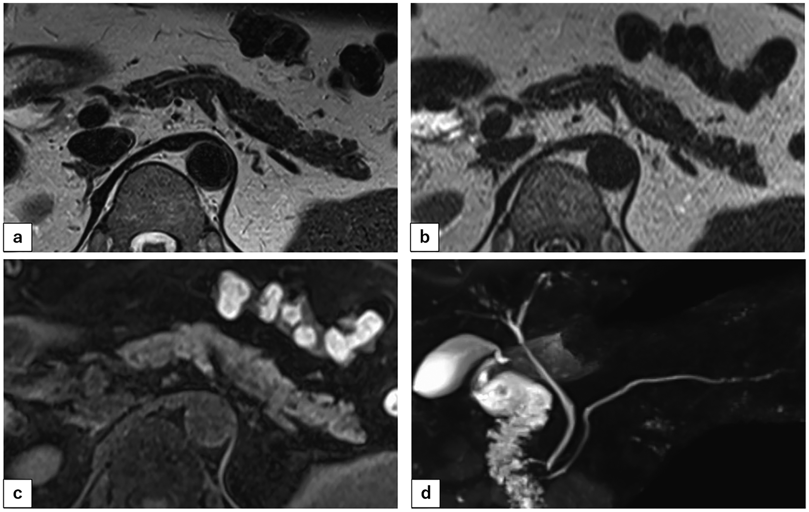
Comparison of images obtained using four different sequences. (a) 3S-TSE-DLR). (b) HASTE. (c) FS-3D-T1W imaging. (d) MRCP. 3S-TSE-DLR, three-shot turbo spin-echo deep learning-based reconstruction; FS-3D-T1W, fat-suppressed 3D T1-weighted; HASTE, half-Fourier acquisition single-shot turbo spin-echo; MRCP, magnetic resonance cholangiopancreatography.
Sequence parameters.

3S-TSE-DLR, three-shot turbo spin-echo with deep learning-based reconstruction; BH, breath-holding; CS, compressed sensing; FS-3D-T1W, fat-suppressed 3D T1-weighted; HASTE, half-Fourier acquisition single-shot turbo spin-echo; MRCP, magnetic resonance cholangiopancreatography; PACE, prospective acquisition correction; PAT, parallel acquisition technique; TE, echo time; TR, repetition time.
Image analysis
Two board-certified diagnostic radiologists (with 17 and 6 years of experience, respectively) evaluated the images. They independently scored the following items using a 5-point system: motion artifacts; sharpness of the pancreatic margin; the ability to identify the MPD using 3S-TSE-DLR, HASTE, and FS-3D-T1W imaging sequences; and the ability to identify the MPD using MRCP. For MRCP, maximum-intensity projection images were used for evaluation. Details of the evaluation methods are shown in Table 2. The scoring criteria were determined in consultation with two other diagnostic radiologists (with 20 and 10 years of experience, respectively), who scored the training sets for the 11 participants and presented and explained their scores to the evaluators. Figs. 4–6 show some of the training set images presented to the evaluators as examples illustrating how to score each item. The 2D imaging method produced strong artifacts in some slices; as shown in Table 2, the motion artifact score was 1–3 depending on the percentage of slices with strong artifacts. Margins were assessed on the basis of the score of the dominant slices, and if there were no dominant slices, the score was set to 1 less than the highest score.
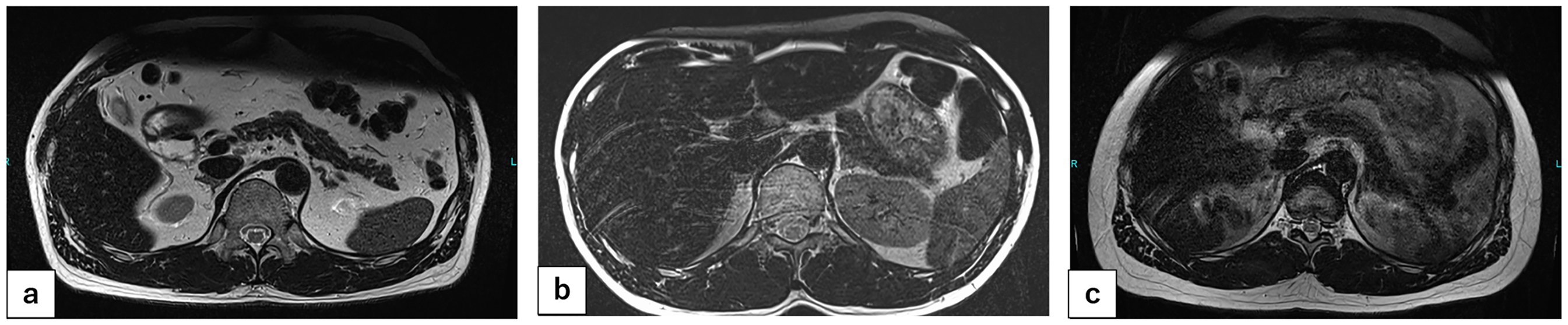
Example images from the training set used for scoring motion artifacts. 3S-TSE-DLR images are shown. (a) Score of 5 = no artifacts. (b) Score of 4 = slight artifacts. Artifacts were assessed according to the extent to which they affected evaluation of the pancreas. (c) Score of ≤3. Slices with artifacts that strongly affect pancreatic evaluation. Scores of 1–3 were awarded depending on the percentage of slices with strong artifacts. 3S-TSE-DLR, three-shot turbo spin-echo with deep learning-based reconstruction.
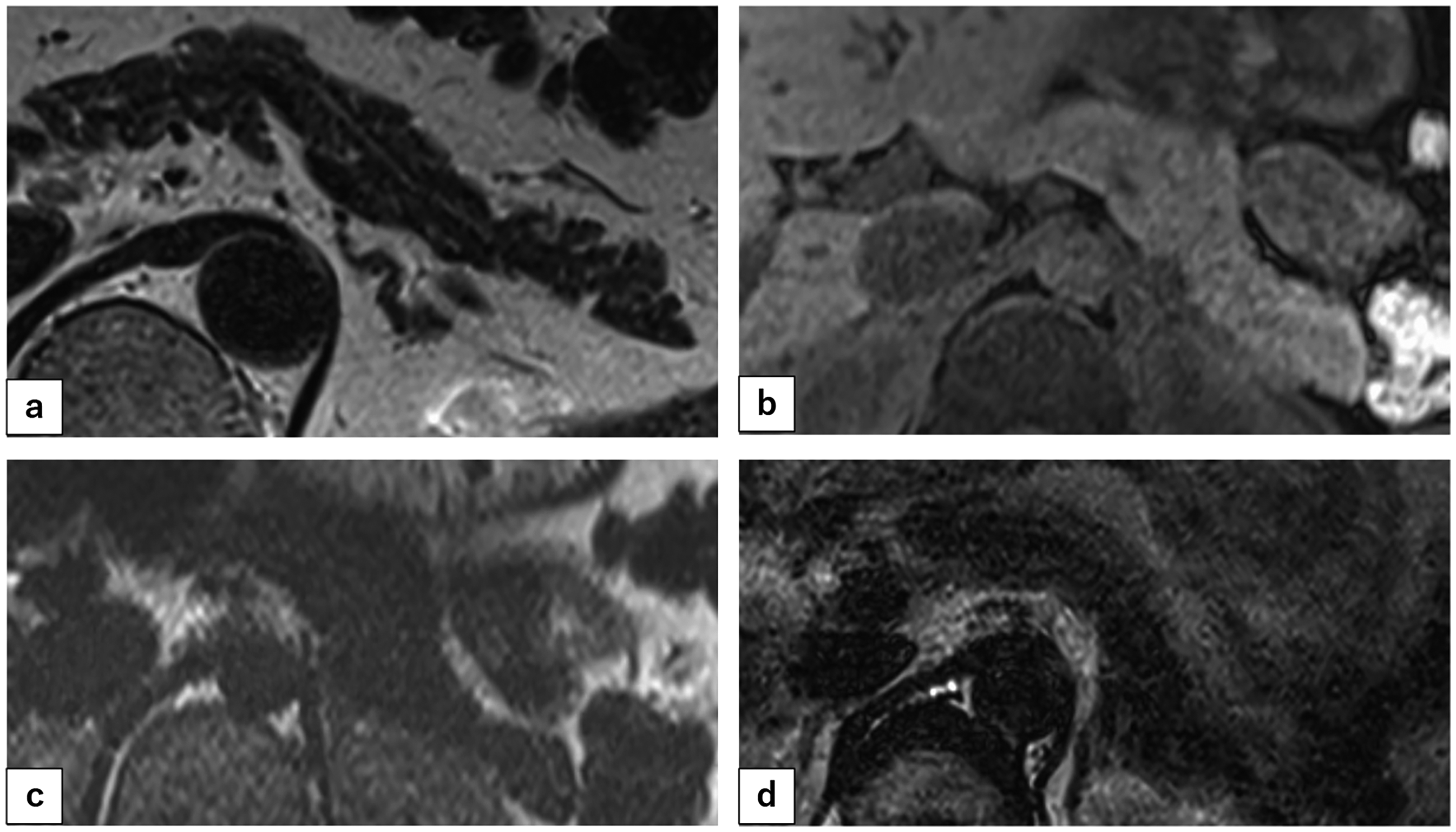
Example images from the training set used for scoring margin sharpness. (a) 3S-TSE-DLR image. Score of 5 = entire margin clearly delineated. Even the shape of the pancreatic surface is clear. (b) FS-3D-T1W image. Score of 4 = most of the margin is clearly delineated. The border is largely clear, with only mild blurring. (c) HASTE image. Score of 3 = sightly unclear. There is blurring and noise. (d) 3S-TSE-DLR image. Score of 2 = very unclear. There is strong blurring and noise. 3S-TSE-DLR, three-shot turbo spin-echo deep learning-based reconstruction; FS-3D-T1W, fat-suppressed 3D T1-weighted; HASTE, half-Fourier acquisition single-shot turbo spin-echo.

Example images from the training set used for scoring the conspicuity of the main pancreatic duct. (a) MRCP image. Score of 5 = clearly delineated in all segments. (b) 3S-TSE-DLR image. Score of 5. (c) FS-3D-T1W image. Score of 2 = very unclear. 3S-TSE-DLR, three-shot turbo spin-echo deep learning-based reconstruction; FS-3D-T1W, fat-suppressed 3D T1-weighted; MRCP, magnetic resonance cholangiopancreatography.
Detailed scoring arrangements.

Margin sharpness is judged by dominant slices. If there are no dominant slices, the score shall be minus 1 from the higher score.
MRCP images were scored only for conspicuity of the MPD.
MPD, main pancreatic duct; MRCP, magnetic resonance cholangiopancreatography.
Statistical analysis
All statistical analyses were performed using commercially available statistical software (SPSS Statistics version 29.0; IBM Corp., Armonk, NY, USA). Data are expressed as mean ± standard deviation. Friedman's ranking test was used to determine significant differences in scores between 3S-TSE-DLR and the other sequences. A P value <0.05 was considered statistically significant. Differences among conventional sequences (HASTE, FS-3D-T1W imaging, MRCP) were not analyzed because this was not the purpose of our study.
Cohen's weighted κ-coefficient was calculated to quantify the agreement between the two evaluators, as follows: <0 = no agreement; 0.00–0.20 = poor agreement; 0.21–0.40 = fair agreement; 0.41–0.60 = moderate agreement; 0.61–0.80 = substantial agreement; and 0.81–1.00 = almost perfect agreement.
Results
Scores for each item and evaluator, P values for the comparisons between 3S-TSE-DLR and other sequences, and Cohen's weighted κ-values are shown below and Table 3. The 3S-TSE-DLR artifact scores of both evaluators (4.1 ± 0.9 and 3.7 ± 0.8, κ = 0.448) were significantly higher than those for HASTE (3.6 ± 0.8 P = 0.009 and 3.2 ± 0.9; P = 0.006, κ = 0.262) and significantly lower than those for FS-3D-T1W imaging (4.8 ± 0.4; P <0.001 and 4.7 ± 0.5; P <0.001, κ = 0.359). The 3S-TSE-DLR margin sharpness scores of both evaluators (4.6 ± 0.5 and 4.7 ± 0.5, κ = 0.545) were significantly higher than those for HASTE (3.0 ± 0.2; P < 0.001 and 3.0 ± 0.0; P <0.001 [κ could not be calculated]) and FS-3D-T1W imaging (4.0 ± 0.0; P = 0.002 and 4.0 ± 0.0; P <0.001 [κ could not be calculated]). The 3S-TSE-DLR MPD identification scores of the two evaluators were 4.3 ± 1.1 and 4.1 ± 1.1 (κ = 0.544). The score for HASTE was significantly higher for one evaluator but not for the other evaluator (3.6 ± 1.3; P = 0.002 and 3.9 ± 1.2; P = 0.230, κ = 0.636). The 3S-TSE-DLR MPD identification scores of both evaluators were significantly higher than those for FS-3D-T1W imaging (2.2 ± 1.3; P < 0.001 and 2.4 ± 0.8; P < 0.001, κ = 0.430). By contrast, the 3S-TSE-DLR MPD identification scores were as high as those of MRCP (4.3 ± 1.1; P = 0.938 and 4.4 ± 1.1; P = 0.188, κ = 0.791).
Scoring results in two evaluators.

Values are given as mean ± standard deviation unless otherwise indicated. κ < 0, no; 0.00–0.20, poor; 0.21–0.40, fair; 0.41–0.60, moderate; 0.61–0.80, substantial; and 0.81–1.00, almost perfect.
P < 0.05 in Friedman's test.
Almost all cases were matched, so kappa could not be calculated.
3S-TSE-DLR, three-shot turbo spin-echo with deep learning-based reconstruction; FS-3D-T1W, fat-suppressed 3D T1-weighted; HASTE, half-Fourier acquisition single-shot turbo spin-echo; MPD, main pancreatic duct; MRCP, magnetic resonance cholangiopancreatography; N/A, not available.
Discussion
We developed a novel imaging sequence for the pancreas, 3S-TSE-DLR, and compared it with three conventional sequences. For this study, we used a qualitative evaluation method based on evaluator scoring. Although this method is subjective, we focused on how radiologists perceived these images obtained with different sequences of each parameter.
The motion artifact score for 3S-TSE-DLR was intermediate between those of FS-3D-T1W imaging and HASTE. On the basis of this score, we consider 3S-TSE-DLR to be as stable as existing pancreatic sequences in clinical practice. 3S-TSE-DLR can provide more stable images than HASTE, which uses the same T2W imaging system. One factor possibly contributing to the higher scores for 3S-TSE-DLR than HASTE is abdominal wall signal suppression, which should have been applied to HASTE; however, the purpose of this study was only comparison with the sequence that we normally use. The agreement between the two evaluators was poor for HASTE and FS-3D-T1W imaging, but the overall score trends were similar.
Margin sharpness, necessary for detailed assessment of pancreatic morphology, was significantly improved with 3S-TSE-DLR in this study. The agreement between the two evaluators was fair for 3S-TSE-DLR, and the HASTE and FS-3D-T1W imaging scores were in close agreement (although κ was not calculable). We expect 3S-TSE-DLR to capture localized atrophy of the pancreas more sensitively than conventional sequences. Although DLR is a major contributor to the increased resolution achieved herein, adjustments of the original pulse sequence are also considered important, as shown by the 3S-TSE-DLR development-stage images in Fig. 1. In addition, the purpose of this study was not to evaluate the effectiveness of DLR alone, but rather to evaluate the entire sequence created by incorporating DLR. There are reports of improved upper abdominal image quality using prototype or only denoising DLR methods for HASTE (and the single-shot fast spin-echo technique) (13–15). However, these were not available on our commercial systems at the time of the study, and one of our goals (i.e. to increase image resolution) was not likely to be achieved by denoising alone. 3S-TSE-DLR is now available for commercial scanners and DLR methods comparable to ours. Of course, future development of DLR methods for HASTE, 3D sequences, and diffusion-weighted images is expected to improve the overall image quality of the protocol (13–16).
The ability to detect the MPD was comparable between MRCP and 3S-TSE-DLR in this study, although the latter has the advantage of simultaneously evaluating pancreatic morphology at the site of MPD changes. Combined with the aforementioned margin sharpness results, we believe that 3S-TSE-DLR can evaluate pancreatic morphology in more detail than conventional sequences.
3S-TSE-DLR enables detailed evaluation of pancreatic morphology and MPD via 2-mm slices, and it also satisfies the recommended slice thickness (≤2.5 mm) for CT in the Japanese classification of pancreatic carcinoma (12). Comparison with contrast-enhanced CT is an issue for the future, but 3S-TSE-DLR has the advantage of not requiring radiation exposure or contrast administration. Because of these advantages over conventional MRI sequences and contrast-enhanced CT, 3S-TSE-DLR is expected to facilitate the follow-up of patients at risk for pancreatic cancer through high-frequency and detailed pancreatic evaluation.
The present study has some limitations. First, as mentioned above, the study design was based on the evaluator's subjective assessment. However, when comparing completely different sequences, we focused more on qualitative evaluations of how the images looked to the evaluator than on quantitative evaluations. Second, this is a study of healthy, young volunteers. Clinical research with patients is the next task. Third, our results were obtained using a single vendor's DLR technology. It is conceivable that MRI scanners from other vendors with similar technology could also be used, but confirming this was outside the scope of this study. It should be noted that 3S-TSE-DLR is not designed for research purposes, but rather for immediate use. Finally, only a 3-T scanner was used in this study, although we showed that 3S-TSE is feasible for 3-mm slices acquired using the 1.5-T MRI scanner (MAGNETOM Sola; Siemens Healthcare) at our institution.
In conclusion, 3S-TSE-DLR is expected to provide better image sharpness than conventional sequences, can identify MPD equally well or better than HASTE, and has identification performance comparable to that of MRCP. Therefore, 3S-TSE-DLR can be considered useful in the evaluation of pancreatic morphology.
Footnotes
Acknowledgments

Declaration of conflicting interests
The authors declared no potential conflicts of interest with respect to the research, authorship, and/or publication of this article.
Funding
The authors received no financial support for the research, authorship, and/or publication of this article.