
Abstract
The American alligator (Alligator mississippiensis) is a keystone species of the southeastern United States. In December of 2022, a free-ranging American alligator was found stuporous and tetraparetic. On postmortem evaluation, lesions were limited to the central nervous system, consisting of prominent perivascular cuffs of lymphocytes and histiocytes that extended into the surrounding neuroparenchyma along with gliosis. Next-generation sequencing of the affected brain identified the presence of a piscichuvirus closely related to the freshwater turtle neurovirus 1 (FTuNV-1) recently reported in an alligator snapping turtle with similar microscopic lesions. In situ hybridization using zz-probes that recognize FTuNV-1 identified widespread hybridization signal in neurons and glial cells in the alligator’s brain and spinal cord. This case represents only the second association of piscichuviruses with vertebrate disease. Moreover, it highlights the potential for disease transmission between different orders (Crocodylia and Testudines) of free-ranging aquatic reptiles that share similar habitats in the United States.
Keywords
Case Report
The American alligator (Alligator mississippiensis) is a keystone reptile species native to the southeastern United States, with ecological, agricultural, and cultural significance. In their natural habitat, which extends from Texas to the coast of North Carolina, free-ranging American alligators function as apex predators and significantly influence the structure, biodiversity, and health of wetland ecosystems. 15 Despite the critical role of American alligators in the environment and agriculture, disease knowledge of the species is limited, particularly regarding diseases of free-ranging alligators.
Nonsuppurative inflammation of the central nervous system (CNS) is uncommonly reported for all members of the order Crocodylia, comprising the families Alligatoridae (alligators and caiman), Crocodylidae (crocodiles), and Gavialidae (gharial and false gharial). Lymphoplasmacytic, histiocytic, and heterophilic meningoencephalitis has been reported in farmed A. mississippiensis naturally infected with West Nile virus in the southeastern United States.8,12 Moreover, encephalitis was reported in Australia in farmed saltwater crocodiles (Crocodylus porosus) naturally infected with crocodyline herpesvirus 2. 6 There is scant data on infectious diseases available for free-ranging crocodilians.
Chuvirids are negative-sense, single-stranded RNA viruses first identified in a 2015 arthropod viral biodiversity metagenomic study. 11 At present, the family Chuviridae contains 16 genera and 43 recognized chuvirid species. 7 Most chuvirids have been identified during metagenomic studies of invertebrates, and, as such, any associations with clinical disease were limited. Recently, however, novel chuvirids in the genus Piscichuvirus were linked to lymphoplasmacytic meningoencephalitis in free-ranging aquatic turtles. 10 Microscopic, genetic, and in situ hybridization analyses colocalized viral nucleic acids to CNS lesions in 2 marine turtles (Kemp’s ridley [Lepidochelys kempii] and loggerhead [Caretta caretta]) and a freshwater alligator snapping turtle (Macrochelys sp.), providing the first definitive evidence that chuvirids are capable of causing clinical disease in vertebrates.
In December of 2022, a free-ranging, 18 kg, 2-m long, subadult, male American alligator (A. mississippiensis) in Clay County, Florida (30.164474° N, −81.751952° W) was identified as a nuisance animal due to inappropriate behavior (remaining in a public location for approximately 2 days without moving). An astute and experienced trapper recognized the alligator’s unique, abnormal behavior, and the animal was transferred alive for clinical examination. General and neurologic examinations identified severe stupor, tetraparesis, and absent proprioception. Lesion localization of either the brainstem or diffusely intracranial was favored. Given the severity of the neurologic disease and poor prognosis, the alligator was euthanized and immediately necropsied.
The alligator exhibited adequate muscling but a markedly atrophied coelomic fat body on gross examination. No significant gross lesions were observed. Representative samples of all visceral organs were obtained, fixed in 10% neutral-buffered formalin, and processed routinely. The brain was removed intact, and representative segments of the spinal cord were collected; 2 brain samples were retained at −80°C, and all remaining brain and spinal cord were fixed and routinely formalin-fixed and processed.
Significant microscopic lesions were restricted to the CNS and present in all regions of the brain, including the olfactory tract, cerebral cortex, corpus striatum, dorsal ventricular ridge, thalamus, hypothalamus, midbrain, optic tectum, cerebellum, and medulla oblongata, as well as the spinal cord. Lesions primarily affected the gray matter but also extended into the white matter. Blood vessels were cuffed by up to 10 or more layers of lymphocytes, macrophages, fewer plasma cells, and rare granulocytes (Fig. 1). Similar inflammatory cells extended into the neuroparenchyma (Figs. 1a, b, c, e). Regions of inflammation were often accompanied by loose aggregates of swollen glial cells and/or dense nodules of glial cells. Scattered neuronal cell bodies displayed central chromatolysis. Rare lymphocytes and histiocytes within perivascular cuffs and lymphocytes, histiocytes, and glial cells within the adjacent neuroparenchyma exhibited shrunken cytoplasm and condensed, pyknotic, or karyorrhectic nuclei. The leptomeninges were moderately expanded by a similar lymphohistiocytic infiltrate, and meningeal vessels exhibited lymphohistiocytic cuffs (Fig. 1a). In the spinal cord white matter, occasional axon sheaths were swollen with digestion chambers. Together, the microscopic changes were interpreted as severe lymphohistiocytic meningoencephalomyelitis.
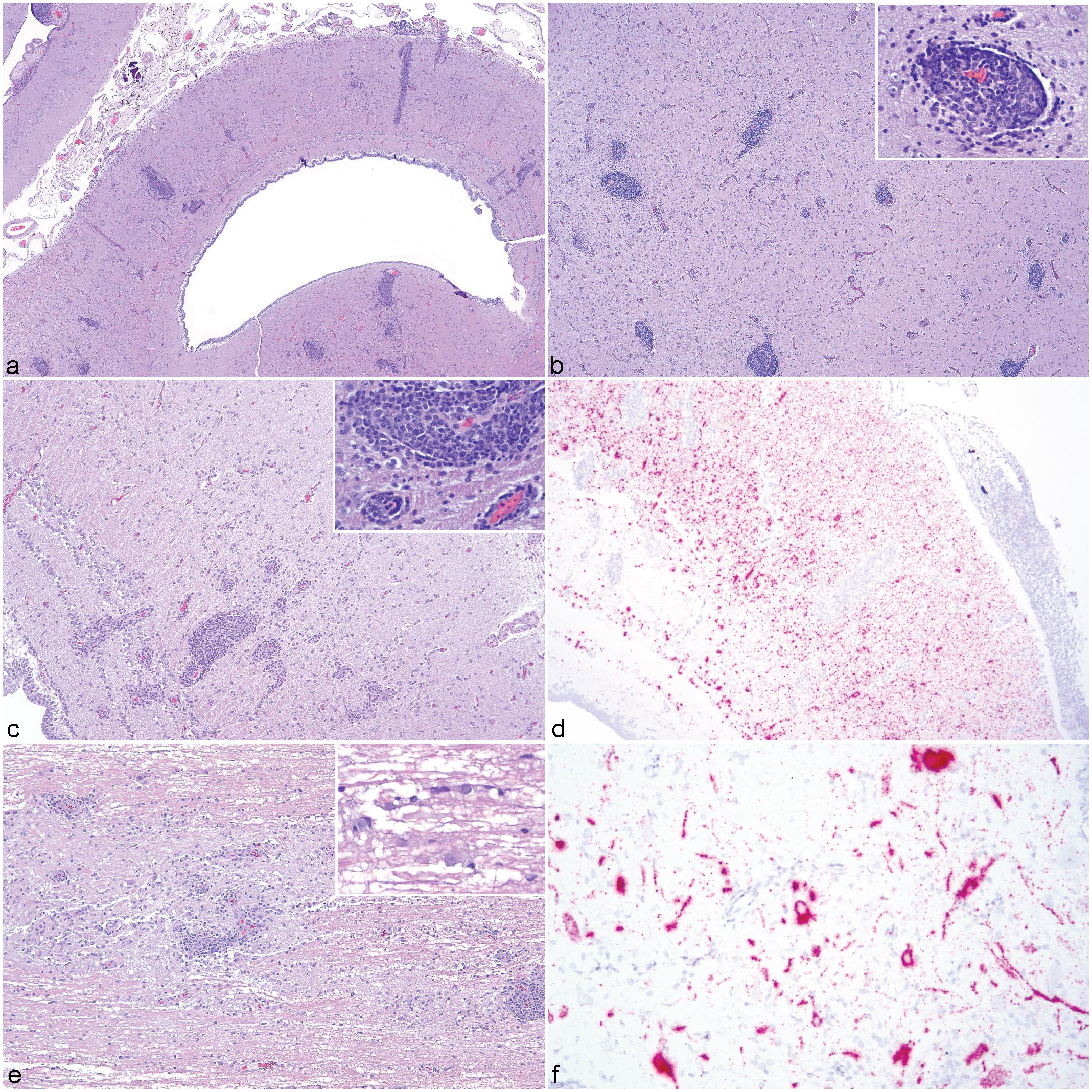
Meningoencephalomyelitis associated with a piscichuviral infection in a free-ranging American alligator (Alligator mississippiensis). (a) Brain, including midbrain, optic tectum, dorsal thalamus, cerebral cortex, and dorsal ventricular ridge. Prominent cuffs of primarily lymphocytes and histiocytes surround vessels throughout the section. Hematoxylin and eosin (HE). (b) Brain, dorsal ventricular ridge. Perivascular cuffs of lymphocytes and histiocytes (inset) are accompanied by widespread gliosis and scattered inflammation throughout the neuroparenchyma. HE. (c) Brain, optic tectum. Lymphocytes and histiocytes are the primary inflammatory infiltrates present within perivascular cuffs (inset) and the neuroparenchyma, accompanied by increased numbers of glial cells. HE. (d) Brain, optic tectum. Numerous cells within the neuroparenchyma, including neurons and glial cells exhibit strong, intracytoplasmic hybridization signal for piscichuviral RNA by in situ hybridization. Scant hybridization signal is present in inflammatory cells forming perivascular cuffs. (e) Spinal cord, gray and white matter. Perivascular cuffs, parenchymal inflammation, and gliosis, similar to that observed in the brain, are also present in the gray matter primarily, and to a lesser degree extending into the white matter. Occasional digestion chambers are present in the white matter (inset). HE. (f) Spinal cord, gray and white matter. Prominent neuronal cell bodies as well as glial cells predominantly in the gray matter exhibit strong, intracellular hybridization signal for piscichuviral RNA by in situ hybridization.
Based on the nature of the microscopic changes, next-generation sequencing was performed on a frozen brain sample to investigate a potential viral cause. RNA was extracted from the frozen brain sample using an RNeasy Mini kit (QIAGEN) according to the manufacturer’s protocol. Then, a cDNA library was generated using a NEBNext Ultra RNA Library Prep kit (New England Biolabs), and sequencing was performed on an Illumina MiSeq sequencer. A. mississippiensis host sequences (GenBank GCA_000281125.3) were removed from sequence data using Kraken v2.0. 16 De novo assembly of the remaining paired-end reads was performed using SPAdes v3.15.3. 1 The assembled contigs were then subjected to BLASTX searches against the NCBI non-redundant protein database using OmicsBox v1.2 (BioBam). De novo assembly and BLASTX analysis resulted in a 10,814 bp genome predicted to encode 4 open-reading frames (Genbank accession PQ373863). Each open-reading frame showed the highest nucleotide identity (92.3%–97.2%; Table 1) to a piscichuvirus from a free-ranging alligator snapping turtle (Genbank OQ547744). Large protein gene (L) amino acid sequences from 48 Chuviridae members were aligned with the alligator virus using MAFFT within Geneious Prime v2022.2.2. 9 A maximum likelihood phylogenetic tree was constructed in IQ-TREE with 1000 bootstraps, 13 generating a well-supported tree that identified the discovered alligator virus as a sister virus to the alligator snapping turtle freshwater turtle neural virus 1 (FTuNV1) isolate (Alligator snapping turtle/Florida/ST0994/2009) within the genus Piscichuvirus (Fig. 2).
Genome annotation of the piscichuvirus detected in the brain of an American alligator (Alligator mississippiensis; isolate alligator/Florida/N22-690/2022).
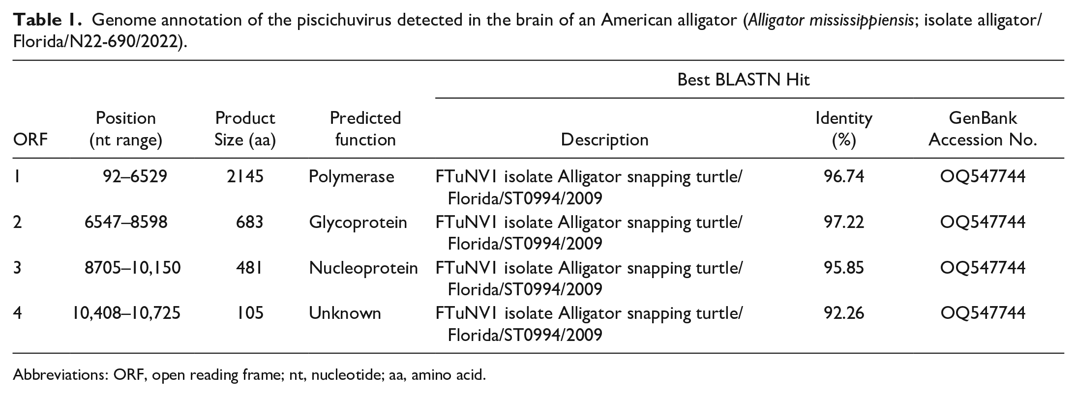
Abbreviations: ORF, open reading frame; nt, nucleotide; aa, amino acid.
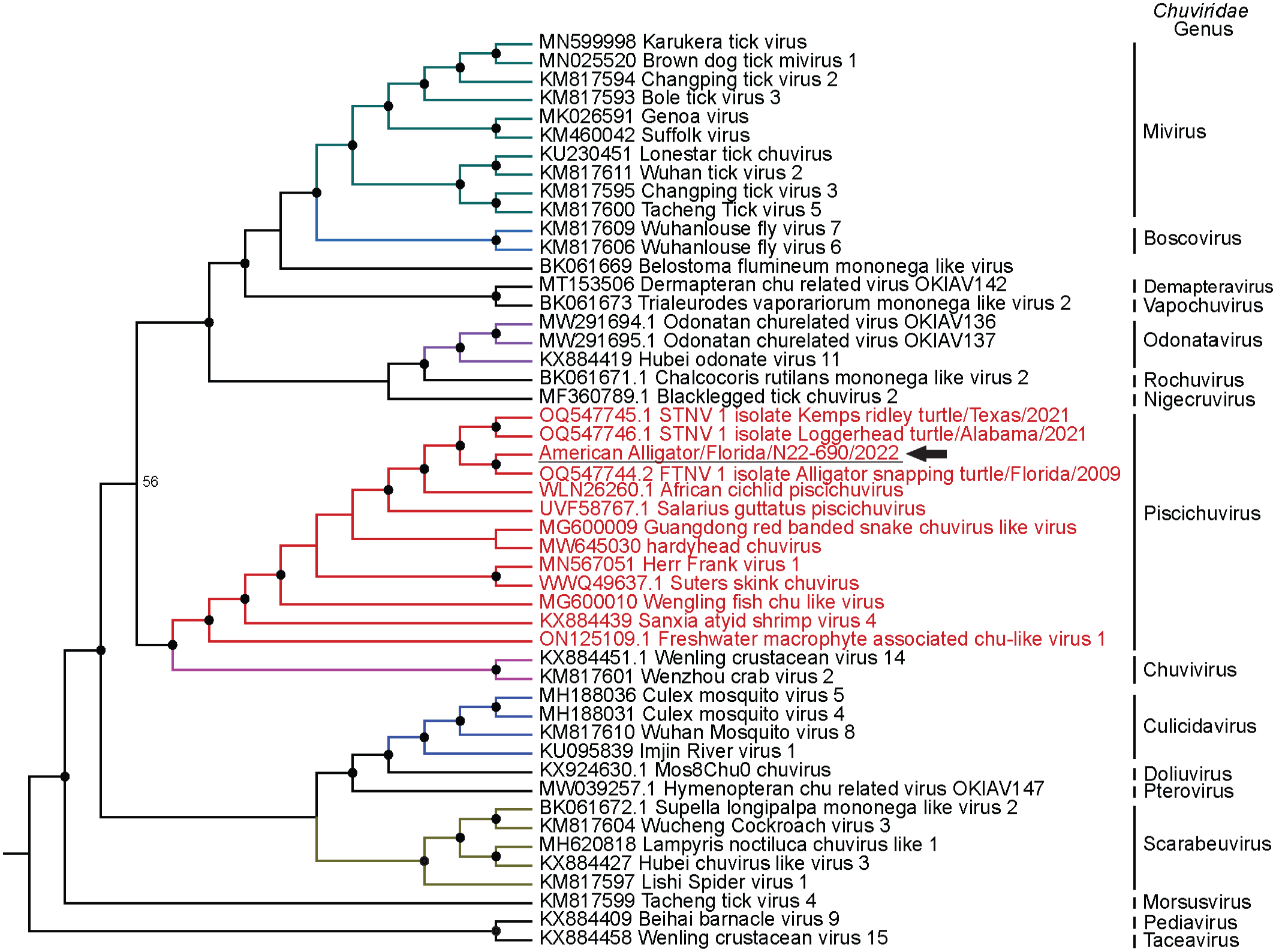
Maximum likelihood cladogram depicting the relationship of the piscichuvirus detected in an American alligator with meningoencephalomyelitis (indicated by a black arrow) to 48 members of the family Chuviridae based on the amino acid alignment of large protein gene (L). Nodes with black circles are supported by bootstrap values ≥80%. STNV = sea turtle neural virus; FTNV = freshwater turtle neural virus.
To screen for piscichuviral RNA in brain and spinal cord lesions, RNAscope in situ hybridization was performed. Based on the high nucleotide similarity of FTuNV1 to the alligator piscichuvirus, zz-probes designed against the large protein gene of FTuNV1 were used in the RNAscope 2.5 HD in situ hybridization (Advanced Cell Diagnostics, Inc.) as previously described. 10 In situ hybridization was performed on CNS sections of the neurologic alligator, an alligator with no CNS disease (negative biological control), and an alligator snapping turtle with FTuNV1-associated meningoencephalomyelitis (positive biological control). A Bacillus subtilis dihydrodipicolinate reductase gene probe was also used (negative probe control). Widespread, strong, intracytoplasmic hybridization signal for piscichuviral viral RNA was detected in neurons, glial cells, and occasionally ependymal cells of the brain and spinal cord of the alligator (Figs. 1d, f). In the brain, viral RNA was detected adjacent to regions of inflammation and scattered throughout the neuroparenchyma, predominantly in the gray matter (Fig. 1d). In the spinal cord, hybridization signal was also most prominent in the gray matter (especially neuronal cell bodies) and sparsely in the white matter (Fig. 1f). No hybridization signal was detected in sections of brain from an unaffected American alligator (data not shown). The hybridization signal for the positive biological control and the absence of signal for the negative probe control were appropriate (data not shown).
This report describes the first documentation of piscichuvirus-associated CNS disease in a crocodilian (free-ranging American alligator) and the second report to identify piscichuviral disease in free-ranging reptiles. To date, reptiles are the only vertebrates where piscichuviral nucleic acids have been co-localized to diseased tissues, though this is likely to change as these viruses receive further study. Both clinical signs and microscopic lesions in the marine and freshwater turtles previously reported, 10 and the alligator in this case were similar. All animals exhibited antemortem signs consistent with CNS disease, and inflammatory changes were restricted to the CNS and characterized by severe lymphoplasmacytic (marine and freshwater turtle cases) or lymphohistiocytic (American alligator case) meningoencephalomyelitis with thick and prominent perivascular cuffs.
While the CNS lesions in this alligator were striking and suggestive of a viral cause, the pattern and nature of the CNS inflammation observed in this crocodilian were similar to those seen in farmed American alligators from the southeastern United States with natural West Nile virus infections and farmed saltwater crocodiles in Australia naturally infected with crocodyline herpesvirus 2.6,8,12 In contrast, Providencia rettgeri CNS infections in farmed American alligators are predominantly characterized by granulocytic infiltrates. 3 Although lymphohistiocytic infiltrates and prominent perivascular cuffs in the CNS of crocodilians may be useful to prioritize a viral cause, such changes likely represent a common response to multiple viral causes requiring molecular diagnostic modalities for definitive diagnosis.
Marine turtle neuroviruses (STuNVs) have been detected in a Kemp’s ridley turtle stranded off the coast of Texas in 2021 and a loggerhead turtle stranded off the coast of Alabama in 2021. Based on species demarcation criteria approved by the International Committee on Taxonomy of Viruses, 4 the previously reported marine (STuNVs) and freshwater turtle (FTuNV) viruses were considered to represent a single viral species. 10 However, the L protein amino acid sequences of the freshwater and marine turtle viruses were different (91.6% amino acid identity) and only slightly greater than the suggested species demarcation (<90%). 4 Perhaps more importantly, ecologically the assignment of those viruses as a single species did not fit with the non-overlapping ecosystems of their hosts. Given the identification of highly similar viruses in an American alligator and an alligator snapping turtle, which inhabit similar freshwater aquatic ecosystems, the viruses found in these 2 hosts likely represent a single viral species. Additional viral sequences and characterization are needed; however, we suspect the freshwater piscichuviruses represent a different species than the marine turtle viruses. Interestingly, the neurologic alligator in this report was just a little over 100 km to the northeast of the alligator snapping turtle, but 13 years later. This finding warrants additional study on the potential impacts of this virus on reptile inhabitants of the wetlands of north Florida.
A unique aspect of this virus is its apparent ability to infect different orders of aquatic reptiles (Crocodilia and Testudines). Few viruses are capable of such broad infectivity, and even fewer are known in aquatic ecosystems. One notable example is frog virus 3 (Ranavirus rana1), which can infect select species of fish, reptiles, and amphibians. 5 Ranaviruses can be transmitted through several methods, including water. 2 Further research will be necessary to elucidate piscichuvirus transmission methods and reservoirs. At a minimum, both transmission by water and ingestion should be considered, as turtles are a routine food item of Florida alligators. 14
This report documents a natural piscichuvirus infection in a free-ranging American alligator exhibiting neurologic signs associated with severe lymphohistiocytic meningoencephalomyelitis. The detected piscichuvirus exhibits a high nucleotide identity with FTuNV1, reported to cause CNS disease in aquatic turtles, and represents only the second documentation of piscichuviral disease in vertebrates. Much remains to be learned of these viruses, but reported cases affirm the inclusion of piscichuviruses as a differential cause of neurologic disease in free-ranging or captive turtles and crocodilians.
Footnotes
Acknowledgements
The authors would like to thank Michael Haley for his incredible astuteness and assessment of the health of this alligator and the need to have it examined by veterinarians, James F.X. Wellehan, Jr. and members of the Zoological Medicine and Neurology Services at the Small Animal Hospital at University of Florida’s College of Veterinary Medicine for examination and euthanasia of this alligator, the staff of the Histology Laboratory at University of Florida’s College of Veterinary Medicine for slide preparation, Steven Tillis for virus isolation attempts, and the Florida Fish and Wildlife Conservation Commission for connecting Mr. Haley with us.
Declaration of Conflicting Interests
The author(s) declared no potential conflicts of interest with respect to the research, authorship, and/or publication of this article.
Funding
The author(s) received no financial support for the research, authorship, and/or publication of this article.